책임저자:이종욱, 서울시 영등포구 영등포동 94-200
150-719, 한림대학교 한강성심병원 성형외과 Tel: 02-2639-5780, Fax: 02-2676-2431
E-mail: [email protected]
Kihyun Cho, M.D., Jongwook Lee, M.D., Janghyu Ko, M.D., Dongkook Seo, M.D., Jaikoo Choi, M.D. and Youngchul Jang, M.D.
Department of Plastic and Reconstructive Surgery, Hangang Sacred Heart Hospital, Hallym University College of Medicine, Seoul, Korea
Purpose: Nowadays importance of growth factors in wound
healing is being focused. Wound healing can be accelerated by various growth factors. Wound healing cascade consists of inflammatory, proliferative, and remodeling phases. Basic fibroblast growth factor (bFGF) helps proliferation of fibro- blast and promotes angiogenesis and formation of gran- ulation tissue through proliferative phase. We investigated the effect of recombinant basic fibroblast growth factor FiblastⓇ (Kaken Pharmaceutical, Japan) on second degree burns.Methods: 57 patients from July 2009 to September 2009
with second degree burn were treated with bFGF. Average age, sex, cause of burn, depth of burn, location of wound, ep- ithelization period and number of operation were studied.Recombinant bFGF was used with spraying. The bFGT was sprayed and wait for 30 seconds and then foam dressing was applied to wounds. The bFGF administration continued until the wound healed.
Results: The average healing time in the bFGF-treated
group was 8.4±2.2 days (4∼14 days). Among 57 patients, 19 patients had superficial second degree burn and the aver- age healing time in the bFGF-treated group was 7.2±1.5 days (4∼9 days), 30 patients had deep second degree burn and the average healing time in the bFGF-treated group was 11.2±1.7 days (9∼14 days). 20 patients had deep second degree burn and were clinically considered to get operation during hospital course but eventually 8 of patients (40%) with deep second degree burn treated with bFGF underwent operation.(Journal of Korean Burn Society 2009;12:115-120) Key Words: Biologic dressing, Basic fibroblast growth factor,
Second degree burn, Growth factor
서 론
창상 치유에서 성장인자를 이용한 드레싱의 중요성이 인 식되면서 이를 이용한 창상 치료가 활발히 이루어지고 있 다. 창상 치유과정은 크게 염증기, 증식기, 재구축기라는 3 가지 단계로 나눌 수 있다
1). 손상된 창상에 즉시 지혈이 되 고, 호중구, 대식세포, 림프구들의 염증세포가 창상 부위로 이동한다. 염증기에는 이들 염증세포에 의해 세균이나 이 물질의 제거가 이루어지며 이와 함께 세포증식인자도 방출 되어 증식기로 이행한다
2). 증식기에 중심적인 역할을 하는 염기성 섬유아세포증식인자(Basic Fibroblast growth fac- tor, bFGF)는 섬유아세포구의 증식과 함께 혈관신생 촉진 및 육아조직 형성을 돕는다
3-5). 재구축기에 육아조직은 콜 라겐섬유나 탄성섬유로 바뀌게 된다.
Fiblast
Ⓡ(Kaken Pharmaceutical, Japan)는 bFGF를 유전 학적으로 재조합한 스프레이 형태로 분무할 수 있는 제재 로 보관과 사용이 용이하며, 또한 환자에 대한 통증이 거의 없는 창상 치유를 촉진하는 성장인자를 이용한 치료제다.
저자들은 Fiblast
Ⓡ를 이용하여 창상치료의 상피화 기간 을 측정하고, 심재성 2도 환자에서 수술을 얼마나 감소시키 는지에 대해 조사했다.
대상 및 방법
1. 재료
염기성 섬유아세포증식인자(Basic Fibroblast growth
factor, bFGF)는 섬유아세포구의 증식과 함께 혈관신생 촉
진 및 육아조직 형성을 돕는다. Fiblast
Ⓡ는 일본 카켄제약에
116
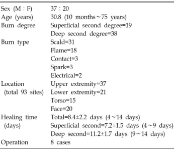
대한화상학회지 Vol. 12, No. 2, 2009Table 1. Patient Profiles
Sex (M:F) 37:20
Age (years) 30.8 (10 months∼75 years) Burn degree Superficial second degree=19
Deep second degree=38 Burn type Scald=31
Flame=18 Contact=3 Spark=3 Electrical=2 Location Upper extremity=37 (total 93 sites) Lower extremity=21
Torso=15 Face=20
Healing time Total=8.4±2.2 days (4∼14 days)
(days) Superficial second=7.2±1.5 days (4∼9 days) Deep second=11.2±1.7 days (9∼14 days) Operation 8 cases
Fig. 1. Fiblast is spray type wound healing product.
서 개발한 세계 최초의 bFGF (섬유아세포 성장인자)제제 로, 섬유아세포의 증식 및 새로운 혈관 생성을 도와 창상 부위의 표피형성 및 치유를 촉진하는 치료제이다(Fig. 1).
2. 대상
2009년 6월부터 2009년 8월까지 2도 화상을 수상한 57명 의 환자를 대상으로 임상적으로 물집이 벗겨졌을 때 혈액 순환이 좋은 진한 분홍색 진피가 노출된 19명을 표재성 2 도, 진피의 그물층 일부까지도 화상을 입어 진피의 색깔이 흰색을 띠거나 가피가 형성된 38명을 심재성 2도로 분류하 였다. 적용부위는 상지 37명, 하지 21명, 얼굴 20명, 몸통 15 명 순으로 많았으며, 57명의 환자 중 남자가 37명, 여자가 20명이었고, 연령분포는 10개월에서 75세(평균 30.8세)였다
(Table 1).
3. 방법
수상 후 4일 이내에 Fiblast
Ⓡ를 적용했으며 2일마다 폼 드레싱을 시행했다. 드레싱 시에는 창상면에 5 cm 정도 떨 어진 거리에서 5회 정도 분무하였으며 30초 정도 기다린 다 음 hydrocolloid나 폼 제재를 이용하여 창상면을 덮었다.
결 과
수술을 받은 8명을 제외한 57명의 환자 중 49예에서 모두 특별한 합병증 없이 Fiblast
Ⓡ를 적용 후 4일에서 14일(평균 8.4±2.2일)사이에 상피화 되었다(Fig. 2, 3). 표재성 2도 화상 인 19명의 환자에서 Fiblast
Ⓡ를 적용한 후 4일에서 9일(평균 7.2±1.5일) 사이에 상피화 되었으며, 심재성 2도 화상에서 수술을 받은 8명을 제외한 30명 환자에서 9일에서 14일(평 균 11.2±1.7일)사이에 상피화 되었다. 임상적으로 초기에 수 술이 필요하다고 판단된 심재성 2도 화상의 20예에서 Fiblast
Ⓡ를 이용하여 드레싱한 결과 최종적으로 8예(40%) 에서만 수술을 시행했으며 이들은 초기에 가피가 형성된 심재성 2도 화상이었다(Fig. 4∼6).
고 찰
성장인자를 이용한 드레싱은 통증완화, 감염방지, 습윤
환경 유지, 창상 치유 촉진 등의 여러 장점이 있다. 최근의
연구에 의하면 외인적으로 rhGH (recombinant human
growth hormone), EGF, FGF를 투약하여 치료할 경우에 조
직의 수복에 중요한 영향을 미치는 것으로 나타났다
6,7).
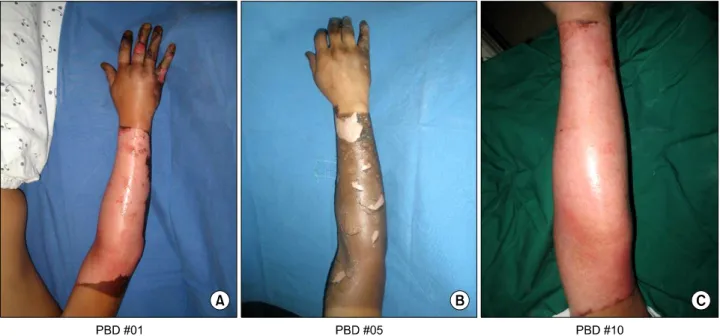
Fig. 2. Patient 1. (A) A 23-month old baby presented with deep second degree scalding burn. (B) There is deep dermis exposure at hand dorsum and 2, 3rd finger. (C) With FiblastⓇ and foam dressing wound is epithelized at 10th day.
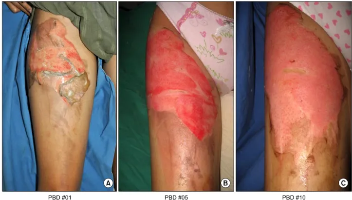
Fig. 3. Patient 2. (A) A 16 year old female presented with deep second degree scalding burn at right thigh. (B) There is deep dermis exposure at thigh. (C) With FiblastⓇ and foam dressing wound is epithelized at 10th day.
118
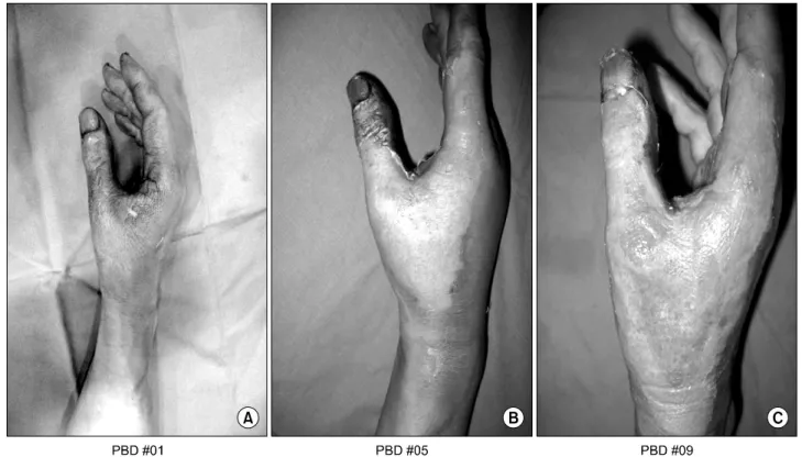
대한화상학회지 Vol. 12, No. 2, 2009Fig. 4. Patient 3. (A) A 54 year old male presented with deep second degree spark burn at right thigh. (B) There is dermis exposure right hand. (C) With FiblastⓇ and foam dressing wound is epithelized at 9th day.
Fig. 5. Patient 4. (A) A 37 year old male presented with deep second degree spark burn at right hand. (B) There is dermis exposure and eschar formation at right hand. (C) With FiblastⓇ and foam dressing wound is epithelized at 9th day.
FGF는 혈관 신생, 상처 치유, 태생기 발달(embryonic de- velopment)에 중요한 역할을 하며 다양한 세포와 조직의 발생과 분화에 중요한 역할을 한다. FGF는 acidic fibroblast
growth factor (FGF1)과 basic fibroblast growth factor
(FGF2, bFGF)로 분리된다. 이들은 내피세포의 증식에 관여
하며 내피세포를 원통 모양으로 배열시켜 기존의 혈관에서
Fig. 6. Patient 5. (A) A 42 year old male presented with deep second degree flame burn at right hand. (B) There is dermis exposure and eschar formation at right hand. (C) With FiblastⓇ and foam dressing wound is epithelized at 10th day.
새로운 혈관이 생성되는 것을 도와준다. FGF1과 FGF2는 vascular endothelial growth factor (VEGF)나 platelet-deri- ved growth factor (PDGF)보다 더욱 강력한 혈관 신생 작 용을 한다. bFGF는 상피화에 관여하며 콜라겐 축적을 촉진 시킨다
8-10).
현재 bFGF를 이용한 연구 결과를 비교할 문헌이 없는 상 태지만 Fu 등
6)이 발표한 상피화 기간인 표재성 2도 화상의 12.4±2.7일, 심재성 2도 화상의 21.2±4.9일과 비교하면 표재 성 2도 화상에서는 5일, 심재성 2도 화상에서는 10일 이상 단축되어 환자의 치료기간을 줄일 수 있었다. 치료 도중에 임상적으로 수술이 필요하다고 판단된 20예에서 Fiblast
Ⓡ를 이용하여 드레싱한 결과 8예에서만 수술이 필요했으며 성장인자를 이용한 드레싱이 수술을 줄이는 데 중요한 역 할을 한다고 판단할 수 있었다.
Fiblast
Ⓡ를 bFGF가 정확하게 어떠한 기전을 통해서 상처 치유를 도와주는지 완전히 밝혀지진 않았다. bFGF는 mes- senger RNA, DNA, 단백질 합성을 자극하여 fibroblast, vascular endothelial cell, keratinocyte의 분열을 촉진하고 granulation tissue와 epidermal regeneration을 촉진시킨다
11). 외인성 bFGF는 EGF, TGF와 같은 다른 성장의 합성을 돕 고, 이러한 성장인자가 macrophage, platelet을 상처 조직 에 불러들이는 것을 간접적으로 촉진시킨다. 이러한 연구 결과를 통해 FGF, EGF, GH 등의 성장 인자들과 그들의 수 용체의 상호작용은 상처 치유를 돕거나 지연시키는 데 중 요한 역할을 할 것으로 판단할 수 있다.
결 론
성장인자를 이용한 Fiblast
Ⓡspray는 스프레이 형태로 분 무할 수 있는 제재로 보관과 사용이 용이하며, 또한 환자에 대한 통증이 거의 없는 창상 치유를 촉진하는 성장인자를 이용한 치료제다.
본원에서는 2도 화상에 Fiblast
Ⓡ를 적용한 결과 상피화 기간을 단축되었고, 수술이 필요한 경우를 줄이고 환자의 치료기간을 줄일 수 있었다.
REFERENCES
1) Engrav LH, Heimbach DM, Reus JL, Harnar TJ, Marvin JA.
Early excision and grafting vs. non-operative treatment of burns of interminant depth: a randomized prospective study.
J Trauma. 1983;23:1001.
2) Iwashita N, Muramatsu H, Toriyama K, Torii S, Muramatsu T. Expression of midkine in normal and burn sites of rat skin.
Burns. 1999;25:119.
3) Galeano M, Altavilla D, Bitto A, et al. Recombinant human erythropoietin improves angiogenesis and wound healing in experimental burn wounds. Crit Care Med. 2006;34:1139.
4) Kibe Y, Takenaka H, Kishimoto S. Spatial and temporal expression of basic fibroblast growth factor protein during wound healing of rat skin. Br J Dermatol. 2000;143:720.
5) Gibran NS, Isik FF, Heimbach DM, Gordon D. Basic fibroblast growth factor in the early human burn wound. J Surg Res.
1994;56:226.
120
대한화상학회지 Vol. 12, No. 2, 20096) Fu X, Shen Z, Chen Y, et al. Randomised placebo-controlled trial of use of topical recombinant bovine basic fibroblast growth factor for second-degree burns. Lancet. 1998;352:1661.
7) Fujii T. Local treatment for extensive deep dermal thickness burn and follow-up study. Acta Chir Plast. 1990;32:46.
8) Steiling H, Werner S. Fibroblast growth factors: key players in epithelial morphogenesis, repair and cytoprotection. Curr Opin Biotechnol. 2003;14:533.
9) Akita S, Akino K, Imaizumi T, Hirano A. A basic fibroblast growth factor improved the quality of skin grafting in burn
patients. Burns. 2005;31:855.
10) Harrison CA, Gossiel F, Bullock AJ, Sun T, Blumsohn A, MacNeil S. Investigation of kertinocyte regulation of collagen I synthesis by dermal fibroblasts in a simple in vitro model.
Br J Dermatol. 2006;154:401.
11) Pereira CT, Herndon DN, Rocker R, Jeschke MG. Liposomal gene transfer of keratinocyte growth factor improves wound healing by altering growth factor and collagen expression. J Surg Res. 2007;139:222.