INTRODUCTION
Coronary artery disease is usually caused by atherosclerosis and associated with traditional risk factors such as smoking, hypertension, diabetes mellitus, aging, and hyperlipidemia (1, 2). Besides these traditional risk factors, recently, several newer risk factors and surrogate markers for the early detec- tion of atherosclerosis including the measurement of endothe- lial dysfunction, arterial wall structure, arterial stiffness, and serologic markers of vascular inflammation were proposed and widely had been studied (3, 4).
Endothelial dysfunction involves every stages of the pro- gression of atherosclerosis (5, 6) and is non-invasively mea- sured by flow-mediated vasodilation (FMD) of the brachial artery (7). Arterial stiffness is now an established cardiovas- cular risk factor and one of the best prognostic indicators for future events in hypertensive population. Pulse wave veloci- ty (PWV) is a simple, reproducible, and non-invasive mea- surement which can be used as a valuable index of both arte- rial stiffness and atherosclerosis in large populations (8). Recent studies have been demonstrated that high sensitivity C-reac- tive protein (hsCRP) and microalbuminuria were significant- ly associated with the presence or severity of atherosclerotic
cardiovascular diseases and could be used as simple surrogates of atherosclerosis (9-12).
In the previous studies, some kinds of drugs, although they had individual differences, such as angiotensin converting en- zyme inhibitor (ACEI), angiotensin receptor blocker, or cal- cium channel blocker (CCB) could improve endothelial func- tion and arterial stiffness, and decrease the level of hsCRP and microalbuminuria (13, 14). However, the effects of CCB mo- no-therapy and CCB plus ACEI combination therapy on these surrogate markers in patients with angina pectoris were poor- ly evaluated.
Therefore, the aim of this study was to evaluate the effects of CCB and ACEI on surrogate markers of atherosclerosis including endothelial function, arterial stiffness, urinary albu- min excretion (UAE), and hsCRP in patients with angina pectoris.
MATERIALS AND METHODS Study design
The study is a single-center, randomized, open clinical trial
223
Kye Hun Kim, Myung Ho Jeong, Sook Hee Cho, Jae Youn Moon, Young Joon Hong, Hyung Wook Park, Ju Han Kim, Youngkeun Ahn, Jeong Gwan Cho, Jong Chun Park, and Jung Chaee Kang
The Heart Center of Chonnam National University Hospital, Gwangju, Korea
Address for correspondence Myung Ho Jeong, M.D.
Heart Center of Chonnam National University Hospital, 8 Hak-dong, Dong-gu, Gwangju 501-757, Korea Tel : +82.62-220-6243, Fax : +82.62-227-3105 E-mail : [email protected]
DOI: 10.3346/jkms.2009.24.2.223
Clinical Effects of Calcium Channel Blocker and Angiotensin Converting Enzyme Inhibitor on Endothelial Function and Arterial Stiffness in Patients with Angina Pectoris
To evaluate the effects of calcium channel blocker (CCB) and angiotensin convert- ing enzyme inhibitor (ACEI) on endothelial function and arterial stiffness in stable angina pectoris (SAP), 87 patients with SAP (57.6±10.0 yr, 52 males) were divid- ed into two groups; CCB group (group I: n=44, 57.9±9.7 yr, 23 males) vs. CCB plus ACEI group (group II: n=43, 57.2±10.5 yr, 29 males). Flow mediated vasodilation (FMD) of the brachial artery, pulse wave velocity (PWV), urinary albumin excretion (UAE), and high sensitivity C-reactive protein (hsCRP) were compared. FMD, PWV, UAE, and hsCRP were not different between the groups at baseline. After 6 months of treatment, FMD were significantly improved in group II (7.5±3.7 to 8.8±2.7%, p<0.001), but not in group I (7.9±2.7 to 8.2±2.8%, p=0.535). Brachial-ankle PWV were significantly improved in both groups (1,621.3±279.4 to 1,512.1±225.0 cm/
sec in group I, p<0.001, 1,586.8±278.5 to 1,434.5±200.5 cm/sec in group II, p<
0.001). However, heart-femoral PWV were significantly improved (1,025.7±145.1 to 946.2±112.2 cm/sec, p<0.001) and UAE were significantly decreased (20.19±
29.92 to 13.03±16.42 mg/g Cr, p=0.019) in group II only. In conclusion, combina- tion therapy with CCB and ACEI improves endothelial function, arterial stiffness, and UAE than CCB mono-therapy more effectively in patients with SAP.
Key Words : Endothelial Function; Arterial Stiffness; Angina Pectoris
Received : 16 August 2007 Accepted : 7 July 2008
to evaluate the effects of CCB and ACEI on surrogate mark- ers of atherosclerosis including endothelial function, arterial stiffness, UAE, and hsCRP in patients with stable angina pectoris (SAP). The study protocol was approved by the Insti- tutional Review Board of Chonnam National University Hos- pital and informed consent was obtained from each patient.
Inclusion criteria were 1) patients with newly diagnosed SAP who underwent coronary angiography for the first time, 2) diameter stenosis more than 50% of target lesion, 3) success- ful, uncomplicated percutaneous coronary intervention with less than 10% residual stenosis, 4) able to give informed con- sent, 5) able to successful follow-up measurement of FMD, PWV, UAE, and hsCRP. Exclusion criteria included 1) prior history of coronary intervention, 2) blood pressure more than 160 mmHg in systole and 100 mmHg in diastole, 3) known intolerance to CCB or ACEI, 4) previous use of angiotensin receptor blocker, 5) known heart failure or ejection fraction
<50%, 6) renal insufficiency with creatinine >2.5 mg/dL, 7) need for warfarin anticoagulation, 8) known hepatic dys- function, 9) current participation in another randomized trial, 10) major life threatening illness, 11) patients refusal during clinical follow-up or incomplete clinical follow-up.
Study subjects
From September 2004 to April 2006, a total of 92 patients with SAP were enrolled initially. Among them, 5 patients were excluded owing to the adverse events (intractable cough:
2 patients in group II, headache: 1 patient in group II, facial flushing: 1 patient in group I, follow-up loss: 1 patient in group I). Although dry cough was developed in 6 patient of group II, the symptom was not severe and tolerable in 4 pa- tients and thus the study was continued except for 2 patients who complained intractable cough. Therefore, a total of 87 patients (57.6±10.0 yr, 52 males) were finished this study and analyzed. The patients were divided into two groups ac- cording to the treatment modalities with 1 to 1 fashion; CCB (cilnidipine 10 mg per day) group (group I: n=44, 57.9± 9.7 yr, 23 males) and CCB (cilnidipine 10 mg per day) plus ACEI (captopril 25 mg per day) group (group II: n=43, 57.2
±10.5 yr, 29 males). FMD, PWV, UAE, and hsCRP were compared at baseline and 6 months after treatment. All param- eters were measured early in the morning following overnight fasting more than 12 hr. Vasoactive medications including long-acting nitrates or intravenous nitrates or CCB or ACEI were withheld and not prescribed for at least 24 hr until the baseline measurements were completed. Only short-acting sublingual nitroglycerin for control of angina and anti-platelet agents were permitted. The doses of CCB or ACEI were not changed during the study periods. Follow-up measurements were also preformed early in the morning after 6 months of medical treatment at more than 12 hr after last dosing.
Measurement of endothelial function
FMD of the brachial artery as the non-invasive parameter of endothelial function was measured according to the guide- line described previously (7). A 8 MHz high resolution linear vascular ultrasound transducer was used to image the brachial artery longitudinally just above the antecubital fossa. The to- urniquet measuring blood pressure was placed on the forearm in order to create shear stress induced by reactive hyperemia.
The diameter of the brachial artery was measured at the onset of the R-wave on electrocardiogram. After baseline measure- ments of the brachial artery diameter, the blood pressure cuff was inflated to at least 50 mmHg above systolic blood pres- sure to occlude arterial flow for 5 min. Subsequent deflation of the cuff induces a brief high flow state through the brachial artery (reactive hyperemia) to accommodate the dilated resis- tance vessels. The resulting increase in shear stress causes the brachial artery to dilate. The brachial artery was imaged for the first 2 min of reactive hyperemia continuously. The flow- mediated dilatory response was used as a measure of endothe- lium dependent vasodilation. After the 10 min of rest to re- establish baseline condition, 0.6 mg of nitroglycerin was ad- ministered sublingually. The brachial artery was imaged for 5 min continuously to measure peak diameter. The dilatory response to nitroglycerin was used as a measure of endothe- lium independent vasodilation.
Measurement of arterial stiffness
Assessment of arterial stiffness was performed non-invasive- ly with the commercially available VP-2000 PWV analysis system (Colins, Komaki, Japan). All PWV measurements were performed by the single well-trained clinical technician at supine position in a quiet, temperature controlled room after at least 10 min of rest. Heart-femoral and brachial-ankle PWV were measured and used for analysis. Brachial-ankle PWV were calculated from the mean value of the right and left brachial- ankle PWV.
Measurements of UAE and hsCRP
UAE was measured by turbidimetric immunoassay method using Olympus 5431 autoanalyzer in a spot urine samples by simultaneously assessing creatinine excretion (mg/g Cr), collected from the first voided mid-stream urine. The level of hsCRP was measured by immunoturbidimetric CRP-Latex (II) assay using Olympus 5431 autoanalyzer. The sample for the measurement of hsCRP was obtained early morning in overnight fasting more than 12 hr before coronary angiog- raphy or intervention.
Study endpoints
Primary endpoints were changes of FMD, PWV, UAE, and
hsCRP at 6 months. Secondary endpoints were major adverse cardiac events including death, myocardial infarction, and tar- get lesion revascularization during the study periods between the groups.
Statistical analysis
Statistical analysis was performed using commercially avail- able software (SPSS for Windows, Version 13.0, Chicago, IL, U.S.A.). All parameters were expressed as the mean±stan- dard deviation. Categorical variables were evaluated using chi- square test. Differences in the mean values between the 2 gro- ups were evaluated using unpaired t-test and changes in the mean values using paired t-test. Numerical correlations were established by a Spearman correlation. A p value of less than 0.05 was considered to be statistically significant.
RESULTS Baseline clinical characteristics
The study population consisted of 87 patients (57.6± 10.0 yr, 52 males) with stable angina pectoris who had newly diagnosed coronary artery disease by coronary angiog- raphy. Baseline clinical characteristics including age, sex, risk factors, and prescribed medications were not different between the groups (Table 1).
Angiographic characteristics
Angiographic characteristics are summarized in Table 2.
Baseline angiographic variables including target lesion, num- ber of diseased vessel, types of implanted stent, lesion mor- phology, and severity of diameter stenosis were not different between the groups.
Baseline measurements of FMD, PWV, UAE, and hsCRP
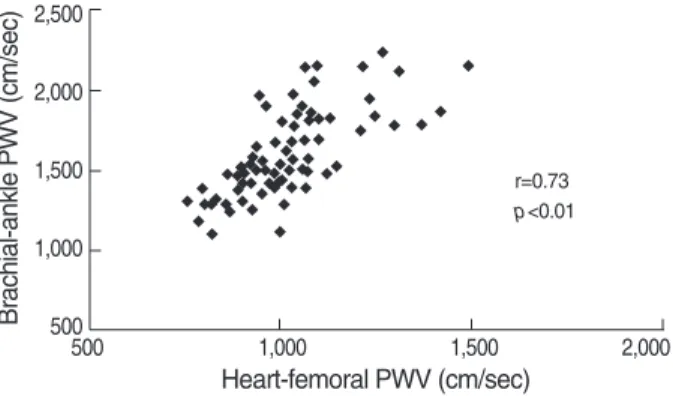
FMD of the brachial artery was 7.7±3.2% and nitroglyc- erin mediated vasodilation was 16.8±4.6%. Heart-femoral PWV was 1,020.3±146.1 cm/sec and brachial-ankle PWV was 1,604.3±277.9 cm/sec (right: 1,598.7±276.1, left:
1,609.7±285.6). UAE was 17.1±24.7 mg/g Cr and the level of hsCRP was 0.23±0.37 mg/dL. Heart-femoral PWV were significantly correlated with brachial-ankle PWV (r=
0.73, p<0.01) (Fig. 1). UAE were significantly correlated with
LDL, low-density lipoprotein; HDL, high-density lipoprotein; LVEF, left ven- tricular ejection fraction.
Group I (n=44) Group II (n=43) p value
Age (yr) 57.9±9.7 57.2±10.5 0.731
Males (%) 23 (52.3) 29 (64.1%) 0.149
Height (cm) 164.1±8.2 166.2±6.5 0.182
Weight (kg) 69.2±10.6 70.1±9.0 0.763
Smoking (%) 20 (45.5) 22 (51.2) 0.594
Hypertension (%) 20 (45.5) 17 (39.5) 0.577
Diabetes mellitus (%) 12 (27.3) 13 (29.6) 0.760 Total cholesterol (mg/dL) 194.7±44.7 192.4±33.3 0.783 LDL cholesterol (mg/dL) 136.8±41.4 134.9±31.4 0.816 HDL cholesterol (mg/dL) 49.7±11.5 46.8±9.8 0.202 Triglyceride (mg/dL) 114.5±43.6 122.7±58.4 0.458 Prescribed medications (%)
Aspirin 44 (100.0) 43 (100.0) 1.000
Clopidogrel 39 (88.6) 40 (93.0) 0.479
Nitrates 33 (76.7) 29 (67.4) 0.436
Statins 25 (56.8) 23 (53.5) 0.755
Beta-blockers 15 (34.1) 12 (27.9) 0.533
LVEF (%) 67.9±7.7 69.7±6.5 0.319
Table 1. Baseline clinical characteristics between the groups
ACC/AHA, American College of Cardiology/American Heart Association;
TIMI, thrombolysis in myocardial infarction.
Group I Group II p value (n=44) (n=43)
Target lesion (%) 0.721
Left anterior descending artery 23 (52.3) 19 (44.2) Right coronary artery 14 (31.8) 15 (34.9) Left circumflex artery 7 (15.9) 9 (20.9)
Number of involved vessel (%) 0.624
Single vessel disease 23 (52.3) 24 (55.8) Two vessel disease 17 (38.6) 13 (30.2) Three vessel disease 4 (9.1) 6 (14.0)
Types of stent (%) 0.670
Drug eluting stent 41 (93.2) 39 (90.7)
Bare metal stent 3 (6.8) 4 (9.3)
ACC/AHA lesion morphology (%) 0.456
Simple lesion (A/B1) 25 (56.8) 21 (48.8) Complex lesion (B2/C) 19 (43.2) 22 (51.2)
TIMI flow (%) 0.730
O 0 (0) 0 (0)
I 0 (0) 0 (0)
II 6 (13.6) 7 (16.3)
III 38 (86.4) 36 (83.7)
Diameter stenosis (%) 89.1±8.6 90.5±10.2 0.276 Table 2. Angiographic characteristics between the groups Fig. 1. Relation between heart-femoral pulse wave velocity (PWV) and brachial-ankle PWV.
Brachial-ankle PWV (cm/sec)
2,500
2,000
1,500
1,000
500
500 1,000 1,500 2,000
Heart-femoral PWV (cm/sec)
r=0.73 p<0.01
heart-femoral PWV (r=0.51, p<0.01) and brachial-ankle PWV (r=0.43, p<0.01) (Fig. 2). However, FMD and hsCRP were not correlated with PWV or UAE. FMD, PWV, UAE, and hsCRP were not different between the groups at baseline (Table 3).
Changes of FMD, PWV, UAE, hsCRP, and hemodynamic parameters
FMD, PWV, UAE, and hsCRP were measured in all pati- ents after 6 months of medical treatment. FMD were signif-
Fig. 2. Relation between urinary albumin excretion (UAE) and pulse wave velocity (PWV).
Heart-femoral PWV (cm/sec)
1,600 1,400 1,200 1,000 800 600 400 200 0
-20 0 20 40 60 80 100 120 140
UAE (mg/g Cr)
Brachial-ankle PWV (cm/sec)
2,500 2,000 1,500 1,000 500
0-20 0 20 40 60 80 100 120 140
UAE (mg/g Cr)
r=0.51 p<0.01
r=0.43 p<0.01
FMD, flow-mediated vasodilation; NMD, nitroglycerin-mediated vasodilation; hfPWV, heart-femoral pulse wave velocity; baPWV, brachial-ankle pulse wave velocity; UAE, urinary albumin excretion; hsCRP, highly sensitive C-reactive protein; SBP, systolic blood pressure; DBP, diastolic blood pressure;
HR, heart rate.
Group I (n=44) Group II (n=43)
Baseline Follow-up p value Baseline Follow-up p value
FMD (%) 7.9±2.7 8.2±2.8 0.535 7.5±3.7 8.8±2.7 <0.001
NMD (%) 17.5±8.6 18.3±9.1 0.082 16.1±10.2 16.4±9.7 0.682
HfPWV (cm/sec) 1,015.1±148.6 1,000.1±132.8 0.060 1,025.7±145.1 946.2±112.2 <0.001 BaPWV (cm/sec) 1,621.3±279.4 1,512.1±225.0 <0.001 1,586.8±278.5 1,434.5±200.5 <0.001
UAE (mg/g Cr) 14.15±18.19 12.80±15.50 0.214 20.19±29.92 13.03±16.42 0.019
HsCRP (mg/dL) 0.19±0.33 0.16±0.20 0.307 0.25±0.42 0.21±0.25 0.111
SBP (mmHg) 136.4±11.2 131.5±9.1 <0.001 135.7±12.2 127.8±9.4 <0.001
DBP (mmHg) 82.2±7.4 79.6±6.7 0.001 83.6±7.8 79.7±6.4 <0.001
HR (rate/min) 65.0±8.2 65.2±7.8 0.547 65.2±9.2 64.8±8.0 0.446
Table 3. FMD, PWV, UAE, hsCRP, and hemodynamic parameters at baseline and at 6 months
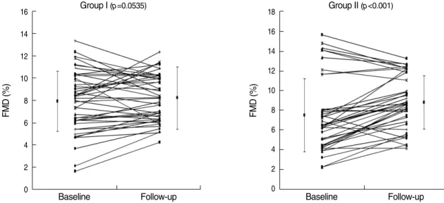
Fig. 3. Changes of flow-mediated vasodilation (FMD) of the brachial artery after 6 months of medical therapy.
FMD (%)
16 14 12 10 8 6 4 2 0
Group I (p=0.0535)
Baseline Follow-up
FMD (%)
18 16 14 12 10 8 6 4 2 0
Group II (p<0.001)
Baseline Follow-up
icantly improved in group II (7.5±3.7 to 8.8±2.7%, p<
0.001), but not in group I (7.9±2.7 to 8.2±2.8%, p=0.535) (Fig. 3). FMD was improved in 31 patients and not changed or decreased in 12 patients of group II. PWV were more de- creased in patients with the improvement of FMD than in patients without improvement of FMD, but it did not reached statistical significance (hfPWV: -93.6±148.6 vs. -58.9± 75.7 cm/sec, baPWV: -127.3±193.7 vs. -75.1±110.4 cm/
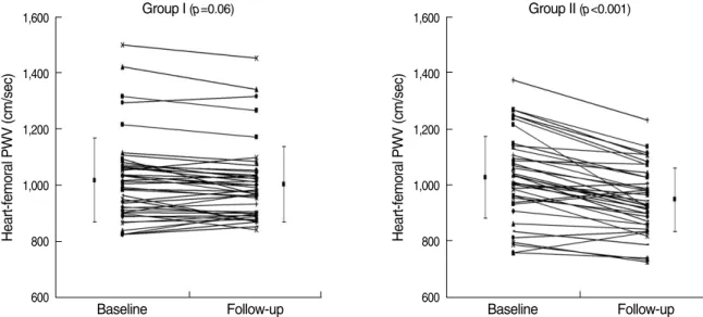
sec, p=ns). Brachial-ankle PWV were significantly improved in both groups (1,621.3±279.4 to 1,512.1±225.0 cm/sec in group I, p<0.001, 1,586.8±278.5 to 1,434.5±200.5 cm/sec in group II, p<0.001) (Fig. 4). However, heart-femoral PWV were significantly improved only in group II (1,025.7
±145.1 to 946.2±112.2 cm/sec, p<0.001), but not in group I (1,015.1±148.6 to 1,000.1±132.8 cm/sec, p=0.06) (Fig.
5). FMD was improved in 31 patients and not changed or
decreased in 12 patients of group II. The improvement of FMD was not associated with the changes of PWV. UAE were significantly decreased only in group II (20.19±29.92 to 13.03±16.42 mg/g Cr, p=0.019), but not in group I (14.15
±18.19 to 12.80±15.50 mg/g Cr, p=0.214). The levels of hsCRP were not changed in both groups (0.19±0.33 to 0.16
±0.20 mg/dL in group I, p=0.307, 0.25±0.42 to 0.21± 0.25 mg/dL in group II, p=0.111). Systolic and diastolic blood pressures were significantly improved in both groups, but were not different between the groups at 6 months. Heart rates were not changed in both groups and were not differ- ent between the groups at 6 months. Changes of FMD, PWV, UAE, hsCRP, and hemodynamic parameters were summa- rized in Table 3. FMD was improved in 31 patients and not changed or decreased in 12 patients of group II. The improve- ment of FMD was not associated with the changes of PWV.
Fig. 5. Changes of brachial-ankle pulse wave velocity (PWV) after 6 months of medical therapy.
Brachial-ankle PWV (cm/sec)
2,400 2,200 2,000 1,800 1,600 1,400 1,200 1,000 800
Group I (p<0.001)
Baseline Follow-up
Brachial-ankle PWV (cm/sec)
2,400 2,200 2,000 1,800 1,600 1,400 1,200 1,000 800
Group II (p<0.001)
Baseline Follow-up
Fig. 4. Changes of heart-femoral pulse wave velocity (PWV) after 6 months of medical therapy.
Heart-femoral PWV (cm/sec)
1,600
1,400
1,200
1,000
800
600
Group I (p=0.06)
Baseline Follow-up
Heart-femoral PWV (cm/sec)
1,600
1,400
1,200
1,000
800
600
Group II (p<0.001)
Baseline Follow-up
Clinical events during follow-up periods
Clinical follow-up were possible in all patients. Death, my- ocardial infarction, or stent thrombosis was not developed in both groups. Follow-up coronary angiography at 6 months was performed in 55 patients (29 patients in group I, 26 patients in group II). Target lesion revascularization were performed in 5 patients (9.1%) due to restenosis and were not different between the groups (3 patients: 10.4% in group I, 2 patient:
7.7% in group II, p=0.763). Baseline and follow-up values of FMD, PWV, UAE, and hsCRP were not associated with angiographic restenosis.
DISCUSSIONS
The main finding of the present study is that the combi- nation therapy with CCB and ACEI more effectively improves endothelial function, arterial stiffness, and UAE than CCB mono-therapy do in patients with stable angina pectoris. Am- erican College of Cardiology/American Heart Association 2002 update guideline for the management of stable angina recommended that ACEI should be used in most cases as a routine secondary prevention for patients with known coro- nary artery disease based on the results of the Heart Outcomes Prevention Evaluation trial (15). However, the precise bene- ficial mechanism of ACEI in angina pectoris was not eluci- dated. Several mechanisms such as anti-proliferative effects, hormonal/vascular effects and anti-atherogenic properties were suggested as a favorable role of ACEI. Although the major adverse cardiac events were not different between the groups, the results of this study also support that ACE inhibition plays a beneficial role in the management of stable angina pectoris by anti-atherogenic effects including improvements of endo- thelial dysfunction, arterial stiffness, and UAE.
Recently, several surrogate markers for the early detection of atherosclerosis including the measurement of endothelial dysfunction, arterial wall structure, arterial stiffness, and sero- logic markers of vascular inflammation were proposed and widely studied (3, 4). Recent studies suggested that these sur- rogate markers for atherosclerosis are associated with the pres- ence of atherosclerotic coronary artery disease and also asso- ciated with future adverse cardiac events (8-12). To reduce the risk of death or disability associated with atherosclerotic coronary artery disease, therefore, early identification and ther- apeutic modification of risk factors or surrogate markers are important.
Endothelial dysfunction measured by FMD of the brachial artery is associated with the presence of coronary artery dis- ease or the severity of the coronary artery stenosis or in-stent restenosis following percutaneous coronary intervention (16, 17). Some kinds of drugs such as ACEI, angiotensin recep- tor blocker, or CCB are known to have beneficial effects on endothelial function in the previous studies. Mancini et al.
(18) reported that ACEI (quinapril) could improve endothe- lial function measured by invasive measurements of coronary diameter change in response to acetylcholine in patients with coronary artery disease. Anderson et al. (19) also reported that ACEI (quinapril) could improve endothelial function mea- sured by FMD of the brachial artery in patients with coronary artery disease, but CCB (amlodipine) could not improve FMD.
In this study, combination therapy with ACEI (captopril) and CCB (cilnidipine) showed significant improvement in endo- thelial function measured by FMD of the brachial artery in patients with coronary artery disease. However, CCB mono- therapy did not improve FMD of the brachial artery. The re- sults of this study and Anderson et al. (19) suggested that ACE inhibition may play more important roles in endothe- lial function than calcium channel blockade in patients with overt coronary artery disease. Although FMD of the brachial artery were improved only in combination therapy group, clinical events were not different between the groups. FMD of the brachial artery were not associated with in-stent resteno- sis in this study. However, Kitta et al. (16) and Patti et al. (17) reported that impaired FMD of the brachial artery was a sig- nificant independent predictor of late in-stent restenosis fol- lowing percutaneous coronary intervention. To date, no pros- pective randomized clinical trial in large population has eval- uated the relationship between endothelial dysfunction and in-stent restenosis. Thus, further clinical trials will be need- ed to elucidate the relationship between endothelial dysfunc- tion and in-stent restenosis.
Arterial stiffness is now an established risk factor for pre- mature coronary artery disease, peripheral vascular disease, and stroke (8). It is a single best prognostic indicator for future cardiovascular events in hypertensive population and also pre- dicts adverse outcomes in patients with documented coronary artery disease (20-24). PWV is a simple, reproducible, and non-invasive measurement which can be used as a valuable index of both arterial stiffness and atherosclerosis in large pop- ulations. The results of the studies including Nigam et al. (25, 26) suggested that large conduit artery stiffness correlates sig- nificantly with FMD of the brachial artery. However, the re- sults of this study failed to demonstrate the relationship be- tween PWV and FMD of the brachial artery at baseline and after 6 months of medical treatment. Although the PWV was more decreased in patients who showed the improvement of FMD, it did not reached statistical significance. Life style mod- ifications and pharmacological therapy could improve arteri- al stiffness measured by PWV. Long-term ACEI therapy has been shown to be superior to diuretics or beta-blockers in im- proving carotid, femoral, or radial arterial compliance and is marginally superior to CCB therapy as well (14). CCB as a mono-therapy or combination therapy with ACEI improved brachial-ankle PWV, but heart-femoral PWV was only im- proved in combination therapy with ACEI and CCB in the present study. This finding suggested that combination ther- apy with ACEI and CCB could have more favorable effects on
cardiovascular disease or events than CCB mono-therapy. The reason why heart-femoral PWV and brachial-ankle PWV showed different response is not clear, but several possible mechanisms would have been involved. Firstly, the improve- ment of heart-femoral PWV in group II is possibly associat- ed with the pleiotropic effect of ACEI such as the favorable impacts on the aortic wall endothelial function or components.
ACEI is known to be decrease collagen content, fibrosis, and thus improve elasticity. Secondly, the improvement of heart- femoral PWV in group II is possibly caused by the simple hemodynamic change of BP. Although the change of BP did not showed significant statistical difference, the drop of BP in group II was about 3 mmHg more than in group I. Fur- thermore, ACEI showed different response on central aortic pressure according to the age group in the previous studies.
According to the study of Morgan et al. (27), ACEI therapy, despite a relatively small effect on peripheral BP, showed a more beneficial effect on central BP in elderly patients with hypertension. We did not checked central aortic pressure in the present study, but the changes of central aortic pressure in group II might be significantly higher than the changes of peripheral brachial BP. Thirdly, the number of the subjects involved in this study is relatively small and thus it also may affect the result of the heart-femoral PWV.
HsCRP is an important surrogate marker for atherosclerot- ic coronary artery disease and associated with cardiovascular events in patients with documented coronary artery disease (9, 10). We want to evaluate the relationship between hsCRP and other surrogate markers for atherosclerosis, and also the effects of pharmacologic treatment with CCB or CCB plus ACEI on the level of hsCRP. Several studies demonstrated that the level of hsCRP were significantly associated with FMD of the brachial artery, PWV, and UAE and improved by pharmacological therapy (28-31). However, the level of hsCRP were not correlated with FMD of the brachial artery or PWV, or UAE and not changed with pharmacologic treat- ments in this study. The correlation among these surrogate markers has not been reported consistently in the previous studies (26, 32, 33). None of the surrogate markers provides highly sensitive and specific recognition of progressive athe- rosclerotic cardiovascular diseases and rather than these sur- rogate markers may have different supplemental action mech- anism in the development of atherosclerotic coronary artery disease (33). Therefore, an attractive strategy has been to use a combination of these surrogates to better define the pres- ence of early disease.
Microalbuminuria measured by UAE is also an important surrogate marker for atherosclerosis and predicts future car- diac events (11, 12). UAE was significantly improved after combination therapy with CCB and ACEI, but not after CCB mono-therapy in the present study. This finding support that the inhibition of renin-angiotensin-aldosterone system can retard or reverse microalbuminuria (34). Although the change of UAE was not compared between the groups, however, the
baseline level of UAE was significantly lower in group I than in group II. Therefore, we cannot exclude that the lower ch- anges of UAE in group I might be caused by the lower base- line level than in group II. Recently, Morimoto et al. (35) re- ported that CCB (cilnidipine) was associated with the impro- vement in UAE besides the improvement of brachial-ankle PWV. This study showed the same finding compared with the present study in that CCB could improve brachial-ankle PWV, but the effect of CCB on UAE was the opposite. Their study also had a limitation that only the small numbers of the study population was involved. Therefore, the effect of cilnidipine on UAE should be considerably re-evaluated by more large randomized controlled trial.
There are some limitations in this study. Firstly, the main limitation of this study was the relatively small sample size and thus some selection bias could present inevitably. Second- ly, although the prescribed medications such as statins or beta- blockers were not different between the groups, these drugs also could affect diversely on FMD, PWV, and UAE. Third- ly, although the degree of the fall of BP did not showed sig- nificant statistical difference between the groups, the greater fall of BP in group II than in group I would have affect the greater improvement of endothelial function and arterial stiff- ness in group II than in group I. Therefore, the degree of BP change between the groups should be controlled at a similar degree in the study design by adding hydrochlothiazide as a placebo in group I. Fourthly, the dose of the used ACEI was fixed (25 mg per day) and thus not reached its full dose. It may not be sufficient to evaluate the effect of ACEI.
In conclusion, combination therapy with CCB and ACEI more effectively improve endothelial dysfunction, arterial stiff- ness, and microalbuminuria than CCB mono-therapy in pa- tients with angina pectoris.
REFERENCES
1. Anderson KM, Odell PM, Wilson PW, Kannel WB. Cardiovascular disease risk profiles. Am Heart J 1991; 121: 293-8.
2. Grundy SM, Pasternak R, Greenland P, Smith S Jr, Fuster V. Assess- ment of cardiovascular risk by use of multiple-risk-factor assessment equations: a statement for healthcare professionals from the American Heart Association and the American College of Cardiology. Circu- lation 1999; 100: 1481-92.
3. Patel SN, Rajaram V, Pandya S, Fiedler BM, Bai CJ, Neems R, Fein- stein M, Goldin M, Feinsetin SB. Emerging, noninvasive surrogate markers of atherosclerosis. Curr Atheroscler Rep 2004; 6: 60-8.
4. Ross R. The pathogenesis of atherosclerosis: a perspective for the 1990s. Nature 1993; 362: 801-9.
5. Gibbons GH. Endothelial function as a determinants of vascular func- tion and structure: a new therapeutic target. Am J Cardiol 1997; 79:
3-8.
6. Selwyn AP, Kinlay S, Creager M, Libby P, Ganz P. Cell dysfunction in atherosclerosis and the ischemic manifestations of coronary artery
disease. Am J Cardiol 1997; 79: 17-23.
7. Corretti MC, Anderson TJ, Benjamin EJ, Celermajer D, Charbonneau F, Creager MA, Deanfield J, Drexler H, Gerhard-Herman M, Her- rington D, Vallance P, Vita J, Vogel R. International Brachial Artery Reactivity Task Force. Guidelines for the ultrasound assessment of endothelial dependent flow mediated vasodilation of the brachial artery: a report of the International Brachial Artery Reactivity Task Force. J Am Coll Cardiol 2002; 39: 257-65.
8. Izzo JL, Shykoff BE. Arterial stiffness: clinical relevance, measure- ment, and treatment. Rev Cardiovasc Med 2001; 2: 29-40.
9. Lagrand WK, Visser CA, Hermens WT, Niessen HW, Verheugt FW, Wolbink GJ, Hack CE. C-reactive protein as a cardiovascular risk factor: more than an epiphenomenon? Circulation 1999; 100: 96-102.
10. Ridker PM, Rifai N, Rose L, Buring JE, Cook NR. Comparison of C-reactive protein and low-density lipoprotein cholesterol levels in the prediction of first cardiovascular events. N Engl J Med 2002; 347:
1557-65.
11. Luft FC, Agrawal B. Microalbuminuria as a predictive factor for car- diovascular events. J Cardiovasc Pharmacol 1999; 33 (Suppl 1): S11- 5 discussion S41-43.
12. Sukhija R, Aronow WS, Kakar P, Garza L, Sachdeva R, Sinha A, Mehta JL. Relation of microalbuminuria and coronary artery dis- ease in patients with and without diabetes mellitus. Am J Cardiol 2006; 98: 279-81.
13. Bonetti PO, Lerman LO, Lerman A. Endothelial dysfunction: a mark- er of atherosclerotic risk. Atherioscler Thromb Vasc Biol 2003; 23:
168-75.
14. Mahmud A, Feely J. Antihypertensive drugs and arterial stiffness.
Expert Rev Cardiovasc Ther 2003; 1: 65-78.
15. Gibbons RJ, Abrams J, Chatterjee K, Daley J, Deedwania PC, Dou- glas JS, Ferguson TB Jr, Fihn SD, Fraker TD Jr, Gardin JM, O’Rourke RA, Pasternak RC, Williams SV, Gibbons RJ, Alpert JS, Antman EM, Hiratzka LF, Fuster V, Faxon DP, Gregoratos G, Jacobs AK, Smith SC Jr. American College of Cardiology; American Heart Asso- ciation Task Force on Practice Guidelines; Committee on the Man- agement of Patients with Chronic Stable Angina. ACC/AHA 2002 guideline update for the management of patients with chronic stable angina: a report of the American College of Cardiology/American Heart Association Task Force on Practice Guidelines. Circulation 2003; 107: 149-58.
16. Kitta Y, Nakamura T, Kodama Y, Takano H, Umetani K, Fujioka D, Saito Y, Kawabata K, Obata JE, Ichiqi Y, Mende A, Kuqiyama K.
Endothelial vasomotor dysfunction in the brachial artery is associ- ated with late in-stent coronary restenosis. J Am Coll Cardiol 2005;
46: 648-55.
17. Patti G, Pasceri V, Melfi R, Goffredo C, Chello M, D’Ambrosio A, Montesanti R, Di Sciascio G. Impaired flow-mediated dilation and risk of restenosis in patients undergoing coronary stent implantation.
Circulation 2005; 111: 70-5.
18. Mancini GB, Henry GC, Macaya C, O’Neill BJ, Pucillo AL, Carere RG, Wargovich TJ, Luscher TF, Klibaner MI, Haber HE, Uprichard AC, Pepine CJ, Pitt B. Angiotensin-converting enzyme inhibition with quinapril improves endothelial vasomotor dysfunction in patients with coronary artery disease. The TREND (Trial on Reversing ENdothe-
lial Dysfunction) Study. Circulation 1996; 94: 258-65.
19. Anderson TJ, Elstein E, Haber H, Charbonneau F. Comparative study of ACE-inhibition, angiotensin II antagonism, and calcium channel blockade on flow-mediated vasodilation in patients with coronary dis- ease (BANFF study). J Am Coll Cardiol 2000; 35: 60-6.
20. van Popele NM, Grobbee DE, Bots ML, Asmar R, Topouchian J, Re- neman RS, Hoeks AP, van der Kuip DA, Hofman A, Witteman JC.
Association between arterial stiffness and atherosclerosis: the Rot- terdam Study. Stroke 2001; 32: 454-60.
21. London GM, Marchais SJ, Guerin AP, Pannier B. Arterial stiffness:
pathophysiology and clinical impact. Clin Exp Hypertens 2004; 26:
689-99.
22. Weber T, Auer J, O’Rourke MF, Kvas E, Lassnig E, Berent R, Eber B. Arterial stiffness, wave reflections, and the risk of coronary artery disease. Circulation 2004; 109: 184-9.
23. Imanishi R, Seto S, Toda G, Yoshida M, Ohtsuru A, Koide Y, Baba T, Yano K. High brachial-ankle pulse wave velocity is an independent predictor of the presence of coronary artery disease in men. Hyper- tens Res 2004; 27: 71-8.
24. Chirinos JA, Zambrano JP, Chakko S, Veerani A, Schob A, Willens HJ, Perez G, Mendez AJ. Aortic pressure augmentation predicts ad- verse cardiovascular events in patients with established coronary ar- tery disease. Hypertension 2005; 45: 980-5.
25. Nigam A, Mitchell GF, Lambert J, Tardif JC. Relation between con- duit vessel stiffness (assessed by tonometry) and endothelial function (assessed by flow-mediated dilatation) in patients with and without coronary heart disease. Am J Cardiol 2003; 92: 395-9.
26. Kobayashi K, Akishita M, Yu W, Hashimoto M, Ohni M, Toba K.
Interrelationship between noninvasive measurements of atherosclero- sis: flow-mediated dilation of brachial artery, carotid intima-media thickness and pulse wave velocity. Atherosclerosis 2004; 173: 13-8.
27. Morgan T, Lauri J, Bertram T, Anderson A. Effect of different anti- hypertensive drug classes on central aortic pressure. Am J Hypertens 2004; 17: 118-23.
28. Fichtlscherer S, Rosenberger G, Walter DH, Breuer S, Dimmeler S, Zeiher AM. Elevated C-reactive protein levels and impaired endothe- lial vasoreactivity in patients with coronary artery disease. Circula- tion 2000; 102: 1000-6.
29. Nagano M, Nakamura M, Sato K, Tanaka F, Segawa T, Hiramori K.
Association between serum C-reactive protein levels and pulse wave velocity: a population-based cross-sectional study in a general pop- ulation. Atherosclerosis 2005; 180: 189-95.
30. Tsioufis C, Dimitriadis K, Taxiarchou E, Vasiliadou C, Chartzoulakis G, Tousoulis D, Manolis A, Stefanadis C, Kallikazaros I. Diverse as- sociations of microalbuminuria with C-reactive protein, interleukin 18 and soluble CD 40 ligand in male essential hypertensive subjects.
Am J Hypertens 2006; 19: 462-6.
31. Vitale C, Cerquetani E, Wajngarten M, Leonardo F, Silvestri A, Mer- curo G, Fini M, Ramires JA, Rosano GM. In patients with coronary artery disease endothelial function is associated with plasma levels of C-reactive protein and is improved by optimal medical therapy. Ital Heart J 2003; 4: 627-32.
32. Yufu K, Takahashi N, Hara M, Saikawa T, Yoshimatsu H. Measure- ment of the brachial-ankle pulse wave velocity and flow-mediated di-
latation in young, healthy smokers. Hypertens Res 2007; 30: 607-12.
33. Cohn JN, Quyyumi AA, Hollenberg NK, Jamerson KA. Surrogate markers for cardiovascular disease: functional markers. Circulation 2004; 109: IV31-46.
34. Jerums G, MacIsaac RJ. Treatment of microalbuminuria in patients
with type 2 diabetes mellitus. Treat Endocrinol 2002; 1: 163-73.
35. Morimoto S, Yano Y, Maki K, Iwasaka T. Renal and vascular pro- tective effects of cilnidipine in patients with essential hypertension. J Hypertens 2007; 25: 2178-83.