https://doi.org/10.14734/PN.2017.28.3.103 pISSN 2508-4887•eISSN 2508-4895
Soohyun Ri, MD1, Jeonghee Shin, MD1, Bo-Kyung Je, MD, PhD2, Byung Min Choi, MD, PhD1
1Department of Pediatrics, Korea University College of Medicine, Seoul, Korea
2Department of Radiology, Korea University College of Medicine, Seoul, Korea
Angiotensin receptor blockers are contraindicated drugs during pregnancy because of their potential fetotoxicity, including oligohydramnios, anuria, renal tubular dysplasia, limb contractures, cranial ossification defects, prolonged patent ductus arteriosus, and even in utero death. It is believed that these abnormalities are associated with drug-mediated fetal hypotension. We report a case of skull ossification defects, pneumothorax, renal parenchymal disease, congenital calcaneovalgus foot, and inferior vena cava thrombosis in a neonate born to a hypertensive woman who had continued use of an angiotensin receptor blocker during her pregnancy.
Key Words: Congenital defect of skull, Angiotensin receptor blocker, Neonate
Introduction
Use of angiotensin converting enzyme inhibitors or angiotensin receptor antagonists (ARBs) in the second and third trimesters of pregnancy has been associated with oligo
hydramnios, anuria, renal tubular dysplasia, limb contractures, cranial ossification defects, prolonged patent ductus arteriosus, and even in utero death. It is believed that these ab
normalities are associated with drugmediated fetal hypotension.16
We report a case of skull ossification defects, pneumothorax, renal parenchymal disease, congenital calcaneovalgus foot, and inferior vena cava (IVC) thrombosis in a neonate born to a hypertensive woman who had continued use of ARBs during her pregnancy.
Case
A newborn infant (birth weight 3.2 kg, expected Ballard score gestational age [GA] 40 weeks, spontaneous vaginal delivery) was transferred to our hospital for evaluation of im
palpable skull bones.
His mother was a 35yearold woman with chronic hypertension treated with valsartan (an ARB) and had moderate mental retardation. She was not aware of her pregnancy and had continued use of her antihypertensive drugs throughout her pregnancy and visited an emergency room with abdominal pain, which turned out to be labor pain. A male infant was born through spontaneous vaginal delivery and his expected Ballard score was GA 40 weeks.
On neonatal intensive care unit admission, the baby showed a heart rate of 113 bpm, re
spiratory rate of 61 bpm, blood pressure of 65/35 mmHg, and body temperature of 36.6℃, Received: 5 May 2017
Revised: 21 June 2017 Accepted: 22 August 2017 Correspondence to Jeonghee Shin, MD
Department of Pediatrics, Korea University College of Medicine, 123 Jeokgeum-ro, Danwon-gu, Ansan 15355, Korea
Tel: +82-31-412-6557 Fax: +82-31-405-8591 E-mail: [email protected]
Copyright© 2017 by The Korean Society of Perinatology
This is an Open Access article distributed under the terms of the Creative Com- mons Attribution Non-Commercial License (http://creativecommons.org/
license/by-nc/4.0/), which permits unrestricted non-commercial use, distribution, and reproduction in any
Hypocalvaria of Newborn Infant: Intrau
terine Exposure to an Angiotensin Re
ceptor Blocker
(1025th percentile). His skull was not palpable over the fron
tal, temporal, parietal, and occipital areas, and these clinical findings were confirmed on skull ra diography (Fig. 1A). A brain computed tomography (CT) scan showed a membranous neu
ro cranium, but with underdevelop ed ossification (Fig. 2). A neurosurgeon recommended using a helmet to avoid trau ma, but the mother was unable to comply due to economic problems.
Spontaneous and mild pneumothorax but no lung hypoplasia was seen on the chest radiography and respiratory distress was treated by noninvasive respiratory support for 2 days (Fig.
3). Urine output was fair, but hyponatremia (Na 133 mmol/ L), elevated blood urea nitrogen (38.75 mg/dL), elevat ed creatinine (1.05 mg/dL), and mild metabolic acidosis (capillary blood gas analysis; pH 7.311, pCO2 29.3 mmHg, pO2 67 mmHg, HCO3 14.8
mmol/L, BE –11 mmol/L) were observed. Proteinuria was also noted (spot urine protein/creatinine ratio=9.17). Sonographic examination revealed the kidneys to be increased in size and increased renal cortical echogenicity. The laboratory and ima
ge findings suggested parenchymal renal disease. After con
servative management such as oral sodium and sodium bicar
bonate replacement, the laboratory test results had improved.
Because IVC thrombosis was detected accidently by abdo
minal ultrasonography, abdominal CT and inferior vena cavo
graphy was performed. The common iliac vein was not ob
served and the IVC was completely obstructed, and therefore all of the venous flow from the lower extremities was draining to the azygos vein through collateral vessels to the superior vena cava (Fig. 4). Owing to his asymptomatic status and the
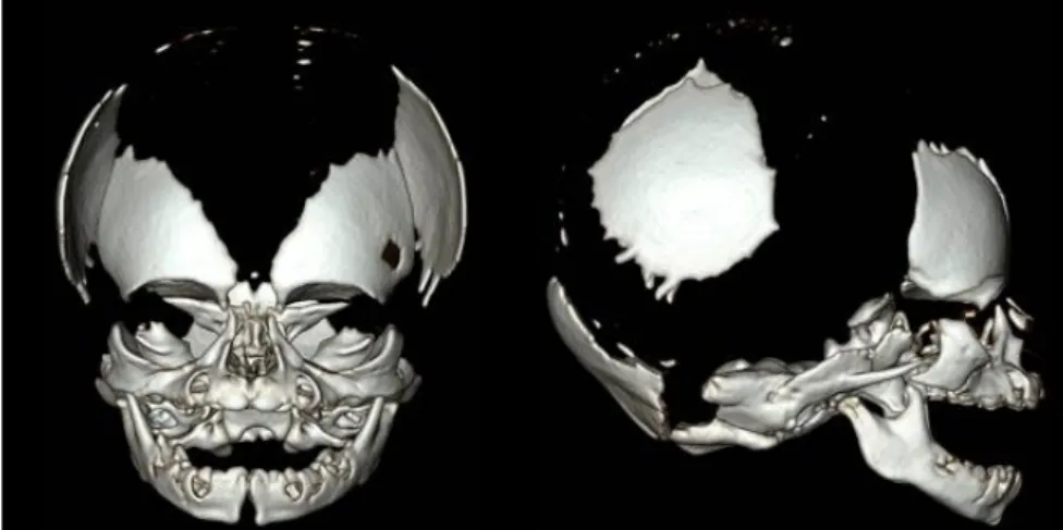
Fig. 2. Three-dimensional computed tomography volume rendering images of the skull showing poor ossification of the frontal, temporal, parietal, and occipital bones. Wide cranial sutures are noted.
Fig. 1. Left lateral plain skull radiograph. (A) Poor ossification of frontal, temporal, parietal, and occipital bones at birth. (B) Fully developed skull bones at 2 months’ of age.
A B
months, his skull bones were noted to be fully developed (Fig.
1B), and he showed correction of his abnormal laboratory findings. The patient continued to visit the outpatient clinic in our hospital for periodic reevaluation of his kidney func tion and IVC thrombosis. It should be noted that he was asympto
matic and his laboratory findings had become normal. How
ever, increased renal cortical echogenicity and IVC throm bosis continued. His body profile measurements at his 24 months’
of age were weight 9.7 kg (<3rd percentile), height 81.8 cm (5th percentile), and head circumference 46.5 cm (5−10th percentile). On the Korean Development Screening Test for Infants & Children (KDST), his development was generally delayed for his age.
Discussion
ARBs are contraindicated drugs during pregnancy because of their potential fetotoxicity. They are known to reduce fetal renal blood flow and cause ischemic kidney injury, such as renal tubular dysgenesis, anuria, oligohydramnios, and anhy
dramnios.5 Anuriaassociated oligohydramnios may cause fetal limb contractures, craniofacial deformation, and pulmonary hypoplasia.7 Barr and Cohen6 found possible links between calvarial underdevelopment, especially the membranous skull bones, and renal tubular dysgenesis. They suggested that this
“kidneyskull connection” caused by angio tensin converting enzyme (ACE) inhibition may be due to intrauterine hypoten
sion and/or chronic hypoxia. Hypocalvaria can be secondary to poor perfusion of platelike bone structures in the skull.
For other cases of exposure to ACE inhibitors or ARBs dur
ing the second and third trimesters of pregnancy, complications such as oligohydramnios, renal failure, neonatal hy potension, bony deformity, and skull ossification defects have been re
ported. Also, limb contracture, patent ductus arterio sus, respi
ratory distress syndrome, lung hypoplasia and ap nea, and even in utero death have been seen. In most cases, treatment was conservative therapy for each symptom.8
In our case, the fetus was exposed to ARBs throughout the pregnancy, and the newborn showed various ARBrelated symptoms, such as skull ossification defects, pneumothorax, respiratory distress, and renal parenchymal disease. His con
risks associated with treatment of thromboembolism, we deci
ded to closely observe the patient rather than actively treating his IVC thrombosis.
Following conservative management and observation for 2
Fig. 3. Mild spontaneous pneumothorax is visible on the chest radio- graph. The arrows show the pleural line.
Fig. 4. Abdominal computed tomography and inferior vena cavogra- phy. Complete obstruction of the iliac vein and inferior vena cava are visible. The arrow shows the thrombus of the inferior vena cava.
genital calcaneovalgus foot, which was not reported in pre
vious cases, is thought to result from oligohydramnios due to ARB use during pregnancy. These suggest that maternal use of ARBs throughout pregnancy has a significantly large effect on fetal development. Although the IVC thrombosis confirmed by abdominal CT is thought to be unrelated to ARB use, further studies are needed to determine the association between IVC thrombosis and the use of ARBs.
Although these fetotoxic effects of ARBs are well known and several cases of hypocalvaria have been reported, the distinc tiveness of our study was the imaging we performed. Ours was the first case in which hypocalvaria in the patient was con
firmed using a skull CT, that was further reconstructed using 3dimensional graphics.
Usually, physicians educate pregnant women to discontinue the use of ARBs when pregnancy is confirmed. However, in our case, the fetus was exposed to ARBs throughout pregnancy.
Both his parents had moderate mental retardation, and were not aware of the patient’s mother’s pregnancy. She therefore continued the use of antihypertensive drugs including valsar
tan (an ARB). They were economically disadvantaged. With limitedtono knowledge about medical care, they were easily exposed to the risks of an unplanned pregnancy and did not seek medical attention during the mother’s pregnancy. The patient’s mother received medical treatment at a clinic for hy
pertension through a drug prescription, but her physician did not recognize her pregnancy. Physicians need to reflect on the fact that this unfortunate event happened in a medically ad
vanced country like Korea, and therefore they need to obtain a careful and detailed medical history while treating vul nerable classes, such as the disabled and economically disadvantaged young adults.
Because the sexuality of disabled women is suppressed so
cially, sex education provided to them is limited. Therefore, it is necessary to accept the sexual and reproductive needs of disabled women and provide comprehensive sex education.
ccording to the ‘Research on Pregnancy and Childbirth Support Policy for the Promotion of Maternal Rights for Women with Disabilities’,9 intellectually disabled women have low under
standing of books on sex education, pregnancy, and childbirth, and have limited understanding of the content of the medical consultations. Therefore, it is necessary to develop an easy
toread book that meets the needs of women with intel lec tual disabilities, and additional customized education is needed to achieve a clear understanding on their part.
In addition, it has been identified that the average number of women accessing antenatal care among the intellectually disabled women is less than that in the general population in Korea.9 This is thought to be due to a lack of understanding of the importance of antenatal care and economic difficulties.
In the UK, a ‘midwife system’ is used for antenatal care at the national level for every woman, and if additional care is required, such as for maternal complications or disability, a more specialized medical system consisting of an obstetrician and other physicians is available. In this way, advanced coun
tries have wellequipped human resources and economic sup
port systems for vulnerable women, and Korea needs to move in the right direction.
At the age of the infant was 24 months, weight, height, and head circumference of the patient were all below the 10th per
centile, and overall developmental delay was evident on the K
DST. It is unclear whether this is a genetic effect, a drug effect, or an effect of inadequate parenting.
In conclusion, it is our opinion that greater attention, sex education, and protection of the socially vulnerable women are needed at the individual physician level and at the national level. Also, attention needs to be directed not only toward an
tenatal consultations during pregnancy but also towards proper child rearing by intellectually disabled women.
References
1) Diav-Citrin O, Shechtman S, Halberstadt Y, Finkel-Pekarsky V, Wajnberg R, Arnon J, et al. Pregnancy outcome after in utero exposure to angio- tensin converting enzyme inhibitors or angiotensin receptor blockers.
Reprod Toxicol 2011;31:540-5.
2) Pryde PG, Sedman AB, Nugent CE, Barr M Jr. Angiotensin-converting enzyme inhibitor fetopathy. J Am Soc Nephrol 1993;3:1575-82.
3) Easterling TR, Carr DB, Davis C, Diederichs C, Brateng DA, Schmucker B.
Low-dose, short-acting, angiotensin-converting enzyme inhibitors as rescue therapy in pregnancy. Obstet Gynecol 2001;97:956-61.
4) Boix E, Zapater P, Picó A, Moreno O. Teratogenicity with angiotensin II receptor antagonists in pregnancy. J Endocrinol Invest 2005;28:1029- 31.
5) Barr M Jr. Teratogen update: angiotensin‐converting enzyme inhibitors.
Teratology 1994;50:399-409.
6) Barr M Jr, Cohen MM Jr. ACE inhibitor fetopathy and hypocalvaria: the kidney-skull connection. Teratology 1991;44:485-95.
7) Schaefer C. Angiotensin II‐receptor‐antagonists: further evidence of feto toxicity but not teratogenicity. Birth Defects Res A Clin Mol Teratol 2003;67:591-4.
8) Shotan A, Widerhorn J, Hurst A, Elkayam U. Risks of angiotensin-con-
verting enzyme inhibition during pregnancy: experimental and clinical evidence, potential mechanisms, and recommendations for use. Am J Med 1994;96:451-6.
9) Seo HJ, Bae SH, Lee KM. Research on pregnancy and childbirth support policy for the promotion of maternal rights for women with disabilities.
Korea: Korea Disabled People's Development Institute; 2016.