https://doi.org/10.4174/astr.2021.100.4.193 Annals of Surgical Treatment and Research
A randomized controlled trial to compare the efficacy of regenerated and non-regenerated oxidized cellulose gauze for the secondary treatment of local bleeding in patients undergoing hepatic resection
Chengshuo Zhang, Dazhi Fu, Fengshan Wang, Xinping Zhong, Lei Yang, Gang Wu, Baifeng Li, Jialin Zhang
Department of Hepatobiliary Surgery, The First Hospital of China Medical University, Shenyang, China
INTRODUCTION
Intraoperative bleeding is common in patients undergoing hepatic resection [1]. Secondary adverse events significantly increase when bleeding occurs during surgical procedures.
Increasing evidence shows that blood loss and its related transfusion is an independent risk factor for recurrence of
malignant tumors [2,3]. Therefore, the control of bleeding is a major concern for surgeons performing hepatectomy.
Many distinct surgical techniques have been used to control bleeding in patients undergoing hepatectomy. These techniques include controlled low central venous pressure strategies [4], diverse hepatic vascular occlusion techniques [5,6], and the use of various physical parenchyma transection and hemostatic
Received September 10, 2020, Revised November 28, 2020, Accepted January 5, 2021
Corresponding Author: Jialin Zhang
Department of Hepatobiliary Surgery, The First Hospital of China Medical University, 155 Nanjingbei Street, Heping District, Shenyang 110001, Liaoning, China
Tel: +86-24-83283310, Fax: +86-24-83282997 E-mail: [email protected]
ORCID: https://orcid.org/0000-0003-2617-2735
Copyright ⓒ 2021, the Korean Surgical Society
cc Annals of Surgical Treatment and Research is an Open Access Journal. All articles are distributed under the terms of the Creative Commons Attribution Non- Commercial License (http://creativecommons.org/licenses/by-nc/4.0/) which permits unrestricted non-commercial use, distribution, and reproduction in any medium, provided the original work is properly cited.
Purpose: Oxidized cellulose is available in many forms, but manufactured using either a regenerated or non-regenerated process. In this study, we evaluated the effects of 2 different hemostatic agents for the treatment of local bleeding in patients undergoing hepatic resection.
Methods: This was a monocentric, parallel-group, randomized, and controlled clinical trial to compare oxidized regenerated cellulose gauze (ORCG) with oxidized non-regenerated cellulose gauze (ONRCG) in patients undergoing hepatectomy. The primary endpoint was the time to hemostasis at the target bleeding site. The secondary endpoints were the postoperative drainage volume on the first 2 days after surgery and the hospital stay.
Results: There was no significant difference between the ORCG and ONRCG groups in time to hemostasis from column analysis (238.8 ± 121.6 seconds vs. 193.7 ± 85.3 seconds, P = 0.068), and there were no differences in the rates of hemostatic success between the 2 groups at 120 seconds (18.4% vs. 24.3%; odds ratio [OR], 0.703; 95% confidence interval [CI], 0.231–2.136) and 300 seconds (71.1% vs. 89.2%; OR, 0.298; 95% CI, 0.085–1.041). However, the ONRCG group was superior to the ORCG group in hemostasis according to the survival analysis (log-rank test, P = 0.044). Moreover, there were also no significant differences between the 2 groups in postoperative drainage volume on the first 2 days (P = 0.436, P
= 0.381) and hospital stay (P = 0.537, P = 0.200).
Conclusion: ONRCG was not inferior to ORCG as a hemostatic agent in patients undergoing liver resection.
[Ann Surg Treat Res 2021;100(4):193-199]
Key Words: Hepatectomy, Hemostasis, Oxidized cellulose, Surgicel, Traumastem
apparatus [7-10]. In addition, hemostatic, sealant, and adhesive products are also widely applied to control surgical bleeding when standard techniques are insufficient.
Topical hemostats are agents that stop bleeding by inducing blood to clot. Oxidized cellulose, a sort of mechanical hemostatic material, predominantly forms a block to stop the blood flow and provides a surface to induce more rapid clotting [11]. It was marketed for the first time in 1945 and has been widely used for its convenience, biocompatibility, and bactericidal properties [12]. It is currently available in many commercial products and manufactured using either a regenerated or non-regenerated process. The physicochemical properties and hemostatic efficacy of oxidized regenerated cellulose gauze (ORCG) and oxidized non-regenerated cellulose gauze (ONRCG) have been well documented using in vitro tests and animal models, and ONRCG is seemingly superior to ORCG in terms of hemostasis [13]. However, no clinical study has been performed to verify this. Therefore, the objective of this prospective randomized study was to assess the hemostatic efficacy of ORCG vs. ONRCG for hemostasis of local bleeding in patients undergoing hepatic resection.
METHODS
Study population
In this prospective randomized study, 81 patients (18–75 years old) with masses undergoing hepatic resection for any potential disease in the First Hospital of China Medical University, Shenyang, P.R. China, during a 10-month period from August 2017 to May 2018 were allocated randomly to apply ORCG (Surgicel Original, Ethicon Inc., San Angelo, TX, USA) or ONRCG (Traumastem TAF, Bioster a.s., Prague, Czech Republic) for the treatment of local bleeding. Patients were randomized to groups using a web-based calculator available at http://www.
lvsezhifei.com.
Oxidized cellulose gauze
The ORCG (Surgicel Original) is white to pale yellow. In this study, 5.1 × 7.6-cm sized patches were used for each patient.
The ONRCG (Traumastem TAF) is white to light yellow. A pattern of 7 × 5-cm product was used for each patient in this study. Both oxidized celluloses are sterile absorbable knitted fabrics with a faint caramel-like smell. Both of these products are strong and can be cut or sutured without fraying. When the gauzes are soaked with blood, they then turn into a black gelatinous mass that aids in the formation of a clot to control local hemorrhage.
Inclusion and exclusion criteria
Patients who gave informed consent, aged 18–75 years, with selective removal of an equivalent tissue volume of at least
1 anatomical hepatic segment qualified for inclusion in this study. Precise liver resection was performed with a physical parenchyma transection apparatus, a cavitron ultrasonic surgical aspirator. The topical hemostatic gauzes were necessary when minor-to-moderate amounts of bleeding (oozing/diffuse) continued at the surgical site after the end of the primary hemostatic procedures to control arterial pulsating bleeding and venous hemorrhage using vascular clips, sutures, ligatures, point electrocautery, and argon beam coagulation at the end of the surgery. Exclusion criteria were surgical contraindication or indication for emergency surgery, participation in other clinical trials in the past 3 months, obvious hematologic disorders, brain disease or mental disorder (abnormal judgment) that prevents cooperation, severe cardiac disease, severe metabolic disease or endocrine disorders, asthma or allergies, immunodeficient patients (acquired immune deficiency syndrome), skin infection in the targeted incisional area, pregnancy, or breastfeeding females or fertility planning within 1 year after the surgery. Intraoperative exclusion criteria included unresectable liver lesions and the application of other topical hemostatic materials.
Ethical issues
This study was a monocentric, pragmatic, parallel-group, prospective, randomized, and controlled clinical trial. It was approved by the Ethics Committee of the First Hospital of China Medical University, Shenyang, P.R. China with the institution review board number 2017QL013-KS-1 (registered under ClinicalTrials.gov Identifier no. NCT03489070). Written informed consent from every patient was collected before enrollment, and the patients were entitled to withdraw from the trial at any time.
Objectives
The primary endpoint of this study was the time to hemostasis. Specifically, a representative liver cut surface (prominent bleeding site) was applied with 2 felts of hemostatic gauze with even finger gentle pressing under aseptic conditions, then it was left untouched and observed for 10 minutes.
Vascular inflow occlusion of the liver was allowed if necessary, but it was open for evaluation of hemostatic efficacy. Hemostatic time was measured starting from when the hemostatic gauze was applied. Time to hemostasis was recorded in seconds, and the maximum time to hemostasis was 600 seconds. Hemostasis success was achieved based on the following requirements.
First, there was no visible bleeding or minimal ooze from the observational site. Second, sterile dry gauze was bloodless after wiping the hemostatic gauze repeatedly. Third, a local scar was formed when the hemostatic gauze was gently removed from the wound.
The secondary endpoints were the postoperative drainage
volume on the first 2 days after surgery, the hospital stay, and the postoperative hospital stay.
Blinding
Blinding of the surgeons was impossible due to the difference in gross appearance of the 2 agents (texture and size). Those performing the postoperative evaluation were completely blinded as to which hemostatic gauze was used for each patient.
Statistical analysis
The sample size of 78 patients (39 in each group) was set to ensure 95% power to access a statistically significant result at the 5% level, assuming that 30% of the patients in the ORCG group and 67% of the patients in the ONRCG group achieved hemostasis within 10 minutes. The sample size calculation was based on the statistics of continuity corrected chi-square (36 patients per group) and assumed to approximate a 5% dropout rate.
IBM SPSS Statistics ver. 22.0 (IBM Corp., Armonk, NY, USA) was used for statistical analyses in this study. Quantitative variables with normal distribution are given as mean ± standard deviation, otherwise as median and range. Comparisons were performed between the 2 groups using independent Student t-test or Mann-Whitney test for quantitative variables, and qualitative variables were compared by chi-square test. The primary endpoint was analyzed using curve analysis, and the result was compared using the log-rank test. Statistical differences were considered significant when P < 0.05 (2-sided significance testing).
RESULTS
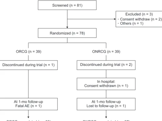
In this study, a total of 81 potential patients undergoing hepatic resection were screened, 78 of whom were enrolled and randomized, and 75 finally completed treatment with ORCG (n
= 38) or ONRCG (n = 37) for local hemostasis at the late stage of surgery. A study flowchart is shown in Fig. 1.
The baseline characteristics of patients between the 2 groups are listed in Table 1. The median age of all patients was 56 years (range, 25–73 years), and 53.3% (40 of 75) were male, and liver cirrhosis was present in 26.7% of patients. Hepatocellular carcinoma and hepatic hemangioma (62.7%, 47 of 75) were the most common reason for liver resection. Traditional laparotomy, laparoscopy, and robotic surgery were used to perform liver resection according to the characteristics, size, and location of the lesions, and the median operative time was 170 minutes (60–448 minutes). The preoperative and intraoperative parameters were similar between the 2 groups.
There was no significant difference between the ORCG and ONRCG groups in time to hemostasis from column analysis (238.8 ± 121.6 seconds vs. 193.7 ± 85.3 seconds, P = 0.068) (Table 2, Fig. 2A). In addition, there were no differences in the rates of hemostatic success between the 2 groups (odds ratio [OR], ORCG/ONRCG) at 120 seconds (18.4% vs. 24.3%; OR, 0.703;
95% confidence interval [CI], 0.231–2.136) and 300 seconds (71.1%
vs. 89.2%; OR, 0.298; 95% CI, 0.085–1.041) (Table 2, Fig. 2B).
However, the ONRCG group seemed to be superior to the ORCG group in hemostasis from a curve analysis (log-rank test, P = 0.044) (Fig. 2C).
Discontinued during trial (n = 1)
At 1-mo follow-up Fatal AE (n = 1)
Discontinued during trial (n = 2)
At 1-mo follow-up Lost to follow-up (n = 1)
In hospital:
Consent withdrawn (n = 1)
ORCG, completed (n = 38) ONRCG, completed (n = 37)
ORCG (n = 39) ONRCG (n = 39)
Excluded (n = 3) Consent withdraw (n = 2) Others (n = 1)
Randomized (n = 78) Screened (n = 81)
Fig. 1. Flowchart of the patients.
ORCG, oxidized regenerated cellulose gauze; ONRCG, oxidi
zed nonregenerated cellulose gauze; AE, adverse events.
Table 1. Baseline characteristics of patients
Characteristic ORCG ONRCG Pvalue
No. of patients 38 37
Basic characteristic
Age (yr) 60.5 (30–73) 56 (25–72) 0.213
Sex, male:female 21 (55.3):17 (44.7) 19 (51.4):18 (48.6) 0.734
Body mass index (kg/m2) 23.80 ± 3.44 24.33 ± 3.90 0.541
Cirrhosis 10 10 >0.999
Pathology of tumor
Benign disease 16 (42.1) 12 (32.43) 0.387
Malignant disease 22 (57.9) 25 (67.57)
Hepatic hemangioma 7 8 0.729
Intrahepatic bile duct lithiasis 4 2 0.695
Hepatic focal nodular hyperplasia 2 1 >0.999
Other primary benign tumor 3a) 1b) 0.629
Hepatocellular carcinoma 14 18 0.301
Intrahepatic cholangiocarcinoma 2 2 >0.999
Hilar cholangiocarcinoma 1 2 0.981
Other primary malignant tumor 2c) 1d) >0.999
Metastasis liver tumor 3e) 2f) >0.999
Intraoperative characteristic
Blood loss (mL) 200 (30–2,100) 300 (50–3,000) 0.789
Surgery time (min)
Mean time 160 (78–448) 175 (60–397) 0.974
≥180 18 (47.4) 18 (48.7) 0.912
Surgery approach
Laparotomy 32 (84.2) 30 (81.1) 0.720
Laparoscopy 5 (13.2) 3 (8.1) 0.738
Robot 1 (2.6) 4 (10.8) 0.339
Values are presented as number only, median (range), number (%), or mean ± standard deviation.
ORCG, oxidized regenerated cellulose gauze; ONRCG, oxidized nonregenerated cellulose gauze.
a)Hepatic angiomyolipoma, 1; hepatic cyst, 1; and hepatic vascular tumor, 1. b)Hepatobiliary cystadenoma. c)Gallbladder carcinoma.
d)Hepatic sarcomatoid carcinoma. e)Liver metastasis of colorectal cancer, 2 and liver metastasis of ovarian carcinoma 1. f)Liver metastasis of colorectal cancer, 1 and liver metastasis of gastrointestinal stromal tumor, 1.
Table 2. Postoperative profiles
Variable ORCG (n = 38) ONRCG (n = 37) Pvalue
Hemostatic efficacy
Hemostatic time (sec) 238.80 ± 121.60 193.70 ± 85.30 0.068
Hemostatic success
At 120 sec 7 (18.4) 9 (24.3) 0.533
At 300 sec 27 (71.1) 33 (89.2) 0.050
At 600 sec 38 (100) 37 (100)
Postoperative drainage (mL)
POD 1 68 (0–885) 75 (0–500) 0.436
POD 2 20 (0–570) 40 (0–1,160) 0.381
Hospital stay (day)
Total 17 (9–39) 18 (8–47) 0.537
Postoperative 9 (5–22) 9 (4–26) 0.200
Values are presented as mean ± standard deviation, number (%), or median (range).
ORCG, oxidized regenerated cellulose gauze; ONRCG, oxidized nonregenerated cellulose gauze; POD, postoperative day.
The median postoperative drainage volume was 68 mL (0–885 mL) in the ORCG group and 75 mL (0–500 mL) in the ONRCG group on the first day after surgery. On the second day after surgery, the median postoperative drainage volume was 20 mL (0–570 mL) in the ORCG group and 40 mL (0–1,160 mL) in the ONRCG group. There were no significant differences between the 2 groups on the first 2 days after the operation (P = 0.436, P
= 0.381) (Table 2).
The total hospital stay was 17 days (9–39 days) in the ORCG group and 18 days (8–47 days) in the ONRCG group. There was no significant difference between the groups (P = 0.537). The postoperative hospital stay was 9 days (5–22 days) in the ORCG group and 9 (4–26) in the ONRCG group. The difference was also not significant between the groups (P = 0.200) (Table 2).
Surgical complications were evaluated by the Clavien-Dindo classification [14], and a surgical complication with a score higher than grade II was not found in this study. Furthermore, the parameters of laboratory examinations including blood routine, liver function, renal function, coagulation function, and infectious markers did not show any significant differences between the 2 groups perioperatively (Supplementary Table 1).
DISCUSSION
The liver is predisposed to bleed diffusely because of its extensive vessels and inferior vascular contractility [15].
Postoperative morbidity and mortality are closely related to blood loss during hepatic resection [16]. It is therefore challenging to control bleeding after the resection of liver parenchyma intraoperatively. In the past 3 decades, new surgical techniques, apparatus, and products with hemostatic, sealant, and adhesive qualities have been used to substantially reduce bleeding and its related complications during liver resection.
Among these, oxidized cellulose including regenerated and non- regenerated hemostatic agents are widely used [13,17].
It has been demonstrated using scanning electron microscopy that different fiber structures exist between ORCG and ONRCG, while they have similar pH in human pooled plasma and undifferentiated bactericidal effectiveness according to colony- forming unit assays. Oxidized non-regenerated cellulose seems superior to oxidized regenerated cellulose in animal hepatic models in terms of hemostasis, which is likely due to its frayed fiber structure and greater material density [18]. The greater surface area of oxidized non-regenerated cellulose can facilitate
ORCG ONRCG
Hemostatictime(sec)
600
400
200
0
300 600
Hemostaticsuccess(%)
100
80
60
40
20
0
After treatment (sec)
ORCG ONRCG
200 600
Probability(%)
100
80
60
40
20
Time to hemostasis (sec)
ORCG ONRCG
120
0 400
P = 0.044 P = 0.068
A B
C
Fig. 2. Hemostatic time in patients undergoing hepatectomy between the 2 groups. (A) The scatter plot of the time to hemostatic in patients undergoing hepatectomy. (B) Hemostatic success at 120, 300, and 600 seconds after treatment in patients undergoing hepatic resection. (C) Time to hemostasis (KaplanMeier) in patients undergoing hepatectomy. ORCG, oxidized regenerated cellulose gauze;
ONRCG, oxidized nonregenerated cellulose gauze.
bleeding sites to clot more rapidly. However, no clinical study has been performed to evaluate the efficacy of regenerated and non-regenerated oxidized cellulose hemostatic agents in controlling bleeding for patients undergoing hepatic resection.
In this study, we set the time to hemostasis as the primary endpoint, and postoperative drainage volume and duration of hospital stay as the secondary endpoints for comparing the efficacy of these 2 gauzes. There was no significant difference between the ORCG and ONRCG groups in time to hemostasis from column analysis; however, ONRCG was slightly superior to ORCG for achieving hemostasis in patients undergoing hepatic resection from a curve analysis. Moreover, there were also no significant differences between the 2 groups in postoperative drainage volume on the first 2 days or in hospital stay.
The Surgicel Original absorbable hemostat has been proven effective and safe for more than 50 years. However, the absorption of oxidized cellulose can be lessened in cauterized areas, and adverse events from foreign body reactions have been reported in patients using oxidized cellulose [19]. The removal of any excess gauze before surgical closure has been taken as a precaution to facilitate absorption and minimize the possibility of foreign body reactions. Traumastem TAF is an agent used to stop capillary, venous, and very minor arterial bleeding, and has been adopted successfully to reduce postoperative bleeding in patients who have undergone thoracic surgery, neurosurgery, and liver resection [20-22]. It has been reported to fully biodegrade by 8 days; however, the residual reticulum of oxidized cellulose can be detected in some individuals by ultrasound at postoperative 1-month follow- up. The absorption of oxidized non-regenerated cellulose will depend on several factors, including the felts used, wound bed, and degree of saturation with blood [23]. Therefore, care should be taken not to apply the gauze too tightly around the first and second hepatic portal area, as it may cause a stenotic effect on vessels and ducts after liver resection.
The vascular intervention or reoperation for postoperative bleeding is rare occurrence in patients undergoing liver resection in our department. There was no delayed bleeding or complication due to postoperative bleeding in any group from this clinical trial. In addition, we set some objective indicators and did not find any differences between the 2 groups in terms of postoperative drainage volume, hospital stay, or laboratory examinations indexes such as blood routine, liver function, renal function, coagulation function, or infectious markers.
This study does have some limitations that need to be considered when interpreting the findings. First, while our findings indicate relatively superior results for ONRCG in comparison to ORCG, the difference was not obvious. The sample size in our study was relatively limited, which could weaken the statistical power of our conclusions. Furthermore, the span of postoperative observation points between
individuals was large. The rationality of the study design and the compliance of the patient follow-up should be improved.
In addition, other indexes of gauze quality such as flexibility, biocompatibility, absorbability, adhesiveness, and cost were not compared in this study, and this data should be supplemented by future studies.
In conclusion, the results of this study indicate slight discrepancies between the 2 groups, and ONRCG was not inferior to ORCG as a secondary hemostatic agent of operative sites in patients undergoing liver resection.
SUPPLEMENTARY MATERIALS
Supplementary Table 1 can be found via https://doi.
org/10.4174/astr.2021.100.4.193.
ACKNOWLEDGEMENTS
Fund/Grant Support
This research was funded by the China Postdoctoral Science Foundation (No. 2018M641740) and Clinical Medicine Discipline Promotion Plan-Cultivate Discipline Construction Support Plan of China Medical University (No. 111–3110118051).
Conflict of Interests
The authors have no conflict of interests.
ORCID iD
Chengshuo Zhang: https://orcid.org/0000-0001-9679-5271 Dazhi Fu: https://orcid.org/0000-0002-6213-4499 Fengshan Wang: https://orcid.org/0000-0002-1182-8597 Xinping Zhong: https://orcid.org/0000-0003-0480-974X Lei Yang: https://orcid.org/0000-0003-2708-5940 Gang Wu: https://orcid.org/0000-0003-0412-1144 Baifeng Li: https://orcid.org/0000-0003-1271-701X Jialin Zhang: https://orcid.org/0000-0003-2617-2735
Author Contribution
Conceptualization: CZ, JZ Formal Analysis: LY
Investigation: DF, FW, XZ, GW, BL, JZ Methodology: CZ
Project Administration: JZ
Writing – Original Draft: DF, FW, XZ, LY, GW, BL Writing – Review & Editing: CZ, JZ
Additional Contributions
The hemostatic gauzes Surgicel Original and Traumastem TAF used in this study were solely provided by Beijing Zhongxing Kangtai Biotechnology Limited Company without influence on the study design; on the collection, analysis, or interpretation
of data; on the writing of the manuscript; or on the decision to submit the manuscript for publication.
REFERENCES
1. Kubo S, Takemura S, Yamamoto S, Hai S, Ichikawa T, Kodai S, et al. Risk factors for massive blood loss during liver resection for hepatocellular carcinoma in patients with cirrhosis. Hepatogastroenterology 2007;54:830-3.
2. Lei Z, Chang L, Fan-Di M, Qi-Fei W, Feng T, Ming-Hui T, et al. Exploration on surgical- related factors influencing HCC patients prognosis. Hepatogastroenterology 2012;59:1541-3.
3. H a r a d a N, S h i r a b e K , M a e d a T, Kayashima H, Ishida T, Maehara Y.
Blood transfusion is associated with recurrence of hepatocellular carcinoma after hepatectomy in Child-Pugh class A patients. World J Surg 2015;39:1044-51.
4. Li Z, Sun YM, Wu FX, Yang LQ, Lu ZJ, Yu WF. Controlled low central venous pressure reduces blood loss and trans- fusion requirements in hepatectomy.
World J Gastroenterol 2014;20:303-9.
5. Lau WY, Lai EC, Lau SH. Methods of vascular control technique during liver resection: a comprehensive review.
Hepatobiliary Pancreat Dis Int 2010;9:473- 81.
6. Lee N, Cho CW, Kim JM, Choi GS, Kwon CH, Joh JW. Application of temporary inf low control of the Glissonean pedicle method provides a safe and easy technique for totally laparoscopic hemi hepatectomy by Gl issonean approach. Ann Surg Treat Res 2017;92:383- 6.
7. Doklestic K, Karamarkovic A, Stefanovic B, Stefanovic B, Milic N, Gregoric P, et al. The efficacy of three transection techniques of the liver resection: a randomized clinical trial. Hepatogastroenterology 2012;59:1501-6.
8. Wolf RF, Xie H, Petty J, Teach JS, Prahl
SA. Argon ion beam hemostasis with albumin after liver resection. Am J Surg 2002;183:584-7.
9. Rau HG, Wichmann MW, Schinkel S, Buttler E, Pickelmann S, Schauer R, et al.
Surgical techniques in hepatic resections:
Ultrasonic aspirator versus Jet-Cutter. A prospective randomized clinical trial.
Zentralbl Chir 2001;126:586-90.
10. Arita J, Hasegawa K, Kokudo N, Sano K, Sugawara Y, Makuuchi M. Randomized clinical trial of the effect of a saline- linked radiofrequency coagulator on blood loss during hepatic resection. Br J Surg 2005;92:954-9.
11. Spotnitz WD, Burks S. State-of-the- art review: hemostats, sealants, and adhesives II. Update as well as how and when to use the components of the surgical toolbox. Clin Appl Thromb Hemost 2010;16:497-514.
12. Frantz VK, Lattes R. Oxidized cellulose- absorbable gauze (cellulosic acid). JAMA 1945;129:798-801.
13. Lewis KM, Spazierer D, Urban MD, Lin L, Redl H, Goppelt A. Comparison of regenerated and non-regenerated oxidized cellulose hemostatic agents. Eur Surg 2013;45:213-20.
14. Dindo D, Demartines N, Clavien PA.
Classification of surgical complications: a new proposal with evaluation in a cohort of 6,336 patients and results of a survey.
Ann Surg 2004;240:205-13.
15. Genyk Y, Kato T, Pomposelli JJ, Wright JK Jr, Sher LS, Tetens V, et al. Fibrin sealant patch (TachoSil) vs oxidized regenerated cellulose patch (Surgicel Original) for the secondary treatment of local bleeding in patients undergoing hepatic resection:
a randomized controlled trial. J Am Coll Surg 2016;222:261-8.
16. Cescon M, Vet rone G, Gra z i GL , Ramacciato G, Ercolani G, Ravaioli M, et al. Trends in perioperative outcome after hepatic resection: analysis of 1,500 consecutive unselected cases over 20 years. Ann Surg 2009;249:995-1002.
17. Wang Z, Liao BY, Qiu SJ, Sun HC, Yang XR, Zhou J, et al. Oxidized regenerated cel lulose reduces t he amount of fluid drainage after liver resection: a randomized prospective clinical trial.
Hepatogastroenterology 2015;62:951-4.
18. Lewis PB, Wilson ST, Kentala DR, Barry J, Lewis KM. Computed tomographic characterization of Traumastem: a new oxidized cellulose hemostatic agent.
Tomography 2016;2:175-8.
19. Badenes D, Pijuan L, Curull V, Sánchez- Font A. A foreign body reaction to Surgicel® in a lymph node diagnosed by endobronchial ultrasound-guided transbronchial needle aspiration. Ann Thorac Med 2017;12:55-6.
20. Habal P, Simek J, Stĕtina M. [Improving of treatment safety in emergency thoracic surgery]. Rozhl Chir 2010;89:261-4. Czech.
21. Sefr R, Silák J, Ondrák M, Fiala L. [Use of local hemostyptic drugs in liver resections]. Rozhl Chir 2009;88:337-41.
Czech.
22. Haghpanah S, Zahedi Z, Parand S, Karimi M. An experience of using Traumastem P in control of spontaneous nose bleeding in patients with inherited bleeding disorders in southern Iran. Haemophilia 2014;20:e79-80.
23. Young ST, Paulson EK, McCann RL, Baker ME. Appearance of oxidized cellulose (Surgicel) on postoperative CT scans:
similarity to postoperative abscess. AJR Am J Roentgenol 1993;160:275-7.