139
Comparison of Anti-allergenic Activities of Various Polyphenols in Cell Assays
Sang Sik Yun1, Mi Young Kang2, Jun Cheol Park3, and Seok Hyun Nam1*
1Department of Biological Science, Ajou University, Suwon 443-749, Republic of Korea
2Department of Food Science and Nutrition, Kyungpook National University, Daegu 702-701, Republic of Korea
3National Institute of Animal Science, Rural Development Administration, Suwon 441-706, Republic of Korea Received January 27, 2010; Accepted September 16, 2010
The inhibitory effects of 25 polyphenols against in vitro allergic reactions were compared using biochemical and cell assays. Three polyphenols including curcumin, gallic acid, and quercetin suppressed the release of β-hexosaminidase from ionophore A23187-stimulated RBL-2H3 cells more effectively (>50% inhibition at 100µM concentration). They were found to have potencies in suppressing the release of histamine not only from ionophore A23187-, but also from immuno- globulin E (IgE)-stimulated RBL-2H3 cells. Moreover, such suppressive effects of the three polyphenols were also observed in A23187 plus PMA-costimulated rat peritoneal mast cells. The extent of inhibition were quantified as the respective polyphenol concentration that inhibit 50%
(IC50) of β-hexosaminidase or histamine release, showing an inhibition tendency with decreasing order of curcumin>gallic acid>quercetin. Down-regulation of Ca2+ influx was suggested as the cause of the inhibition of β-hexosaminidase and histamine releases in these cells. The immune process inhibition was confirmed by the observed reduction in the gene expressions and release of pro- inflammatory cytokine tumor necrosis factor (TNF)-α, interleukin (IL)-1β, and IL-4, due probably to antioxidant activity of the polyphenols. These findings illustrate that curcumin, gallic acid, and quercetin may be beneficial against allergic inflammatory diseases.
Key words: allergy, β-hexosaminidase, histamine, peritoneal mast cell, polyphenol, proinflammatory cytokines, RBL-2H3 cell
Allergies are a result of an overreaction of the immune system towards foreign substances called allergens, which are ordinarily harmless but perceived to be potentially harmful by the immune system [Nauta et al., 2008]. When these allergens, such as pollens, mold spores, peanut protein, and house dust mites, are inhaled, ingested, or come into contact with the skin, they activate the basophilic and mast cells, the central early effector cells of various allergic diseases and of immune responses through high-affinity IgE (immunoglobulin E) receptor (FcεRI)- mediated responses [Oliver et al., 2000; Stone et al., 2010]. The activation of basophils and mast cells triggers the release of inflammatory mediators including histamine and cytokines, which are responsible for the clinical manifestation of allergic reactions, such as tissue swelling, hay fever, and asthma.
Epidemiological studies revealed that the incidence of allergic diseases is increasing worldwide in epidemic proportions [Pawankar et al., 2008]. With this rising trend in the global
prevalence of allergies, concerted efforts have been directed in developing therapeutic measures against the disease. Scientists and researchers have attempted to find inhibitors of the FcεRI signaling pathway with the intention of blocking the IgE- mediated allergic reaction through inhibition of basophil and mast cell degranulation [Oliver et al., 2000]. Scientific evidences have indicated that inhibition of antigen-induced release of histamine and β-hexosaminidase, biomarkers of mast cell degranulation, is necessary to elicit an anti-allergenic response.
Moreover, suppression of antigen-induced production of pro- inflammatory cytokines, such as tumor necrosis factor-α (TNF- α) and interleukin (IL), and blockading the cellular Ca2+ influx critical to histamine release also provide for a potential therapeutic target for the prevention and treatment of allergic disorders [Beaven et al., 1984; Nauta et al., 2008].
Polyphenols are a group of highly hydroxylated phenolic compounds that are widely distributed in plants and the most abundant antioxidants in the diet [Scalbert et al., 2005]. These compounds are classified based on the number of phenol rings that they contain and of the structural elements that bind these rings to one another [Manach et al., 2004]. Various types of polyphenols, such as phenolic acids, tannins, and flavonoids,
*Corresponding author
Phone: +82-31-219-2619; Fax: +82-31-219-1615 Email: [email protected]
doi:10.3839/jabc.2010.026
exhibit a wide range of pharmacological properties including anti-carcinogenic, anti-mutagenic, and cardioprotective effects as a consequence of their strong antioxidant properties [Urquiaga and Leighton, 2000]. Some polyphenols were also reported to have anti-inflammatory activity and inhibitory effect against histamine release [Benavente-Garcia et al., 1997;
Yamada et al., 1999; Kim et al., 2004]. While the antioxidant properties of polyphenols and their role in the prevention of degenerative diseases, particularly cardiovascular diseases and cancers, have been studied extensively, the comparative anti- allergenic activities of different phenolic compounds are not well documented. The rat basophilic leukemia (RBL-2H3) cell line has been widely used as a cell assay model for allergy and immunological research [Passante and Frankish, 2009]. In this study, the anti-allergenic activities of 25 polyphenols were evaluated and compared. Using RBL-2H3 basophilic leukemia and rat peritoneal mast cells, the capacity of these polyphenols to inhibit the release of histamine and β-hexosaminidase and suppress the genetic expressions and production of pro- inflammatory cytokines were examined in vitro and ex vivo.
Materials and Methods
Materials. The 1,1-Diphenyl-2-picrylhydrazyl (DPPH), o- phthalaldehyde (OPT), ρ-nitrophenyl-N-acethyl-β-glucosaminide, phorbol-12-myristate-13-acetate (PMA), ionophore A23187, guanidium thiocyanate, dimethyl sulfoxide (DMSO), and other chemicals including 25 polyphenols were purchased from Sigma Chemicals (St. Louis, MO, USA). All reagents were of analytical grade and used without further purification. The RPMI 1640 medium, Hank’s balanced salt solution, fetal bovin serum (FBS), and other cell culture reagents were obtained from Hyclone Laboratories (Logan, UT, USA). The anti-dinitrophenyl (DNP) immunoglobulin E (IgE) and dinitrophenyl-bovine serum albumin (DNP-BSA) were also purchased from Sigma Chemicals. The cytokine quantification kits using enzyme- linked immunoabsorbent assay (ELISA) were obtained from Biosource (Camarillo, CA, USA). The AMV reverse transcriptase, Taq DNA polymerase, and dNTP mix were products of Takara Bio (Kyoto, Japan). All PCR primers were custom-synthesized and purified by Bioneer (Daejon, Korea).
Cell culture and preparation of rat peritoneal mast cells.
The RBL-2H3 cells (Japan Health Science Resource Bank, Osaka, Japan) were maintained in RPMI 1640 medium containing 10% heat-inactivated FBS with 100 units/mL penicillin and 100µg/mL streptomycin at 37oC in a humidified air containing 5% CO2. The cells were detached with a trypsin- EDTA solution. The cells were then washed and resuspended in the medium or in an appropriate buffer for subsequent experiments.
Isolation and purification of rat peritoneal mast cells were performed according to the slightly modified method of Lu et al.
[2004]. Male Sprague-Dawley rats (Orient, Seoul, Korea), aged 7 weeks, were first anaesthetized with chloroform, followed by gavaging of the peritoneal cavity with 20 mL of Tyroid B buffer (137 mM NaCl, 2.7 mM KCl, 12 mM NaHCO3, 0.3 mM NaHPO4, 5.3 mM glucose, 0.1% gelatin, pH 7.2). Repeated washings were pooled and centrifuged at 200×g for 10 min at 4oC. The exudated cell pellet was then resuspended in 1 mL of Tyroid A buffer (10 mM HEPES, 130 mM NaCl, 5 mM KCl, 1.4 mM CaCl2, 1 mM MgCl2, 5.6 mM glucose, 0.1% BSA, pH 7.2) and layered onto 2 mL of Histopaque-1119 (Sigma Chemicals).
The cell suspension was then centrifuged at 400×g for 15 min at 4oC. The recovered cells in the pellet were resuspended in Tyroid A buffer. The cell number and viability were determined by trypan blue staining.
β-Hexosaminidase and histamine secretion assay. To evaluate the level of mast cell degranulation, β-hexosaminidase was determined by the method of Jeong et al. [2002] with some modifications. The RBL-2H3 cells or peritoneal mast cells were suspended in Tyroid A buffer. For stimulation of cells with A23187, the cells were seeded into a 96-well plate (1×10⁵ cells/
well), and mixed with appropriate amount of polyphenols for 15 min at 37oC. For stimulation of cells with DNP-BSA, the same number of cells was preloaded at 37oC overnight with 1µg of anti-DNP IgE, and the cell suspension was then mixed with appropriate amount of polyphenols for 15 min at 37oC. For priming cell functions, following the treatment of the polyphenols, ionophore A23187 and DNP-BSA were added to the cell suspension to a final concentration of 10µM and 20 µg/mL, respectively. Simultaneous treatment of PMA and ionophore A23187 (10µM each) was performed to elicit maximum priming of rat peritoneal mast cells. Incubation was continued for another 20 min. The cell suspension was then centrifuged at 300×g for 10 min to recover the supernatant, which contained the released β-hexosaminidase. The recovered supernatant (50 µL) was added with the same volume of 1 mM ρ-nitrophenyl-N- acethyl-β-glucosaminide solution (pH 5.2). The sample was then incubated at room temperature for 1 h with continuous shaking.
The reaction was terminated by adding 0.2 mL of 1 M sodium carbonate buffer (pH 10.2). The plate was read at 405 nm using a microplate reader (Model 550, Bio-Rad, Hercules, CA, USA).
The assay for histamine secretion was performed according to the method of Kawasaki et al. [1994] with some modifications.
The RBL-2H3 cells or peritoneal mast cells were suspended in Tyroid A buffer and then divided into a 24-well plate (1×10⁶ cells/well). The preloading of the polyphenols and cell-priming with each stimulant was performed with the same procedure as described above for β-hexosaminidase. The histamine level was measured using a fluorometric assay as described by Shore et al.
[1959]. In this assay, 1 mL of the recovered supernatant, 0.2 mL of 1 N NaOH, and 0.1 mL of 1% OPT were mixed and the sample was incubated at room temperature for 5 min. The reaction was terminated by adding 0.2 mL of 1 N HCl. The fluorescence intensity was then measured using a spectro- fluorometer (Model RF-550, Shimazu, Kyoto, Japan) at excitation and emission wavelengths of 360 and 405 nm, respectively.
Measurement of cell viability. The effects of the polyphenols on cell viability were determined using MTT colorimetric assay according to the method of Mosmann [1983]. Briefly, the RBL- 2H3 cells were seeded in a 96-well plate at a density of 1×10⁵ cells per well. The cells were loaded with appropriate doses of the polyphenols and cultivated for 48 h at 37oC under humidified air containing 5% CO2. Following removal of culture media, 0.1 mL of 5 mg/mL MTT solution was added to each well and the incubation was continued for another 3 h. The DMSO was then added onto the supernatant-drained cell layer to dissolve the intracellular chromogen by incubating for 30 min.
The absorbance of the supernatant was read in a microplate reader at 570 nm with a reference wavelength of 650 nm.
Measurement of DPPH scavenging activity. For the determination of the antioxidant activity of the polyphenols, the DPPH scavenging activity was measured based from the method of Cavin et al. [1998]. Briefly, 0.5 mL of ethanolic solution of 0.5 mM DPPH was mixed with 1 mL mixture of polyphenol solution and 0.1 M sodium acetate buffer (pH5.5), and 1 mL ethanol. The absorbance of the mixture, measured against ethanol, at 517 nm was determined using UV/VIS spectrophotometer (Model V-550, Jasco, Tokyo, Japan).
Controls containing DPPH solution and DMSO instead of the polyphenol solution and blanks containing ethanol alone instead of DPPH solution were also prepared. The inhibition of the DPPH radical by the polyphenols was calculated according to the following formula: DPPH scavenging activity (%)=[1−(Abs.
of sample−Abs. of blank)/Abs. of control]×100. BHT was used as positive control.
Determination of intracellular Ca2+ level. The intracellular Ca2+ responses were assessed using Fluo-3-AM with the aid of confocal microscope according to the method of Matsubara et al. [2004]. The RBL-2H3 cells were cultivated at a density of 1×10⁴ cells/mL on a dish used exclusively for confocal microscope cell imaging. The cells were suspended in Ca2+
buffer (137 mM NaCl, 2.7 mM KCl, 1.8 mM CaCl2, 1 mM MgCl2, 1 mg/mL glucose, 1 mg/mL BSA, 20 mM HEPES, pH 7.4). They were then loaded with Fluo-3-AM to a final concentration of 10µM and incubated at 37oC for 30 min. After washing with the same buffer to remove the excess dye, the
polyphenols appropriately diluted with Ca2+ buffer were added to the cells and incubated for 15 min. After washing, the cells were immediately placed onto the confocal scanning laser microscope (Model LSM510, Carl Zeiss, Thornwood, NY, USA) and stimulated with 10µM ionophore A23187 for 1 min.
The change in fluorescence of Fluo-3-AM was monitored at an excitation wavelength of 488 nm and an emission wavelength of 540 nm. Using the analyst software, consecutive images of cells were captured at 2 s interval over the time course of 300 s after treatment with ionophore A23187.
Reverse transcription-polymerase chain reaction (RT- PCR) analysis. The total RNA was prepared from 1×10⁷ cells/
mL of RBL-2H3 cells by acid phenol guanidium thiocyanate- chloroform extraction procedure of Chomczynski and Sacchi [1987]. The single stranded cDNA synthesis from 1µg of total RNA was performed using an AMV reverse transcriptase and oligo dT18 as primer. DNA amplification was primed in a reaction mixture containing 400µM of dNTP mix, 2.5 U of Taq polymerase and 20µM each of the primer sets representing the target genes as follows: TNF-α (sense) 5'-TAC TGA ACT TCG GGG TGA TCG GTC C-3', (anti-sense) 5'-CAG CCT TGT CCC TTG AAG AGA ACC-3'; 1L-1β (sense) 5'-GTA GCC CAC GTC GTA GCA AA-3', (anti-sense) 5'-CCC TTC TCC AGC TGG GAG AC-3'; IL-4 (sense) 5'-ACC TTG CTG TCA CCC TGT TC-3'; (anti-sense) 5'-TTG TGA GCG TGG ACT CAT TC-3'; β-actin (sense) 5'-GTG GGG CGC CCC AGG CAC CA-3', (anti-sense) 5'-GTC CTT AAT GTC ACG CAC GAT TTC-3'. PCR was conducted using a thermocycler (Model PTC-200, MJ Research, Reno, NV, USA) with 1 cycle of 5 min at 94oC, followed by 30 cycles of 30 s at 94oC, 45 s at 58oC, and 45 s at 72oC, and finally 1 cycle of 5 min at 72oC. All PCR products were subjected to 1.5% agarose electrophoresis. The intensity of separated bands under DNA was quantified by a gel documentation system (Model LAS-1000CH, Fuji Film, Tokyo, Japan).
ELISA of cytokines. After stimulating the RBL-2H3 cells preloaded with polyphenols, the culture supernatants were recovered and the cytokines TNF-α, IL-β and IL-4 in the resultant supernatants were measured using ELISA assay kits as described according to the manufacturer’s instructions. The absorbance of the final reaction mixture at 420 nm was measured using a microplate reader.
Statistical analysis. Statistical analysis was accomplished with SAS software package (SAS Inst., Cary, NC, USA).
Significant differences between means were determined by ANOVA procedure test and a p value of <0.05 was considered to be statistically significant. The results are expressed as the mean±SD from triplicate test data.
Results and Discussion
Effect of various polyphenols on β-hexosaminidase release in rat basophilic leukemia RBL-2H3 cells. The inhibitory effects of polyphenols on the β-hexosaminidase release in A23187-stimulated RBL-2H3 cells are presented in Table 1.
Among the 25 polyphenols analyzed, curcumin, at 10µM concentration, showed the highest inhibition of β-hexo- saminidase release with 48%, followed by gallic acid (21%), while naringin, hesperitin, 2-hydroxyquinoline, synaptic acid, syringic acid, and hesperidin showed no significant inhibitory effect. Likewise, at a concentration of 100µM, curcumin was found to have the highest potential to suppress β-hexosaminidase release. Inhibition of β-hexosaminidase release above 50% was also observed in gallic acid (77%) and quercetin (56%). On the other hand, lowest inhibition (<10%) was obtained in synaptic acid, syringic acid, naringin, and rutin.
Effect of various polyphenols on cell viability of RBL-2H3 cells. The viabilities of RBL-2H3 cells exposed to polyphenols are shown in Table 2. At 10µM concentration, p-courmaric
acid, ferulic acid, synaptic acid, benzoic acid, vanillic acid, cinnamic acid, curcumin, syringic acid, chrysin, naringenin, naringin, hesperetin, and rutin did not significantly affect the cell viability. Exposure with the rest of the polyphenols resulted in a 5-10% decrease in cell viability. Even at 100µM concentration, the polyphenols examined, except p-coumaric acid and chlorogenic acid, did not significantly change the viability of RBL-2H3 cells compared to that of untreated control (above 90% cell viability after 48 h exposure to the polyphenols). The results indicate that the polyphenols analyzed in this study were mostly not cytotoxic at concentrations up to 100 µM.
Antioxidant activity of various polyphenols. Phenolic compounds have ideal structural chemistry for ROI (reactive oxygen intermediates)-scavenging activities [Rice-Evans et al., 1997] and their antioxidant activity is mainly due to their redox properties, which play an important role in adsorbing and neutralizing free radicals, quenching oxygen, or decomposing peroxides [Karou et al., 2005]. In the present study, the antioxidant activities of various polyphenols were evaluated by determining their DPPH radical-scavenging abilities and the results are shown in Table 3. Gallic acid and quercetin, at 10 Table 2. Cell viability changes in RBL-2H3 basophilic leukemia cells measured using MTT assay
Sample Cell Viability (%)
10 µM 100 µM
Control (-A23187) 100.00±2.17a 100.00±2.17a m-Coumaric acid 90.77±1.64i 94.66±1.91bcd o-Coumaric acid 93.85±3.74h 95.24±1.07bcd p-Coumaric acid 99.81±1.36ab 87.07±1.98fg Caffeic acid 95.12±0.98efgh 92.17±2.57cde Ferulic acid 100.69±1.17a 89.87±0.51ef Synaptic acid 99.45±0.85abc 93.79±0.19bcde Gallic acid 95.59±0.34defgh 93.67±1.14bcde Benzoic acid 98.73±0.46abc 92.26±3.17cde Vanillic acid 98.62±0.32abc 91.59±3.22de Chlorogenic acid 97.62±1.91bcde 85.78±2.83g Cinnamic acid 99.60±1.21ab 90.77±1.64def Rosmarinic acid 93.93±1.00gh 91.85±3.74de
Curcumin 100.55±1.15a 93.91±0.65bcde
2-Hydroxyquinoline 94.57±1.29fgh 99.81±1.36a Esculetin 94.29±1.05gh 92.78±1.34bcde Ellagic acid 95.29±1.78efgh 97.19±0.52ab Purpurin 96.67±1.44cdefg 91.95±1.89cde Syringic acid 98.26±3.15abcd 91.43±0.76de
Chrysin 98.87±0.92abc 96.36±2.02abc
Hesperidin 97.51±1.06bcde 90.03±1.23ef Naringenin 98.75±0.81abc 94.98±3.52bcd Quercetin 97.16±0.05bcdef 90.78±2.87def
Naringin 98.89±1.82abc 92.14±4.72cde
Hesperetin 101.11±0.39a 91.34±1.60de
Rutin 98.48±1.22abc 93.49±2.96bcde
Values with the same superscript within the column are not significantly different at p<0.05.
Table 1. Effects of various polyphenols on ionophore A23187- stimulated β-hexosaminidase release from RBL-2H3 basophilic leukemia cells
Sample Inhibition of β-hexosaminidase release (%)
10 µM 100 µM
Control (-A23187) 100.00±1.02a 100.00±1.02a Control (+A23187) 00.00±0.44m 00.00±0.44p m-Coumaric acid 18.11±0.70cd 32.17±0.25gh o-Coumaric acid 12.56±2.02ef 30.57±4.51h p-Coumaric acid 14.16±1.06de 21.65±4.10i Caffeic acid 18.90±2.20c 22.60±0.17i Ferulic acid 10.13±1.27efgh 21.88±2.52i Synaptic acid 01.29±1.78lm 05.56±1.12no
Gallic acid 21.22±1.25c 77.03±3.09c
Benzoic acid 12.49±1.30ef 21.71±1.11i Vanillic acid 11.94±5.75ef 34.59±4.52fg Chlorogenic acid 08.53±3.08fghi 14.74±1.51jk Cinnamic acid 06.98±0.73ghij 11.63±0.61klm Rosmarinic acid 11.29±0.73efg 24.39±0.73i
Curcumin 48.41±3.27b 84.06±1.87b
2-Hydroxyquinoline 00.98±4.77lm 16.60±0.74j Esculetin 10.50±1.77efgh 12.44±0.69kl Ellagic acid 06.43±2.03hijk 35.72±2.89f
Purpurin 11.94±4.43ef 45.08±0.20e
Syringic acid 02.30±3.82klm 08.24±2.55mn
Chrysin 17.44±1.74cd 21.08±0.47i
Hesperidin 03.74±2.34jklm 10.06±1.84lm
Naringenin 16.82±2.74cd 22.90±0.31i
Quercetin 11.28±1.50efg 56.09±2.23d
Naringin 00.03±1.54m 03.86±1.88o
Hesperetin 00.04±1.01m 11.15±1.05klm
Rutin 04.76±0.67ijk 05.27±0.57no
Values are expressed as mean±SD (n=3). Means with the same superscript within the column are not significantly different at p<0.05.
µM, exhibited the highest scavenging activity with 78.69% and 74.76%, respectively. Lowest activities (<10%) were observed in m-coumaric acid, o-coumaric acid, 2-hydroxyquinoline, chrysin, hesperidin, naringenin, narigin, and hesperetin. On the other hand, at higher concentration (100 µM), purpurin showed the highest DPPH-scavenging activity (103%), followed by curcumin (90%). Naringenin, 2-hydroxyquinoline, chrysin, naringin, m-coumaric acid, o-coumaric acid, and benzoic acid exhibited scavenging abilities lower than 10%. This variation in the antioxidant activities of polyphenols could be attributed to their differences in chemical structures. It was previously reported that the antioxidant activities of polyphenols are dependent on their structures, which define the relative abilities of these compounds to scavenge free radicals [Rice-Evans et al., 1997].
Effect of selected polyphenols on β-hexosaminidase and histamine release in basophilic leukemia and rat peritoneal mast cells. Among the 25 polyphenols investigated for inhibition of β-hexosaminidase release and DPPH radical scavenging activity, curcumin, gallic acid, and quercetin were selected for further analysis on anti-allergic activities on the
basis of their high inhibitory activity against β-hexosaminidase release in RBL-2H3 cells and relatively high antioxidant activity.
Ellagic acid was also used in the succeeding experiments as an internal control with relatively strong antioxidant activity, but with weak inhibitory activity against β-hexosaminidase release.
Table 4 shows the inhibitory effects of selected polyhenols on β- hexosaminidase and histamine releases in RBL-2H3 basophilic leukemia cells. Curcumin exhibited the highest maximum inhibition (94%) for β-hexosaminidase release, while gallic acid and quercetin showed the highest suppression (90%) for histamine release. Curcumin also showed the lowest IC50, the concentration needed to achieve 50% inhibition, for β- hexosaminidase (15µM) and histamine (50 µM). In rat peritoneal mast cells, quercetin and curcumin exhibited the highest maximum inhibition and lowest IC50, respectively, for β- hexosaminidase release, while gallic acid showed the highest maximum inhibition and lowest IC50 for histamine release (Table 5). When the RBL-2H3 cells stimulated via FcεRI with an anti- DNP IgE were exposed to polyphenols at 10µM and 100 µM concentrations, the highest inhibition for β-hexosaminidase release was also found in curcumin-treated cells (Table 6).
Between gallic acid and quercetin, the latter showed significantly greater suppression of β-hexosaminidase release. These results demonstrate that the three polyphenols are highly effective in blockading mast cell degranulation in both in vitro and ex vivo assay systems. Of the three polyphenols analyzed, curcumin appeared to be the most potent inhibitor. Previous investigations have also revealed that curcumin could inhibit the histamine Table 3. Antioxidant activities of polyphenols evaluated by electron
donating ability to DPPH radical
Polyphenols Electrons Donating Ability (%)
10 µM 100 µM
BHT 86.88±0.34a 87.19±0.78cde
m-Coumaric acid 05.17±0.66o 09.13±1.36l o-Coumaric acid 05.67±0.24o 09.97±1.10l p-Coumaric acid 12.08±0.06k 23.36±2.54k Caffeic acid 42.27±2.71f 86.83±1.43cde Ferulic acid 24.88±1.77i 85.46±0.28ef Synaptic acid 35.82±2.72g 82.95±1.08g
Gallic acid 78.69±2.63b 86.32±0.74de
Benzoic acid 11.34±1.72kl 09.63±0.24l Vanillic acid 20.84±0.04j 62.16±0.84h Chlorogenic acid 35.89±0.33g 88.78±1.08bc Cinnamic acid 28.14±0.33h 87.28±0.58cde Rosmarinic acid 61.45±0.21d 88.05±0.11bcd
Curcumin 36.11±2.42g 90.16±0.24b
2-Hydroxyquinoline 06.69±0.90mno 06.60±1.03m
Esculetin 50.12±0.78e 87.15±0.38cde
Ellagic acid 69.55±2.91c 84.10±0.41fg
Purpurin 37.61±0.85g 103.31±0.84a
Syringic acid 30.31±1.34h 88.68±0.65bcd
Chrysin 08.87±2.43lmn 06.71±0.41m
Hesperidin 09.19±1.17lm 25.75±1.69j
Naringenin 06.19±0.62no 06.61±2.89m
Quercetin 74.76±0.83b 86.89±1.64cde
Naringin 05.93±2.37o 07.86±2.58lm
Hesperetin 07.95±0.43mno 32.56±0.86i
Rutin 60.92±1.51d 86.56±0.81cde
Values are expressed as mean±SD (n=3). Means with the same superscript within the column are not significantly different at p<0.05.
Table 4. Inhibitory activities of selected polyphenols against β- hexosaminidase and histamine releases from ionophore A23187- stimulated RBL-2H3 basophilic leukemia cells
Sample
β-hexosaminidase release Histamine release Maximum
inhibition (%) IC50 (µM) Maximum
inhibition (%) IC50 (µM)
Curcumin 94.54 15.49 80.41 50.10
Gallic acid 91.25 25.87 90.42 56.45
Quercetin 81.77 92.92 90.53 87.33
Ellagic acid 47.22 ND 69.32 142.960
ND: not determined
Table 5. Inhibitory activities of selected polyphenols against β- hexosaminidase and histamine releases from ionophore A23187 and PMA-costimulated rat peritoneal mast cells
Polyphenols
β-hexosaminidase release Histamine release Maximum
inhibition (%) IC50 (µM) Maximum
inhibition (%) IC50 (µM)
Curcumin 76.44 51.52 80.93 53.15
Gallic acid 75.33 63.89 88.45 47.50
Quercetin 85.32 52.75 69.41 74.01
Ellagic acid 37.55 ND 40.51 ND
ND: not determined
release from rat peritoneal mast cells [Yano et al., 2000a] and has shown anti-allergic activities in animal models [Yano et al., 2000b]. Suzuki et al. [2005] suggested that the hydroxy groups of curcumin-related compounds play a significant role in exerting both the antioxidative and anti-allergic activities. Likewise, gallic acid was found to reduce mast cell-derived inflammatory allergic responses by blocking the histamine release and pro- inflammatory cytokine expressions [Kim et al., 2006]. Quercetin was reported to inhibit histamine release from rat connective tissue mast cells and mucosal mast cells [Pearce et al., 1984], as well as from human lung and intestinal mast cells [Fox et al., 1988]. In addition, quercetin also showed inhibitory effect against ragweed antigen-induced basophil histamine release in subjects with hay fever [Middleton et al., 1981], implying that the inhibitory effects of these three polyphenols against mast cell degranulation may in some part be related to their antioxidant activities.
Inhibitory effects of selected polyphenols on the production and expression of pro-inflammatory cytokines.
Aside from the suppression of β-hexosaminidase and histamine releases, the inhibition of pro-inflammatory cytokine production
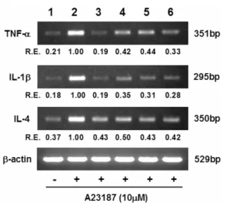
is another key indicator of reduced allergic reactions. Hence, the selected polyphenols were examined whether they could modulate gene expression and release of the pro-inflammatory cytokines including TNF-α, IL-1β, and IL-4. Figure 1 shows the results of exposure of basophilic leukemia cells to polyphenols on the pro-inflammatory cytokine gene expressions obtained with RT-PCR. Curcumin, quercetin, and even ellagic acid with low inhibitory activity toward mast cell degranulation, were able to suppress the transcription of these cytokine genes in stimulated RBL-2H3 cells. Likewise, the release of pro- inflammatory cytokines was also inhibited by these polyphenols (Fig. 2). It is widely known that the expression of pro- Table 6. Effects of selected polyphenols on anti-DNP IgE-
stimulated β-hexosaminidase release from RBL-2H3 basophilic leukemia cells
Polyphenols Inhibition of β-hexosaminidase release (%)
10 µM 100 µM
Control (-DNP-BSA) 100.00±2.26a 100.00±2.26a Control (+DNP-BSA) 00.00±4.34d 00.00±4.34e
Curcumin 63.70±4.34b 91.11±2.26b
Gallic acid 38.77±6.47c 78.52±3.08c
Quercetin 40.99±5.46c 87.16±3.85b
Ellagic acid 11.85±5.36d 34.81±4.93d Values are expressed as mean±SD (n=3). Means with the same superscript within the column are not significantly different at p<0.05.
Fig. 1. Semi-quantitiative RT-PCR analysis of the effect of selected polyphenols on the pro-inflammatory cytokine gene expression in A23187-stimulated RBL-2H3 cells. Lane 1, unstimulated control; lane 2, stimulated control; lane 3, curcumin; lane 4, gallic acid; lane 5, quercetin; lane 6, ellagic acid (internal control as a polyphenol with weak inhibitory activity). Relative expression (R.E.) indicates the relative expression values (cytokine/β-actin) based on the intensity of the DNA band amplified from each cytokine gene. The agarose gel electrophoresis patterns represent results from triplicate experiments.
Fig. 2. ELISA of selected polyphenols on the proinflammatory cytokine releases from A23187-stimulated RBL-2H3 cells. Lane 1, unstimulated control; lane 2, stimulated control; lane 3, curcumin; lane 4, gallic acid; lane 5, quercetin; lane 6, ellagic acid. Each bar represents the average of separate triplicate determination with error bars showing the standard error of the mean. Bars not sharing common letters are significantly different at p<0.05.
inflammatory cytokine genes is regulated at transcription level by NF-κB, and that ROI is mandatorily required for NF-κB activation [Ling et al., 1998; Reynaert et al., 2006]. Therefore, the current finding strongly suggests that the polyphenol-induced attenuation of the pro-inflammatory cytokine expressions is attributed to ROI-scavenging activity of these polyphenols. The cytokines including TNF-α and IL-1β were reported to contribute to the late-phase allergic reactions and allergic inflammation through recruitment of immune cells into the site of inflammation [Musoh et al., 1998; Kang et al., 2008]. Earlier studies revealed the inhibitory effects of curcumin and quercetin against the release and gene expressions of TNF-α and interleukins from peritoneal macrophages, bone marrow-derived cultured murine mast cells, and human mast cells [Kimata et al., 2000; Park et al., 2008; Viswanath and Barrios, 2008]. Results of the current study suggest that curcumin is most effective against late-phase allergic reactions.
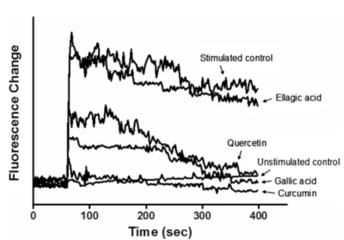
Effect of selected polyphenols on the intracellular calcium level in RBL-2H3 cells. Intracellular calcium ions are known to play a critical role in the degranulation of mast cells. The release of histamine is believed to be a result of an influx of calcium into the mast cell due to permeability changes in the plasma membrane. In the current study, the effect of polyphenols on the calcium ion levels in RBL-2H3 cells was examined using confocal laser microscopy. As shown in Fig. 3, the suppression of Ca2+ influx was highest in cells exposed to curcumin, with calcium ion level similar to that of the unstimulated control.
Gallic acid and quercetin also showed decreased level of intracellular calcium ion. On the other hand, ellagic acid exhibited no inhibitory effects against Ca2+ influx. These findings further substantiate the anti-allergic potential of the three polyphenols, particularly curcumin, through attenuating the degranulation of inflammatory mediators.
Taken together, results of this study demonstrate that curcumin, gallic acid, and quercetin attenuate degranulation of
inflammatory mediators (histamine and β-hexosaminidase) and release of pro-inflammatory cytokines (TNF-α, IL-1β and IL-6) from stimulated mast cell. The attenuation of mast cell degranulation appears to be closely associated with the reduction in calcium ion influx. Curcumin was found to be the most potent inhibitor, followed by gallic acid and quercetin. However, these three polyphenols, as well as ellagic acid, have capacities to suppress the pro-inflammatory cytokines release from activated mast cell probably through their antioxidant actions. Therefore, curcumin, gallic acid, and quercetin may be useful in the treatment of allergic and inflammatory diseases such as bacterial endotoxin-triggered inflammation.
Acknowledgments
This work received grant support from the Agenda Program (No. 200901OFT113068122), Rural Development Administration, Republic of Korea. We thank Dr. Catherine W. Rico for careful correction of the manuscript.
References
Beaven MA, Rogers J, Moore JP, Hesketh TR, Smith GA, and Metcalfe JC (1984) The mechanism of the calcium signal and correlation with histamine release in 2H3 cells. J Biol Chem 259, 7129-7136.
Benavente-Garcia O, Castillo J, Marino FR, Ortuño A, and Del Rio JA (1997) Uses and properties of citrus flavonoids. J Agric Food Chem 45, 4505-4515
Cavin A, Hostettmann K, Dyatmyko W, and Potterat O (1998) Antioxidant and lipophilic constituents of Tinospora crispus.
Planta Med 64, 393-396.
Chomczynski P and Sacchi N (1987) Single-step method of RNA isolation by acid guanidium thiocyanate-phenol-chloroform extraction. Anal Biochem 162, 156-159.
Fox CC, Wolf EJ, Kagey-Sobotka A, and Lichtenstein LM (1988) Comparison of human lung and intestinal mast cells. J Allergy Clin Immunol 81, 89-94.
Jeong HJ, Hong SH, Lee DJ, Park JH, Kim KS, and Kim HM (2002) Role of Ca(2+) on TNF- and IL-6 secretion from RBL- 2H3 mast cells. Cell Signal 14, 633-639.
Kawasaki T, Toyoda M, Teshima R, Sawada J, Hayashi T, Arasawa M, Shimizu M, Inouye S, and Saito Y (1994) In vitro antiallergic activity of flavonoisa in histamine release assay using rat basophilic leukemia (RBL-2H3) cells. J Food Hyg Soc Jpn 35, 497-503.
Kang NI, Kim HK, Ko HM, Kim JH, You HJ, Choi IW, Im SY, and Lee HK. (2008) Tumor necrosis factor-alpha develops late anaphylactic reaction through cytosolic phospholipase A(2) activation. Int Arch Allergy Immunol 147, 315-322.
Karou D, Dicko MH, Simpore J, and Traore AS (2005) Antioxidant and antibacterial activities of polyphenols from ethnomedicinal plants of Burkina Faso. Afr J Biotechnol 4, 823-828.
Fig. 3. Fluorescence assay of the effect of selected polyphenols on the cellular Ca2+ responses using Fluo-3AM with a confocal microscopic fluorometric imaging in A23187-stimulated RBL-2H3 basophilic leukemia cells.
Kim HP, Son KH, Chang HW, and Kang SS (2004) Anti- inflammatory plant flavonoids and cellular action mechanisms.
J Pharmacol Sci 96, 229-245.
Kim SH, Jun CD, Suk K, Shoi BJ, Park S, Lee SH, Shin HY, Kim DK, and Shin TY (2006) Gallic acid inhibits histamine release and pro-inflammatory cytokine production in mast cells. Toxicol Sci 91, 123-131.
Kimata M, Inagaki N, and Nagai H (2000). Effects of luteolin and other flavonoids on IgE-mediated allergic reactions. Planta Med 66, 25-29.
Ling L, Cao Z, and Goeddel DV (1998) NF-κB-inducing kinase activates IKK-α by phosphorylation of Ser-176. Proc Natl Acad Sci USA 95, 3792-3797.
Lu YB, Wu M, and Zhou HL (2004) Changes in phospholipase D activity of rat peritoneal mast cells in degranulation. Acta Pharmacol Sin 25, 104-109.
Manach C, Scalbert A, Morand C, Remesy C, and Jimenez L (2004) Polyphenols: food sources and bioavailability. Am J Clin Nutr 79, 727-747.
Matsubara M, Masaki S, Ohmori K, Karasawa A, and Hasegawa K (2004) Differential regulation of IL-4 expression and degranulation by antiallergic olopatadine in rat basophilic leukemia (RBL-2H3) cells. Biochem Pharmacol 35, 497-503.
Middleton E, Drzewiwcki G, and Krishnarao D (1981) Quercetin:
an inhibitor of antigen-induced human basophil histamine. J Immunol 127, 546-550.
Mosmann T (1983) Rapid colorimetric assay for cellular growth and survival: application to proliferation and cytotoxicity assays. J Immunol Methods 65, 55-63.
Musoh K, Nakamura N, Ueda Y, Inagaki N, and Nagai H. (1998) Possible role of nitric oxide in IgE-mediated allergic cutaneous reaction in mice. Int Arch Allergy Immunol 115, 91-96.
Nauta AJ, Engels F, Knippels LM, Garssen J, Nijkamp FP, and Redegeld FA (2008) Mechanisms of allergy and asthma. Eur J Pharmacol 585, 354-360.
Oliver JM, Kepley CL, Ortega E, and Wilson BS (2000).
Immunologically mediated signaling in basophils and mast cells: finding therapeutic targets for allergic diseases in the human FcεRI signaling pathway. Immunopharmacology 48, 269-281.
Park HH, Lee S, Son HY, Park SB, Kim MS, Choi EJ, Singh TSK, Ha JH, Lee MG, Kim JE, Hyun MC, Kwon TK, Kim YH, and Kim SH (2008) Flavonoids inhibit histamine release and expression of proinflammatory cytokines in mast cell. Arch Pharm Res 31, 1303-1311.
Passante W and Frankish N (2009) The RBL-2H3 cell line: its provenance and suitability as a model for the mast cell.
Inflamm Res 58, 737-745.
Pawankar R, Baena-Cagnani CE, Bousquet J, Canonica GW, Cruz AA, Kaliner, MA, and Lanier BQ (2008) State of world allergy report 2008: allergy and chronic respiratory diseases. WAO J 1, S4-S17.
Pearce FL, Befus AD, and Bienenstock J (1984) Mucosal mast cells. III. Effect of quercetin and other flavonoids on antigen- induced histamine secretion from rat intestinal mast cells. J Allergy Clin Immunol 73, 819-823.
Reynaert NL, van der Vliet A, Guala AS, McGovern T, Hristova M, Pantano C, Heintz NH, Heim J, Ho YS, Matthews DE, Wouters EF, and Janssen-Heininger YM (2006) Dynamic redox control of NF-κB through glutaredoxin-regulated S- glutathionylation of inhibitory κB kinase β. Proc Natl Acad Sci USA 29, 13086-13091.
Rice-Evans CA, Miller JM, and Paganga G (1997) Antioxidant properties of phenolic compounds. Trends Plant Sci 2, 152-159.
Scalbert A, Johnson IT, and Saltmarsh M (2005) Polyphenols:
antioxidants and beyond. Am J Clin Nutr 81, 215S-2157S.
Shore PA, Burkhalter A, and Cohn VH (1959) A method for the fluorometric assay of histamine in tissue. J Pharmacol Exp Ther 127, 182-186.
Stone KD, Prussin C, and Metcalfe DD (2010) IgE, mast cells, basophils, and eosinophils. J Allergy Clin Immunol 125, S73- S80.
Suzuki M, Nakamura T, Iyoki S, Fujiwara A, Watanabe Y, Mohri K, Isobe K, Ono K, and Yano S (2005) Elucidation of anti- allergic activities of curcumin-related compounds with a special reference to their anti-oxidative activities. Biol Pharm Bull 28, 1438-1443.
Urquiaga I and Leighton F (2000) Plant polyphenol antioxidants and oxidative stress. Biol Res 33, 55-64.
Viswanath PK and Barrios CS (2008) Immunomodulatory effects of cucurcumin in allergy. Mol Nutr Food Res 52, 1031-1039.
Yamada K, Shoji K, Mori M, Ueyama T, Matsuo N, Oka A, Nishiyama K, and Sugano M (1999) Structure-activity relationship of polyphenols on inhibition of chemical mediator release from rat peritoneal exudates cells. In Vitro Cell Dev Biol-Animal 35, 169-174.
Yano S, Terai M, Shimizu KL, Futagami Y, Sekine T, Takamoto K, Saito K, Ueno K, and Watanabe K (2000a) Antiallergic activity of Curcuma longa. (II). Features of inhibitory actions on histamine release from mast cells. Nat Med 54, 325-329.
Yano S, Terai M, Shimizu KL, Horie S, and Futagami Y (2000b) Antiallergic activity of Curcuma longa. (I). Effectiveness of extracts containing curcuminoids. Nat Med 54, 318-324.