INTRODUCTION
As the increase of the demand for esthetic dental restorations, all-creamic crowns became a general choice of prosthesis. Since Land1first introduced the porcelain jacket crown using white gold foil in 1886, various all-ceramic restorations have been established and developed to obtain satisfactory clinical results. In early 1980s, the CAD/CAM (computer-assisted design/computer-assisted manufa- cturing) system was first brought into the dental field. Since then, various CAD/CAM systems for fabrication of the all- ceramic restorations were developed and used in dental clinic.
Dental CAD/CAM systems use scanning, design and machining process to custom-shape copings from industrially pre-fabricated ceramic blocks. The methods of scanning are categorized into direct method using an intraoral camera, and indirect method using a made stone model. The indirect method is classified by its sensing method: the non-contact type using a probe and the contact type using a laser scanner or a camera.2Among various CAD/CAM systems, Procera3system accepts contact type, and Lava and Cerec inLab4systems accept non-contact type.
The marginal fidelity of prosthesis is an important factor of the successful prosthodontic treatment. In the case of subgingival or ill-fitted margin, it is possible to cause
hypersensitivity, dental caries, plaque accumulation, and gingivitis as well as periodontitis and alveolar bone loss which bring the loss of teeth. When the marginal gap is large, the surface of cement is exposed which induces dissolution of the cement by saliva.5-10It is important to improve the fidelity of restorations and to reduce the thickness of cement film since the marginal leakage is influenced by them.8
In the study on the fidelity of metal ceramic crowns and restorations fabricated with CAD/CAM system, Yeo et al.11 reported that mean gap dimensions and standard deviations at the marginal opening for the incisor crowns were 87±34
㎛ for metal ceramic crown, 83±33 ㎛ for Celay In- Ceram, 112±55 ㎛ for conventional In-Ceram, and 46±16
㎛ for IPS Empress 2 layering technique. In the study of marginal adaptation and microleakage of Procera AllCeram crowns with four cements, Albert and El-Mowafy3reported that Procera AllCeram copings had a significantly larger mean marginal gap (54 ㎛) compared to metal ceramic (29
㎛). In the study of clinical fit of all-ceramic three-unit fixed partial dentures, generated with three different CAD/CAM systems, Reich et al.4reported that the medians of marginal gaps were 75 ㎛ for Digident CAD/CAM system, 65 ㎛ for Cerec inLab system and Lava system, and 54 ㎛ for the conventional FPDs. They also concluded the accuracy of CAD/CAM generated three-unit FPDs is satisfactory for clinical use.
A COMPARISON OF THE FIDELITY BETWEEN VARIOUS CORES FABRICATED WITH CAD/CAM SYSTEMS
Sun-Hee Park1, DDS, MSD, Kyu-Bok Lee2*, DDS, MSD, PhD
1Graduate student, Department of Prosthodontics, School of Dentistry, Kyungpook National University
2Assistant Professor, Department of Prosthodontics, School of Dentistry, Kyungpook National University
Corresponding Author: Kyu-Bok Lee
The fabrication of prostheses fabricated with various CAD/CAM systems using high-strength ceramic material is available currently and the accuracy of fit of crown-copings fabricated with CAD/CAM systems is similar to, or exceeds that produced using the conventional casting technique.8 However, data on such prostheses were often limited and under-studied.
This study compared and analyzed the fidelity between the conventional metal cast core and the cores fabricated with Procera (Nobel Biocare, Gothenburg, Sweden) which uses the contact scanning system, and Lava (3M ESPE, Seefeld, Germany) and Cerec inLab (Sirona Dental System GmbH, Bensheim, Germany) which use the non-contact scanning system.
MATERIALS AND METHODS
A resin model tooth of mandibular right second molar (AG 3, Frasaco, Germany) was prepared by 2566 milling bur (Edenta AG, Switzerland) and milling machine (PFG- 100, Cendres & Metaux SA., Switzerland) (Fig. 1). A prepared tooth had rounded shoulder margin of 1.0 mm diameter, 3.0 mm axial height, and 12�convergence angle.
All angles and apexes were rounded.
The impression of prepared resin model tooth was taken by additional polymerization impression material (Aquasil Ultra LV & XLV, Densply Caulk, Milford, DE, USA). Self- polymerizing acrylic resin (GC Pattern Resin, GC Corp, Tokyo, Japan) was flown into the impression body to make a pattern for fabrication of the metal master model. The acrylic pattern was invested and casted by the alloy (Rexillium-3, Jeneric/Pentron Incorp., Wallingford, USA) to fabricate a metal master model (Fig. 2). The metal master model was used to measure the fidelity of cores.
Additional polymerization impression material (Aquasil Ultra LV & XLV, Densply Caulk, Milford, DE, USA) was used to take 40 impression of metal master model. 40 duplicated model dies were made using die stone (Fujirock EP, GC, Japan).
Using duplicated model dies, 40 cores (10 cores per group) were fabricated; 10 metal cast cores, 10 Procera cores, 10 Lava cores, and 10 Cerec inLab cores were fabricated. The metal cast cores were fabricated by private dental lab technicians (Gaujung Dental Laboratory, Daegu)
(Fig. 3). The Procera core fabrication was requested to a private dental lab (Myungmun Dental Laboratory, Daegu).
10 duplicated model dies were scanned (Procera Scanner Model 50; Jemtab Systems, Akers, Sweden) and the data were sent to the manufacturer (Procera Sandvik AB; Nobel Biocare AB). 10 Al2O3cores with a thickness of 0.6 mm were fabricated by the manufacturer. The Lava core fabrication was requested to Lava milling center. 10 duplicated model dies were scanned (Scan Scanner, 3M ESPE, Germany) followed by designing process. The block (ZrO2specimen, 3M ESPE, Germany) went through milling process (Lava Form Milling Unit, 3M ESPE, Germany) and sintering process (Lava Therm Furnace, 3M ESPE, Germany) to fabricate 10 Lava cores. The Cerec inLab core fabrication was requested to the private dental lab (Yoon, Won-Sang Dental Laboratory, Seoul). 10 duplicated model dies were scanned (inEos scanner, Sirona Dental Sysem GmbH, Germany) followed by designing process. The block (IPS e.max ZirCAD, Ivoclar Vivadent AG., Liechtenstein) went through milling process (Cerec inLab unit, Sirona Dental System GmbH, Germany) and sintering process (Sintramat high-temperature furnace, Ivoclar Vivadent AG., Liechtenstein) to fabricate 10 Cerec inLab cores (Fig. 4).
Fabricated cores were categorized into 4 groups, 10 each.
Metal cast cores were called group 1 as control group.
Procera cores, Lava cores and Cerec inLab cores were called group 2, group 3 and group 4 as experimental group.
To seat a core on metal master model, a special type of device was designed (Fig. 5). A core was seated on metal master model using Torque controller (TorqControl;
anthogyr, Sallanches, France) applying 10 Ncm torque to the top screw of the device (Fig. 6).
The absolute marginal discrepancy was measured using measuring microscope (MM-40, Nikon, Japan) and digital counter (SC-212, Nikon, Japan) (Fig. 7) at ×100 magnification (Fig. 8). The absolute marginal discrepancy of one core was measured at randomly chosen 50 points along the margin. The value was determined by the mean of two measurements at a same point and the mean value of measurements at 50 points was defined as the absolute marginal discrepancy.
The internal gap was measured by surface area of metal master model via Non-contact type contour measuring
Fig. 1. Preparation of resin tooth by using milling machine. Fig. 2. Metal master model.
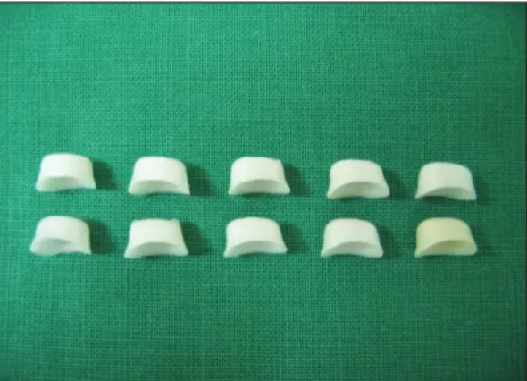
Fig. 3. Metal cast cores. Fig. 4. Cerec inLab cores.
Fig. 5. Loading device with torque controller. Fig. 6. 10Ncm setting of torque controller.
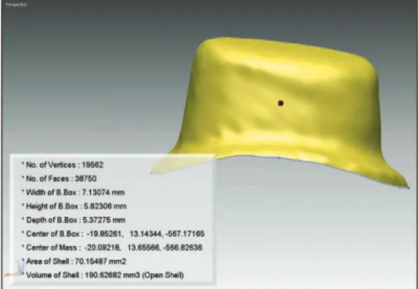
device (VIVID 910, Konica Minolta, Tokyo, Japan), as well as weight and density of silicone paste. Non-contact type contour measuring device was used to measure the surface area of metal master model (Fig. 9). The surface area was 70.15487 mm2. To calculate the density, silicone paste (Fusion Wash Type; GC., Tokyo, Japan) was filled in ring which have a hole with diameter of 1 cm and height of 0.3 cm. The weight and the volume were measured. Density = mass/volume = 0.4118 g/0.2355 cm3= 1.75 g/cm3. The
weight of silicone paste was measured by electron scale (AP210S; Ohaus Corp., Pine Brook, NJ, USA) (Fig. 10).
The electron scale was regulated at zero degree with metal master model and a core. Then, silicone paste was added into a core and the core was seated on metal master model by finger-pressure. Before the completion of polyme- rization, over-filled paste was removed and the weight was measured on electron scale. The internal gap of a core was calculated by the following equation.
Fig. 7. Measuring microscope and digital counter.
Fig. 10. Electron scale.
Fig. 8. Absolute marginal discrepancy of Cerec inLab core (original magnification × 100, white bar represents 100 ㎛).
Fig. 9. Surface area measured using non-contact type contour measuring device.
Thickness (internal gap) = weight/(density × area) The means and standard deviations per group were calculated and statistical inferences among the groups were analyzed using one-way ANOVA test and Tukey’s HSD test at 0.05 level of significance.
RESULTS
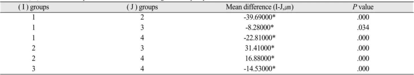
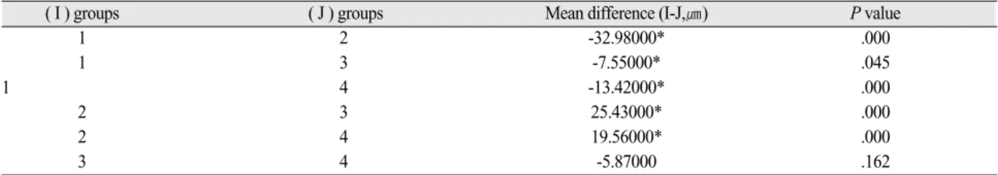
The fidelity of metal cast core showed the smallest gaps, followed by Lava core, Cerec inLab core and Procera core (Table Ⅰ, Ⅳ). When comparing the absolute marginal discrepancies, 3 core groups showed significant differences with the metal cast core group as well as among themselves (P<0.05) (Table Ⅱ, Ⅲ). When comparing the internal gaps, 3 core groups showed significant differences with the metal cast core group. Also, there were significant differences between Procera cores and Lava cores, and between Procera cores and Cerec inLab cores (P<0.05). However, there was no significant difference between Lava cores and Cerec inLab cores (Table Ⅴ, Ⅵ).
DISCUSSION
The crown fidelity is defined variously among researchers. Holmes et al.10defined various types of measurements between the casting surface and the tooth to clarify each term. The angular combination of the marginal gap and the extension error (overextension or underextension) was called the absolute marginal discrepancy. This study accepted the concept of the absolute marginal discrepancy to measure the fidelity of cores.
The clinically acceptable range of the crown fidelity is not yet clearly indicated. Sorensen et al.5reported that small defects less than or equal to 50㎛ were associated with significantly less bone loss than defects exceeding this value, and the prostheses fabricated under a conventional casting method were showed the marginal adaptation below 50 ㎛ when the laboratory condition was optimal.2,6-8 However, as a matter of fact, it is difficult to obtain the marginal adaptation around 50 ㎛. Hung et al.12reported that the practical range for clinical acceptability of fit seems to be approximately 50 to 75 ㎛. McLean and von
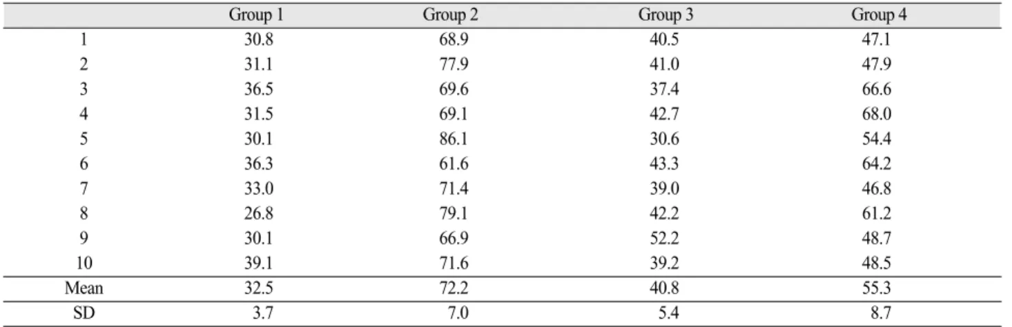
Table I. Mean and standard deviation (SD) of absolute marginal discrepancies in each of 4 core groups (unit: ㎛)
Group 1 Group 2 Group 3 Group 4
1 30.8 68.9 40.5 47.1
2 31.1 77.9 41.0 47.9
3 36.5 69.6 37.4 66.6
4 31.5 69.1 42.7 68.0
5 30.1 86.1 30.6 54.4
6 36.3 61.6 43.3 64.2
7 33.0 71.4 39.0 46.8
8 26.8 79.1 42.2 61.2
9 30.1 66.9 52.2 48.7
10 39.1 71.6 39.2 48.5
Mean 32.5 72.2 40.8 55.3
SD 3.7 7.0 5.4 8.7
Table II. Results of one-way ANOVA test for absolute marginal discrepancy
Sum of squares DF Mean square F P
Between groups 9116.985 3 3038.995 72.106 .000
Within groups 1517.27 36 42.146
Total 10634.255 39
Table III. (ⅰ). Results of Tukey’s HSD test for absolute marginal discrepancy
( I ) groups ( J ) groups Mean difference (I-J,㎛) P value
1 2 -39.69000* .000
1 3 -8.28000* .034
1 4 -22.81000* .000
2 3 31.41000* .000
2 4 16.88000* .000
3 4 -14.53000* .000
* Mean difference is significant at the 0.05 level.
Table III. (ⅱ). Statistical comparisons between groups in absolute marginal discrepancy
Group 1 2 3 4
1 -
2 * -
3 * * -
4 * * * -
* denotes pair of groups significantly different at the 0.05 level.
Table IV. Mean and standard deviation (SD) of internal gaps in each of 4 core groups (unit: ㎛)
Group 1 Group 2 Group 3 Group 4
1 31.8 70.0 40.7 44.8
2 42.4 74.1 50.5 45.6
3 38.3 83.1 46.4 54.6
4 32.6 71.7 39.9 47.2
5 32.6 66.8 42.4 50.5
6 44.8 68.4 44.8 63.5
7 32.6 65.2 35.8 54.6
8 39.1 71.7 61.9 57.8
9 43.2 75.8 50.5 45.6
10 46.4 66.8 46.4 53.8
Mean 38.4 71.4 45.9 51.8
SD 5.7 5.3 7.3 6.2
Table V. Results of one-way ANOVA test for internal gap
Sum of squares DF Mean Square F P
Between groups 5971.287 3 1990.429 52.571 .000
Within groups 1363.021 36 37.862
Total 7334.308 39
Fraunhofer13reported that for a good long-term prognosis, the clinically acceptable marginal gap for a crown is within the range of 120 ㎛. According to numerous studies on the crown fidelity of cast crowns and crowns fabricated with CAD/CAM system, the value of 120 ㎛ was the clinically acceptable marginal gap.3,8,14
According to study results on the crown fidelity of cast crowns, metal margin showed mean marginal opening of 27.5 ㎛15, noble alloy crown showed marginal opening of 25 ㎛16, PFM crowns showed marginal opening of 45-87 ㎛
11-15, and metal-ceramic copings showed mean marginal gap
of 29 ㎛.3
There were many studies on the fidelity of crowns fabricated with CAD/CAM system. May et al.8reported that mean gap dimensions for marginal openings, internal adaptation, and precision of fit for Procera AllCeram crowns were below 70 ㎛. Boening et al.17reported that medians of mean marginal gap widths of Procera AllCeram crowns were between 80 and 95 ㎛ in anterior teeth and between 90 and 145 ㎛ in posterior teeth. Karlsson18
with a range of 3-205 ㎛. Denissen et al.19reported that the marginal gap of Procera cores on the stone dies was 68±53
㎛ and it was a favorable measurement value for a clinically acceptable, strong all-ceramic onlay. Quintas et al.20 reported that the mean values of vertical marginal discrepancy of procera copings were 25 ㎛ before cementation and 44 ㎛ after cementation. Hertlein et al.21investigated the marginal fit of the Lava AllCeramic System for anterior and posterior teeth with a chamfered preparation margin. Under a stereomicroscope, the marginal gap was 38±20 ㎛ and the absolute marginal discrepancy was 72±36 ㎛. In the study on clinical fit of all-ceramic three-unit fixed partial dentures, generated with three different CAD/CAM systems, Reich et al.4reported that the medians of marginal gaps were 65 ㎛ for Lava system and 65 ㎛ for Cerec inLab system. Bindle and Mormann22evaluated the marginal and internal fit of all-ceramic CAD/CAM crown copings with chamfer margin. In the case of Lava system, the marginal gap was 43±23 ㎛, internal mesiodistal gap width was 82±49 ㎛, and internal mid-orobuccal gap width was 114±58 ㎛.
Table VI. (ⅰ). Results of Tukey’s HSD test for internal gap
( I ) groups ( J ) groups Mean difference (I-J,㎛) P value
1 2 -32.98000* .000
1 3 -7.55000* .045
1 4 -13.42000* .000
2 3 25.43000* .000
2 4 19.56000* .000
3 4 -5.87000 .162
* Mean difference is significant at the 0.05 level.
Table VI. (ⅱ). Statistical comparisons between groups in internal gap
Group 1 2 3 4
1 -
2 * -
3 * * -
4 * * NS -
* denotes pair of groups significantly different at the 0.05 level.
NS : not significant
were 32.5±3.7 ㎛, 72.2±7.0 ㎛, 40.8±5.4 ㎛ and 55.3±
8.7 ㎛ respectively. The internal gaps were 38.4±5.7 ㎛, 71.4±5.3 ㎛, 45.9±7.3 ㎛ and 51.8±6.2 ㎛ respectively.
Even though there were general differences in measuring locations, conditions and definition of the crown fidelity, this study showed satisfactory values within the experimental condition based on previous studies. The tested restorations had clinically acceptable fidelity.
The fidelity of restorations is influenced by various factors. In general, tooth preparation, impression body, accuracy of master model, restoration material, processing method, marginal contour and location, type of cement, convergence of axial wall, and luting space have influence on the fidelity of restoraions. Also, in the case of CAD/CAM system, scanning, software design, milling process and shrinkage effect after sintering additionally influences the fidelity.
To obtain the optimal results, several considerations were taken on the experimental design. First, the prepared tooth used in this study had 1.0 mm wide rounded shoulder margin, 3.0 mm axial height and 12�convergence angle.
This design and numerical values are based on data of previous studies. Lin et al.23 evaluated the marginal and internal adaptation of Procera copings using different tooth preparations. Mean external marginal openings were 64 ㎛ for chamfer finish line, 51 ㎛ for 0.8 mm rounded shoulder, and 68 ㎛ for 0.5 mm rounded shoulder. They also reported that the variations in the vertical height of inter-proximal finish lines did not significantly affect marginal opening. In general, less than 12�convergence angle is suggested for the favorable crown retention. However, Nakamura et al.2 reported that it seems appropriate to use the standard 12�
total convergence angle specified by the Cerec system.
Second, because the core mainly determines the overall fit of a veneered crown, in this study, the fit of cores was measured without veneering.20,22,24,25
Third, in this study, the amount of torque applied to the upper screw to seat a thin ceramic core on metal master model was restricted to 10 Ncm, because in an unpublished pilot study most of the ceramic copings were fractured above this limit.20
Forth, Groten et al.26reported that approximately 50 measurements are required for clinically relevant information about gap size regardless of gap definition or
cementation condition. Therefore, based on above data the absolute marginal discrepancy of a core was measured at randomly chosen 50 points along the margin.
Fifth, there was a study result that the type of cement influences the marginal adaptation.3 In this study, the absolute marginal discrepancy was measured without permanent cementation to eliminate the influence of cement on the marginal adaptation.
However, the laboratory testing cannot exactly reproduce the clinical condition. Therefore, the results should be viewed carefully and there are a few limitations in this study. First, in this study, the fidelity of cores was measured without permanent cementation of the core and it could potentially affect the marginal adaptation. To reproduce the clinical condition, Jorgensen27 suggested that any study aimed at determining the marginal adaptation of a crown system requires cementation of the crowns. In the study of the marginal adaptation before and after the permanent cementation, the marginal discrepancy had been increased12,20significantly24after cementation.
Second, due to the limitations of the microscopic imaging, only measurements of absolute marginal discrepancy in vertical dimension could be made. However, the evaluation of vertical discrepancy was chosen as potentially more clinically significant, since this discrepancy affects the exposure of luting agent and the horizontal discrepancy affects cleanability and plaque retension.28
Third, Sorensen9introduced a standardized method for determination of crown margin fidelity: direct view, cross- sectional view, impression technique, and explorer and visual view. This study used direct view to evaluate the absolute marginal discrepancy. The direct view method is convenient, easy, and rapid because the crown is retrievable, unlike the cementation, embedment, and sectioning method, which causes destruction of the crown. However, it is difficult to determine the repeatable measuring point of reference with a rounded margin, and to assess over- contouring of crown margin. Although clinically prepared crown margin seems to be sharp, it is showed rounded under microscope. In this study, it was difficult to determine measuring points of rounded margin and over-contoured margin and also it could affect the results.
Forth, luting space of specimens was not consistent in this
study. There was a study reporting that the crown fidelity was different according to various cementation spaces.2If all cores involved in this study had same luting spaces, the data could have been different. However, this study focused on processing accuracy of cores fabricated with different CAD/CAM systems using same master model. Since each CAD/CAM system accepts different luting spaces as its optimal fabrication condition, inconsistency of the luting spaces did not affect the purpose of this study much.
Nevertheless several limitations mentioned above, all values on the fidelity of cores fabricated with CAD/CAM system were within clinically acceptable range. Especially Lava cores showed the fidelity of below 50 ㎛ and it means that the fidelity of core fabricated with CAD/CAM system is now very close to fidelity of core produced using conventional casting technique.
To obtain more reliable research data on the fidelity of cores fabricated with CAD/CAM system, further evaluation of prostheses in the intraoral condition and more studies considered various factors are needed.
CONCLUSIONS
The results were as follows.
1. The absolute marginal discrepancies were 32.5±3.7 ㎛ for metal cast core, 72.2±7.0 ㎛ for Procera core, 40.8±
5.4 ㎛ for Lava core, and 55.3±8.7 ㎛ for Cerec inLab core. The internal gaps were 38.4±5.7 ㎛ for metal cast core, 71.4±5.3 ㎛ for Procera core, 45.9±7.3 ㎛ for Lava core, and 51.8±6.2 ㎛ for Cerec inLab core.
The fidelity of metal cast core showed the smallest gaps, followed by Lava core, Cerec inLab core and Procera core.
2. When comparing the absolute marginal discrepancies, 3 core groups showed significant differences with the metal cast core group as well as among themselves (P<0.05).
3. When comparing the internal gaps, 3 core groups showed significant differences with the metal cast core group. Also, there were significant differences between Procera cores and Lava cores, and between Procera cores and Cerec inLab cores (P<0.05). However, there
4. The fidelities of 4 core groups were all within the clinically acceptable range (120 ㎛).
REFERENCES
1. Land CH. A new system of restoring badly decayed teeth by means of an enamelled metallic coating. Independent Practitioner 1886;7:407.
2. Nakamura T, Dei N, Kojima T, Wakabayashi K. Marginal and internal fit of Cerec 3 CAD/CAM all-ceramic crowns.
Int J Prosthodont 2003;16:244-8.
3. Albert FE, El-Mowafy OM. Marginal adaptation and mi- croleakage of Procera Allceram crowns with four cements.
Int J Prosthodont 2004;17:529-35.
4. Reich S, Wichmann M, Nkenke E, Proeschel P. Clinical fit of all-ceramic three-unit fixed partial dentures, generated with three different CAD/CAM systems. Eur J Oral Sci 2005;113:174-9.
5. Sorensen SE, Larsen IB, Jorgensen KD. Gingival and alve- olar bone reaction to marginal fit of subgingival crown margins. Scand J Dent Res 1986;94:109-14.
6. Felton DA, Kanoy BE, Bayne SC, Wirthman GP. Effect of in vivo crown margin discrepancies on periodontal health. J Prosthet Dent 1991;65:357-64.
7. Lang NP, Kiel RA, Anderhalden K. Clinical and microbio- logical effects of subgingival restorations with overhanging or clinically perfect margins. J Clin Periodontol 1983;10:563-78.
8. May KB, Russell MM, Razzoog ME, Lang BR. Precision of fit : the Procera Allceram crown. J Prosthet Dent 1998;80:394-404.
9. Sorensen JA. A standardized method for determination of crown margin fidelity. J? Prosthet Dent 1990;64:18-24.
10. Holmes JR, Bayne SC, Holland GA, Sulik WD.
Considerations in measurement of marginal fit. J Prosthet Dent 1989;62:405-8.
11. Yeo IS, Yang JH, Lee JB. In vitro marginal fit of three all- ceramic crown systems. J Prosthet Dent 2003;90:459-64.
12. Hung SH, Hung KS, Eick JD, Chappell RP. Marginal fit of porcelain-fused-to-metal and two types of ceramic crown. J Prosthet Dent 1990;63:26-31.
13. McLean JW, von Fraunhofer JA. The estimation of cement film thickness by an in vivo technique. Br Dent J 1971;131:107-11.
14. Valderrama S, Van Roekel N, Andersson M, Goodacre CJ, Munoz CA. A comparison of the marginal and internal adaptation of titanium and gold-platinum-palladium metal ceramic crowns. Int J Prosthodont 1995;8:29-37.
15. Morris HF. Department of Veterans Affairs Cooperative Studies Project NO. 242. Quantitative and qualitative eval-
No. 147/242. J Prosthet Dent 1992;67:198-204.
16. Leong D, Chai J, Lautenschlager E, Gilbert J. Marginal fit of machine-milled titanium and cast titanium single crowns. Int J Prosthodont 1994;7:440-7.
17. Boening KW, Wolf BH, Schmidt AE, Kastner K, Walter MH. Clinical fit of Procera AllCeram crowns. J Prosthet Dent 2000;84:419-424.
18. Karlsson S. The fit of Procera titanium crowns. An in vitro and clinical study. Acta Odontol Scand 1993;51:129-34.
19. Denissen H, Dozic A, van der Zel J, van Waas M.
Marginal fit and short-term clinical performance of porce- lain-veneered CICERO, CEREC, and Procera onlays. J Prosthet Dent 2000;84:506-13.
20. Quintas AF, Oliveira F, Bottino MA. Vertical marginal dis- crepancy of ceramic copings with different ceramic materi- als, finish lines, and luting agents : an in vitro evaluation. J Prosthet Dent 2004;92:250-7.
21. Hertlein G, Hoscheler S, Frank S, et al. Marginal fit of CAD/CAM manufactured all ceramic zirconia prostheses.
J Dent Res 2001;80:42.
22. Bindl A, Mormann WH. Marginal and internal fit of all-ce- ramic CAD/CAM crown-copings on chamfer prepara-
tions. J Oral Rehabil 2005;32:441-7.
23. Lin MT, Sy-Munoz J, Munoz CA, Goodacre CJ, Naylor WP. The effect of tooth preparation form on the fit of Procera copings. Int J Prosthodont 1998;11:580-90.
24. Beschnidt SM, Strub JR. Evaluation of the marginal accu- racy of different all-ceramic crown systems after simulation in the artificial mouth. J Oral Rehabil 1999;26:582-93.
25. Sulaiman F, Chai J, Jameson LM, Wozniak WT. A com- parison of the marginal fit of In-Ceram, IPS Empress, and Procera crowns. Int J Prosthodont 1997;10:478-84.
26. Groten M, Axmann D, Probster L, Weber H.
Determination of the minimum number of marginal gap measurements required for practical in-vitro testing. J Prosthet Dent 2000;83:40-9.
27. Jorgensen KD. Structure of the film of zinc phosphate ce- ment. Acta Odontol Scand 1960;18:491-501.
28. Akbar JH, Petrie CS, Walker MP, Williams K, Eick JD.
Marginal adaptation of Cerec 3 CAD/CAM composite crowns using two different finish line preparation designs. J Prosthodont 2006;15:155-63.
A COMPARISON OF THE FIDELITY BETWEEN VARIOUS CORES FABRICATED WITH CAD/CAM SYSTEMS
Sun-Hee Park1, DDS, MSD, Kyu-Bok Lee2*, DDS, MSD, PhD
1Graduate student, Department of Prosthodontics, School of Dentistry, Kyungpook National University
2Assistant Professor, Department of Prosthodontics, School of Dentistry, Kyungpook National University
STATEMENT OF PROBLEM: Recently, various all-ceramic crowns fabricated with CAD/CAM systems have come into wide use in dental clinic. However, there are only few domestic studies on CAD/CAM restorations. PURPOSE: Purpose of this study was to compare the fidelity (absolute marginal discrepancy and internal gap) between various cores fabricated with different CAD/CAM systems (Procera system, Lava system, Cerec inLab system) and conventional metal cast core. MATERIALS AND METHODS: 10 cores per each system were fabricated. The absolute marginal discrepancies were measured using measuring microscope and digital counter. The internal gaps were calculated using a silicone paste. The results were statistically analyzed using the one-way ANOVA test and Tukey’s HSD test.
RESULTS: Within the limits of this study the results were as follows. 1. The absolute marginal discrepancies were 32.5±3.7 ㎛ for metal cast core, 72.2±7.0 ㎛ for Procera core, 40.8±5.4 ㎛ for Lava core, and 55.3±8.7 ㎛ for Cerec inLab core. The internal gaps were 38.4±
5.7 ㎛ for metal cast core, 71.4±5.3 ㎛ for Procera core, 45.9±7.3 ㎛ for Lava core, and 51.8±6.2 ㎛ for Cerec inLab core. 2. The fidelity of metal cast core showed the smallest gaps, followed by Lava core, Cerec inLab core, and Procera core. CONCLUSION: The fidelities of 4 core groups were all within the clinically acceptable range (120 ㎛).
KEY WORDS: All-ceramic crown, CAD/CAM system, Absolute marginal discrepancy, Internal gap
Corresponding Author: Kyu-Bok Lee
Department of Prosthodontics, School of Dentistry, Kyungpook National University,
2-188-1 Samduk-dong, Jung-gu, Daegu, 700-412, Korea +82 53 420 5921: e-mail, [email protected] Received May 19, 2008 Last Revison June 2, 2008 Accepted June 20, 2008.