PGHN
Original Article
Stepwise Endoscopy Based on Sigmoidoscopy in Evaluating Pediatric Graft-versus-Host Disease
Kyung Jae Lee, Shin Jie Choi, Hye Ran Yang*, Ju Yuong Chang
†, Hyoung Jin Kang
‡, Hee Young Shin
‡, Gyeong Hoon Kang
§, Jae Sung Ko, and Jin Soo Moon
Department of Pediatrics, Seoul National University College of Medicine, Seoul , *Department of Pediatrics, Seoul National University Bundang Hospital, Seongnam, †Department of Pediatrics, SMG-SNU Boramae Medical Center, Seoul, ‡Department of Pediatrics, Cancer Research Institute, §Department of Pathology, Seoul National University College of Medicine, Seoul, Korea
Purpose: The aim of our study was to establish a safe and convenient diagnostic method for acute gastrointestinal (GI) graft-versus-host disease (GVHD) in children by determining the sensitivity and negative predictive values of upper and lower endoscopic biopsies for children suspected of GI GVHD.
Methods: Patients suspected of GI GVHD who received endoscopic evaluation within 100 days after stem cell trans- plantation and endoscopies between January 2012 and March 2014 in Seoul National University Children’s Hospital were included in our study.
Results: Fifteen patients with a total of 20 endoscopic procedures were included in our study. Sensitivity at the esoph- agus, stomach, and duodenum were 22.2%, 30.0%, and 80.0%, respectively. Negative predictive values at the esophagus, stomach, and duodenum were 22.2%, 30.0%, and 60.0%, respectively. Overall sensitivity and negative predictive values of upper endoscopic biopsy for GVHD were 77.8% and 50.0%, respectively. Overall sensitivity and negative predictive values of lower endoscopic biopsy for GVHD were 88.9% and 66.7%, respectively.
Conclusion: We recommend flexible sigmoidoscopy as a safe and accurate diagnostic tool for GVHD, similar to other studies reported previously. However, if there is no evidence of GVHD on sigmoidoscopy with high index of suspicion of GI bleeding, full colonoscopy and upper endoscopy should be considered.
Key Words: Graft-versus-host disease, Child, Endoscopy
Received:August 28, 2015, Revised:October 8, 2015, Accepted:October 19, 2015
Corresponding author: Jin Soo Moon, Division of Pediatric Gastroenterolgy, Hepatology and Nutrition, Department of Pediatrics, Seoul National University Children’s Hospital, 101 Daehak-ro, Jongno-gu, Seoul 03080, Korea. Tel: +82-2-2072-3627, Fax: +82-2-743-3455, E-mail: mjschj@
snu.ac.kr
Copyright ⓒ 2016 by The Korean Society of Pediatric Gastroenterology, Hepatology and Nutrition
This is an openaccess article distributed under the terms of the Creative Commons Attribution NonCommercial License (http://creativecommons.org/licenses/by-nc/4.0/) which permits unrestricted noncommercial use, distribution, and reproduction in any medium, provided the original work is properly cited.
INTRODUCTION
Acute graft-versus-host disease (GVHD) occurs
within first 100 days after transplantation [1,2].
Severe acute GVHD results in significant morbidity and mortality. It is the major cause of treatment fail-
ure in patients undergoing transplantation for low-risk malignancy [2,3].
Acute GVHD involving intestinal tracts presents with non-specific symptoms such as secretory diar- rhea, nausea, vomiting, anorexia, weight loss, and abdominal pain [1,4]. In severe acute GVHD, diar- rhea can be copious. Bleeding, painful cramping, and ileus may result from mucosal ulceration [2,4,5].
Symptoms of gastrointestinal (GI) GVHD are not specific. Diagnosis of GI GVHD is often inaccurate based on clinical manifestation alone. Therefore, a definite diagnosis of GVHD requires histological con- firmation [1,6,7]. However, there is still controversy on the best diagnostic strategy for GI GVHD in chil- dren [8-10]. Previous studies have reported that rec- tosigmoid biopsy is a preferred procedure for diag- nosing GI GVHD in children [8,10].
The aim of our study was to establish a safe and convenient diagnostic method for acute GI GVHD in children by evaluating the sensitivity and negative predictive values of upper and lower endoscopic bi- opsies for children with suspected GI GVHD.
MATERIALS AND METHODS
Patients
Patients suspected of GI GVHD who received en- doscopic evaluation within 100 days after stem cell transplantation (SCT) and endoscopies between Ja- nuary 2012 and March 2014 in Seoul National University Children’s Hospital (Seoul, Korea) were included in our study. Age, gender, underlying dis- ease, transplantation type, symptom promoting en- doscopic biopsy, endoscopic findings, pathologic findings of endoscopic biopsies, skin biopsies, and liver biopsies were extracted from the patient’s med- ical records. Stool Clostridium difficile toxin examina- tion and stool bacterial culture were performed for some patients. Our study was approved by the Insti- tutional Review Board of our hospital (IRB No.
1501-038-639).
Gastrointestinal endoscopy and biopsy All patient’s blood samples were obtained to de-
termine complete blood cell count, prothrombin time, and activated partial prothrombin time before proce- dures to prevent possibility of bleeding. Prophylactic antibiotics were not used routinely.
During esophagogastroduodenoscopy (EGD), bi- opsies were obtained from the following segments:
esophagus, stomach, and duodenum second portion for all patients except one who did not receive esoph- ageal biopsy. When patients had GI bleeding, we car- ried out EGDs as well as full colonoscopies if bowel preparation was sufficient. We classified lower endo- scopic biopsies into the following three biopsy groups:
cecum, colon, and rectosigmoid colon. Endoscopic findings were classified based on endoscopic diag- nosis of GI GVHD [11].
Histological criteria for intestinal GVHD Diagnostic criterion for GVHD was the presence of epithelial single-cell apoptosis [1,6,7]. This pattern of mucosal damage can be observed in the immedi- ate post transplantation period (within 20 days) due to toxic effect of conditioning chemotherapy on gut epithelium [1]. In our study, a GI pathologist re- viewed all specimens to confirm the diagnosis of GVHD. We considered a case as GI GVHD even if there was just a single endoscopic biopsy result showing a compatible reading with GVHD. Some specimens underwent cytomegaloviral (CMV) immunohisto- chemical (IHC) study.
Statistical analysis
All dichotomous variables were analyzed with Fisher’s exact test. IBM SPSS Statistics version 21.0 for Windows (IBM Co., Armonk, NY, USA) was used for statistical analysis. A p-value less than 0.05 was considered as statistically significant.
We also evaluated the sensitivity and negative pre- dictive values of both upper and lower endoscopic biopsies.
RESULTS
Patient characteristics
A total of 15 patients with suspected GI GVHD
Table 1.Patient Demographic Characteristics (n=15) Characteristic No GVHD (n=3) GVHD (n=12) Sex
Male 2 (66.7) 8 (66.7)
Female 1 (33.3) 4 (33.3)
Age (yr) 2.63 (11 mo-6 yr) 11.25 (4 yr-22 yr) Underlying disease
AML 0 6 (50.0)
ALL 1 (33.3) 3 (25.0)
ALCL 0 1 (8.3)
AMLL 0 1 (8.3)
JMML 0 1 (8.3)
Congenital neutropenia
1 (33.3) 0
SCID 1 (33.3) 0
Transplantation
PBSCT 2 (66.7) 9 (75.0)
HaploPBSCT 0 2 (16.7)
UCBT 1 (33.3) 1 (8.3)
Prophylaxis
CSA+MMF 0 2 (16.7)
Tacrolimus+MTX 2 (66.7) 10 (83.3)
CSA 1 (33.3) 0
Values are presented as number (percentage within group) or median (range).
GVHD: graft-versus-host disease, AML: acute myeloid leukemia, ALL: acute lymphoid leukemia, ALCL: anaplastic large cell lymphoma, AMLL: acute mixed lineage leukemia, JMML: juvenile myelomonocytic leukemia, SCID: severe combined immunode- feciency, PBSCT: peripheral blood stem cell transplantation, HaploPBSCT: haploidentical PBSCT, UCBT: umblical cord blood transplantation, CSA: cyclosporin A, MMF: mycophenolate mofetil,
MTX: methotrexate. Fig. 1. Study flow.
were included in our study. Patient demographic characteristics are summarized in Table 1. Median time of endoscopic biopsies following SCT was 34 (range, 19-61) days. The most common underlying diseases were acute myeloid leukemia (40.0%) and other leukemias 46.7% (1+3+1+1+1/15=46.666%).
There was no statistically significant difference in age, sex, underlying disease, kinds of SCT, or prophy- lactic agents between GVHD group and non-GVHD group (Table 1). Patient numbering and summa- rization of characteristics are attached in supple- mentary material. Of the 15 patients enrolled in our study, three received endoscopies twice.
One patient (Patient 6) received it three times.
Total count of all endoscopic procedures was 20.
GVHD was diagnosed in 12 of 15 (80.0%) patients.
The number of patient who received upper endos- copy, lower endoscopy, and simultaneous upper and lower endoscopies were 2 (10.0%), 7 (35.0%), and 11 (55.0%), respectively (Fig. 1).
The most common symptoms prompting endo- scopic evaluation were diarrhea (100%) and vomit- ing (85.0%). The most common symptoms of GVHD patients was diarrhea (100%) and vomiting (62.5%).
Of skin GVHD patients, there were 6 (85.7%) GI GVHD; of liver GVHD, GI GVHD was 5 (71.4%). Any symptoms of GI GVHD could not be regarded stat- istically different to the non GVHD cases (Table 2).
Sensitivities and negative predictive values of endoscopic biopsies
For those who received upper endoscopy, the sen- sitivity and negative predictive value at the esoph- agus was both at 22.2%. Both the sensitivity and neg- ative predictive value at the stomach was 30%. The sensitivity and negative predictive value at the duo- denum were 80.0% and 60.0%, respectively.
For 18 episodes when lower endoscopy performed, the number of episodes that were intubated at cecum was 5 (27.8%) and hepatic flexure was 2 (11.1%). Of the 5 patients who were intubated at the cecum, two received biopsies, including one who was diagnosed as GVHD. The sensitivity of biopsies obtained in co- lon was 87.5%. The sensitivity and negative pre- dictive value of rectosigmoid biopsy was 86.7% and 60.0%, respectively (Table 3). The sensitivity of co-
Table 3.Sensitivity and Negative Predictive Values of Upper and Lower Endoscopic Biopies
Overall disease Identified GVHD in particular biopsy site p-value Sen (%) NPV (%)
Upper endoscopy Esophagus
(–) (+) 0.757 22.2 22.2
GVHD (–) 2 0
GVHD (+) 7 2
Stomach
(–) (+) 0.42 30 30
GVHD (–) 3 0
GVHD (+) 7 3
Duodenum
(–) (+) 0.035 80 60
GVHD (–) 3 0
GVHD (+) 2 8
Lower endoscopy Cecum
(–) (+) 50
GVHD (+) 1 1 .
Colon
(–) (+) 87.5
GVHD (+) 1 7
Rectosigmoid
(–) (+) 0.012 86.7 60
GVHD (–) 3 0
GVHD (+) 2 13
GVHD: graft-versus-host disease, Sen: sensitivity, NPV: negative predictive value.
Table 2.Association between Presence of GI GVHD and GI Symptoms, Skin GVHD and Liver GVHD
Variable No GI GVHD (n=4) GI GVHD (n=16) Total (n=20) p-value
GI Symptom
Vomiting 1 (25.0) 10 (62.5) 17 (85.0) 0.134
Abdomen pain 2 (50.0) 12 (75.0) 14 (70.0) 0.549
Diarrhea 4 (100) 16 (100) 20 (100) -
Hematemesis 1 (25.0) 1 (6.3) 2 (10.0) 0.368
Hematochezia 1 (25.0) 8 (50.0) 9 (45.0) 0.591
Skin GVHD 1 6 7 0.055
Liver GVHD 2 5 7 0.537
Values are presented as number (percentage within group) or number only.
GI: gastrointestinal, GVHD: graft-versus-host disease.
lonic biopsy was not statistically different from the sensitivity of rectosigmoid biopsy.
Eleven of 20 (55.0%) sessions had simultaneous upper and lower endoscopies. We classified biopsy results into two groups: upper and lower endoscopy.
The sensitivity and negative predictive value of the upper endoscopic biopsy was 77.8% and 50.0%, respectively. The sensitivity and negative predictive value of the lower endoscopic biopsy was 88.9% and
66.7%, respectively (Table 4).
One patient was diagnosed as GVHD only by upper endoscopy. However, he had coincidental CMV col- itis in the lower GI tract. In pathologic review, there was no intact epithelial cell in his colonic biopsy specimen. Therefore, diagnosis of GVHD based on the apoptosis in the epithelial cell could not be made.
However, colonic GVHD cannot be completely ex- cluded also in this case.
Table 5.Associations between Endoscopic Findings and Presence of Graft-versus-Host Disease (GVHD) Endo-
scopic findings
Esophagus Stomach Duodenum Cecum Colon Rectosigmoid
No
GVHD GVHD No
GVHD GVHD No
GVHD GVHD No
GVHD GVHD No
GVHD GVHD No
GVHD GVHD
1 2 (22.2) 5 (50.0) 2 (66.7) 1 (20.0) 2 (25.0) 1 (100) 3 (42.9) 2 (66.7) 2 (15.4)
2 2 (25.0) 1 (100) 3 (42.9) 3 (23.1)
3 1 (11.1) 4 (40.0) 1 (33.3) 1 (14.3) 3 (23.1)
4 3 (33.3) 2 (100) 1 (10.0) 2 (40.0) 3 (37.5) 2 (15.4)
5 3 (33.3)* 2 (40.0)† 1 (12.5) 1 (100)‡ 1 (33.3)§ 3 (23.1)
Total (n) 9 2 10 3 5 8 1 1 1 7 3 13
Values are presented as number (percentage within group) or number only.
1: normal, 2: loss of vascular markings and/or focal mild erythema, 3: moderate edema and/or erythema, 4: edema, erythema, erosions and/or bleeding, 5: ulceration, exudates and bleeding.
*Inflammed granulation tissue, ulcer detritus, acanthotic squamous epithelium; †cytomegaloviral (CMV) duodenitis (Fig. 1B and 1C); ‡CMV typhlitis (Fig. 2B); §CMV sigmoid colitis.
Table 4.Sensitivities and Negative Predictive Values of Concurrent Endoscopic Biopsies
Disease GVHD in particular biopsy site p-value Sen (%) NPV (%)
Upper endoscopy
(–) (+) 0.109 77.8 50.0
GVHD (–) 2 0
GVHD (+) 2 7
Lower endoscopy
(–) (+) 0.055 88.9 66.7
GVHD (–) 2 0
GVHD (+) 1 8
GVHD: graft-versus-host disease, Sen: sensitivity, NPV: negative predictive value.
Gross endoscopic findings, serial endoscopic findings, and coinfections
Table 5 shows endoscopic findings according to the Cruz-Correa’s classification [11] and the pres- ence of GVHD. There were several segments showing normal mucosa (Fig. 2A) in endoscopy. But there were GVHD in pathology results. The percentage of normal finding was 66.7% at the stomach, 25.0% at duodenum, 100% at cecum, 42.8% at colon, and 16.7%
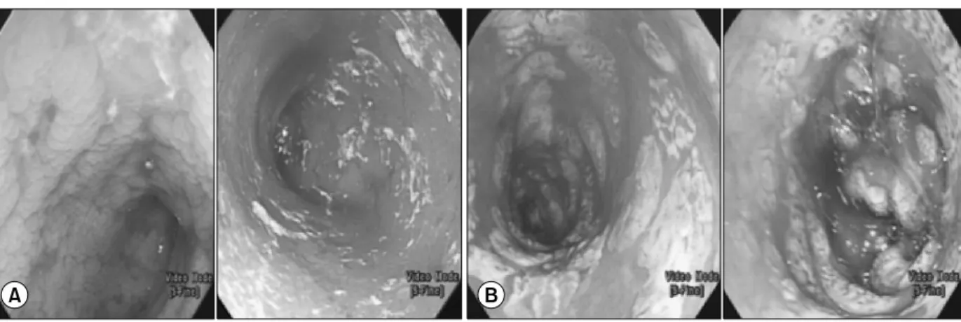
at rectosigmoid. On the other hand, 33.3-100% of non-GVHD patients showed severe ulceration, exu- dates, and bleeding on their endoscopy without pathologic diagnosis of GVHD. The pathologic find- ings in severe ulceration without GVHD was CMV ty- philitis (Fig. 2B), duodenitis (Fig. 3B and 3C), and colitis (Fig. 4B). Other histologic findings in the se- vere ulceration segment were inflammatory gran-
ulation tissue, ulcer detritus, or acanthotic squ- amous epithelium.
There are four patients who received serial endos- copy due to persistent symptoms, of which two (Patient 6 and 9) were initially diagnosed as GVHD and received steroid therapy. But their symptoms were persistent. Therefore, a second endoscopic bi- opsy was performed. Their diagnosis was changed to CMV infection or other nonspecific finding. Therefore, their treatments were also changed. Two patients (Patient 6-2, 12) had coincidental GVHD and CMV infection (upper GI GVHD and lower GI CMV in- fection). One patient showed duodenal bleeding and perforation after duodenal biopsy. No other compli- cations such as sepsis were reported.
Fig. 2. Cecum endoscopic findings of two patients. (A) It showed normal mucosa with graft-versus-host deisease (Pa- tient 2-2). (B) It showed ulcer and exudate at endoscopy (Patient 12). Cytomegaloviral typhilitis was confirmed at histology.
Fig. 3. Duodenum of three patients. They showed grade 5 ulceration, exudate, and bleeding in endoscopy. (A) Graft-versus-host disease with ulcer (Patient 6-2). (B, C) Cytomegaloviral uodenitis (Patients 14 and 13).
Fig. 4. Serial rectosigmoidoscopic findings of the same patient (Patient 6). (A) It showed gross ulcer, exudates, and bleeding.
The patient (Patient 6-1) was diagnosed as graft-versus-host disease (d20). However, steroid therapy failed to relieve his symptoms.
(B) It showed d31 sigmoidoscopic finding of the same patient (Patient 6-2) showing exudates and bleeding. He was diagnosed as cytomegaloviral infection.
DISCUSSION
Our study provides two new points in the endo- scopic diagnosis of GVHD in children. First, we cal- culated the sensitivity and negative predictive value of each biopsy site and compared them by site. There is no data on sensitivity and negative predictive val- ues of each biopsy site in children. Second, we showed important serial endoscopic biopsy results in the same patients. Therefore, we suggest that serial en- doscopy is important in children with the symptoms of GVHD in this complex clinical situation.
Acute GVHD is a major cause of morbidity and mortality in patients undergoing transplantation for low-risk malignancy [2,3,12]. For GI GVHD patients who present with non-specific symptoms such as anorexia, vomiting, abdominal discomfort, and diar- rhea, histologic diagnosis of GVHD is important [1,6,7,13]. However, the best diagnostic strategy for GI GVHD in children remains unclear [8-10].
In our study, 11 patients had received concurrent upper and lower endoscopy. Sensitivity and negative predictive value revealed that lower endoscopic biop- sy was better for diagnosis of GVHD in children than upper endoscopic biopsy. Five patients were diag- nosed as GVHD only by lower endoscopy. There was no change of diagnosis in lower endoscopy by addi- tional upper endoscopy except in one patient. This exceptional patient was diagnosed as GVHD by up- per endoscopy. He also had severe CMV colitis in lower endoscopy, which could not be evaluated for the presence of GVHD due to severe inflammation.
Mortality rate of our GVHD patients was high (40.0%, 6 of 15).
Several previous studies in adults also suggested that biopsy of rectosigmoid colon was the best for di- agnosing GI GVHD [14,15]. A prospective study found that the biopsy site with the highest yield was distal colon [16]. Others insist that sigmoidoscopy is the safest and the most productive method of diag- nosing acute GVHD in children [8,10]. But in 2012, a retrospective study in children found that rectosigmoid combined with upper endoscopic biopsies were equally sensitive for diagnosis of acute GI GVHD in
children [9]. Some authors advocate gastric biopsy and other upper endoscopic biopsies as useful tools for the diagnosis of GI GVHD [4,13,17]. A pro- spective study conducted from August 2002 to February 2006 at Yale Gastrointestinal Procedure Center [18]. They performed 27 cases of concurrent upper and lower endoscopy for suspected acute GVHD. Their results showed diffuse upper and lower GI involvement of acute GVHD with similar diag- nostic yield (sensitivity of stomach, 94.4%; duode- num, 94.4%; colon, 92.8%; rectosigmoid, 88.8%).
Although absolute percentages of sensitivity were the highest at duodenum and stomach, there was no significant difference in sensitivity among segments.
They recommended to use flexible sigmoidoscopy with rectal biopsy alone in patient who were poor candidates for full colonoscopy.
Biopsy of duodenum is known to have high risk of perforation and bleeding [8,19]. One of our patients showed complications of endoscopic biopsy, duode- nal bleeding and perforation. In our study, associa- tion between patient’s symptoms such as nausea, vomiting, diarrhea, hematochezia, hematemesis, and presence of GI GVHD was not statistically significant.
This result is in consistent with results of previous studies [6,10,14]. These symptoms could be present with GI infection, chemotherapy, or medication in- duced side effects [4].
However, in our study, two patients’ histologic findings from the second endoscopic biopsy were differed from those from the first endoscopic biopsy, leading to a change of therapy. A retrospective study has suggested that serial GI endoscopies could affect therapeutic decision-making for patients with per- sistent diarrhea after SCT [20].
There are many results showing that gross endo- scopic findings not correlated with biopsy results [6,7,12,13,17]. However, some studies found high correlation between endoscopic findings and histo- logically proven GI GVHD [11]. In our results, the most common endoscopic finding of diagnosed GVHD had normal mucosa. Some patients had no GVHD, although they had endoscopic severe ulcer- ation and bleeding. These results suggest that dis-
Fig. 5. Stepwise endoscopic approaches for pediatric gastro- intestinal graft-versus-host disease (GVHD). EGD: esophago- gastroduodenoscopy.
crepancy between endoscopic finding and histologic assessment can occur. Histologic confirmation of en- doscopic biopsy is important for the diagnosis of GVHD.
Our study had several limitations. First, this was a retrospective study with a small number of patients.
In addition, CMV IHC was not performed for all cases. Although epithelial single-cell apoptosis can be seen in immediate post transplantation period (within 20 days) due to toxic effect of chemotherapy on gut epithelium [1], one patient had undergone early endoscopy at day 19.
There are many reports that flexible sigmoido- scopy is safe and easy to train [21,22]. In addition, it can be performed without sedation [10,12]. Compli- cation of duodenal biopsy was also reported. Therefore, we also agree that flexible sigmoidoscopy should be regarded as an accurate and safe method for the ini- tial diagnosis of acute GVHD in children.
In conclusion, our results suggest that flexible sig- moidoscopy is a safe and accurate diagnostic tool for GVHD. However, if there is no evidence of GVHD on sigmoidoscopy with high index of suspicion of GI GVHD, full colonoscopy and upper endoscopy should be considered (Fig. 5).
REFERENCES
1. Iqbal N, Salzman D, Lazenby AJ, Wilcox CM. Diagnosis of gastrointestinal graft-versus-host disease. Am J Gastroenterol 2000;95:3034-8.
2. Couriel D, Caldera H, Champlin R, Komanduri K.
Acute graft-versus-host disease: pathophysiology, clin- ical manifestations, and management. Cancer 2004;
101:1936-46.
3. Ferrara JL, Deeg HJ. Graft-versus-host disease. N Engl J Med 1991;324:667-74.
4. Wu D, Hockenberry DM, Brentnall TA, Baehr PH, Ponec RJ, Kuver R, et al. Persistent nausea and anorex- ia after marrow transplantation: a prospective study of 78 patients. Transplantation 1998;66:1319-24.
5. Nevo S, Enger C, Swan V, Wojno KJ, Fuller AK, Alto- monte V, et al. Acute bleeding after allogeneic bone mar- row transplantation: association with graft versus host disease and effect on survival. Transplantation 1999;
67:681-9.
6. Roy J, Snover D, Weisdorf S, Mulvahill A, Filipovich A, Weisdorf D. Simultaneous upper and lower endoscopic biopsy in the diagnosis of intestinal graft-versus-host disease. Transplantation 1991;51:642-6.
7. Xu CF, Zhu LX, Xu XM, Chen WC, Wu DP. Endoscopic diagnosis of gastrointestinal graft-versus-host disease.
World J Gastroenterol 2008;14:2262-7.
8. Khan K, Schwarzenberg SJ, Sharp H, Jessurun J, Gulbahce HE, Defor T, et al. Diagnostic endoscopy in children after hematopoietic stem cell transplantation.
Gastrointest Endosc 2006;64:379-85; quiz 389-92.
9. Sultan M, Ramprasad J, Jensen MK, Margolis D, Werlin S. Endoscopic diagnosis of pediatric acute gas- trointestinal graft-versus-host disease. J Pediatr Gas- troenterol Nutr 2012;55:417-20.
10. Crowell KR, Patel RA, Fluchel M, Lowichik A, Bryson S, Pohl JF. Endoscopy in the diagnosis of intestinal graft-versus-host disease: is lower endoscopy with biop- sy as effective in diagnosis as upper endoscopy com- bined with lower endoscopy? Pediatr Blood Cancer 2013;60:1798-800.
11. Cruz-Correa M, Poonawala A, Abraham SC, Wu TT, Zahurak M, Vogelsang G, et al. Endoscopic findings pre- dict the histologic diagnosis in gastrointestinal graft- versus-host disease. Endoscopy 2002;34:808-13.
12. Yeh SP, Liao YM, Hsu CH, Chen CL, Shen YC, Hsueh CT, et al. Gastric bleeding due to graft-vs-host disease:
discrepancy between endoscopic and histologic assess- ment. Am J Clin Pathol 2004;122:919-25.
13. Velasco-Guardado A, López-Corral L, Alvarez-Delgado
A, Flores-Corral T, Geijo-Martínez F, Caballero-Barrigón D, et al. Endoscopic evaluation and histological findings in graft-versus-host disease. Rev Esp Enferm Dig 2012;104:310-4.
14. Ross WA, Ghosh S, Dekovich AA, Liu S, Ayers GD, Cleary KR, et al. Endoscopic biopsy diagnosis of acute gastrointestinal graft-versus-host disease: rectosig- moid biopsies are more sensitive than upper gastro- intestinal biopsies. Am J Gastroenterol 2008;103:
982-9.
15. Oomori S, Takagi S, Kikuchi T, Utsunomiya K, Yokoyama H, Negoro K, et al. Significance of colono- scopy in patients with intestinal graft-versus-host dis- ease after hematopoietic stem cell transplantation.
Endoscopy 2005;37:346-50.
16. Thompson B, Salzman D, Steinhauer J, Lazenby AJ, Wilcox CM. Prospective endoscopic evaluation for gas- trointestinal graft-versus-host disease: determination of the best diagnostic approach. Bone Marrow Transplant 2006;38:371-6.
17. Ponec RJ, Hackman RC, McDonald GB. Endoscopic and histologic diagnosis of intestinal graft-versus-host dis-
ease after marrow transplantation. Gastrointest Endosc 1999;49:612-21.
18. Aslanian H, Chander B, Robert M, Cooper D, Proctor D, Seropian S, et al. Prospective evaluation of acute graft-versus-host disease. Dig Dis Sci 2012;57:720-5.
19. Ramakrishna J, Treem WR. Duodenal hematoma as a complication of endoscopic biopsy in pediatric bone marrow transplant recipients. J Pediatr Gastroenterol Nutr 1997;25:426-9.
20. Martínez C, Rosales M, Calvo X, Cuatrecasas M, Rodriguez-Carunchio L, Llach J, et al. Serial intestinal endoscopic examinations of patients with persistent di- arrhea after allo-SCT. Bone Marrow Transplant 2012;47:694-9.
21. Volonaki E, Sebire NJ, Borrelli O, Lindley KJ, Elawad M, Thapar N, et al. Gastrointestinal endoscopy and mu- cosal biopsy in the first year of life: indications and outcome. J Pediatr Gastroenterol Nutr 2012;55:62-5.
22. Groveman HD, Sanowski RA, Klauber MR. Training primary care physicians in flexible sigmoidoscopy-- performance evaluation of 17,167 procedures. West J Med 1988;148:221-4.
Supplementary Table. Patients flow DaySymptoms MethodGVHD of segmentEndoscopic findingsGVHDCMV Cx VOMABPDIAHTMHCZESSTDDCCCLRSESSTDDCCCLRSGISkinLiverICH Pt 1d45 (+) Lower (+) 3(+)(+) (–)(–)L Pt 2-1d48(+)(+)(+)Both(–)(–)(+)(+)4232(+)(–)(–)D Pt 2-2d32 (+) Both(–)(–)(–)(+)(+)(+)423012(+) (–)(–) Pt 3d19(+)(+)(+) (+)Both(+)(–)(+) (+)323 3(+) (–)L Pt 4d61(+)(+)(+) (+)Upper (+)(+) 001 (+)(+)(+)(–)(–)L Pt 5-1d20(+)(+)(+)Both(+)(+)(+)(+)(+)321411(+)(+)(–)(–)D Pt 5-2d40(+)(+)(+) (+)Lower (+) 4(+) (–)(–) Pt 6-1d20(+)(+)(+)(+)Lower(+)4(+)(+)(–)(–)D Pt 6-2d31(+)(+)(+)(+)Both(–)(–)(+)(–)0044(+) (+)(+)RS(–) Pt 6-3d52(+)(+)(+) (+)Lower (+) 4(+) (–)(–) Pt 7d25(+)(+)(+) Lower (+)(+) 22(+)(+)(+)(–)(–)D Pt 8d28(+)(+)(+) Lower (+)(–) 000(+)(+) (–)L Pt 9-1d29(+)(+)Both(–)(–)(–)(+)(+)00000(+)(–)(–)L Pt 9-2d53 (+)(+) Both(–)(–)(–) (–)403 1 (–) (–)(–) Pt 10d26(+) (+) Both(–)(–)(+) (+)(+)323 00(+)(+)(+)(–)(+) Pt 11d23 (+)(+) Both(–)(+)(+) (–)(+)200111(+) (–)(–)L Pt 12d27 (+)(+) Both(–)(–)(+)(–)(+)(–)300411(+) (+)CC(–)L Pt 13d35 (+)(+) Both(–)(–)(–) (–)304 0(–)(+)(+)(+)(–)L Pt 14d36(+) (+)(+)(+)Upper (–)(–) 34 (–) (+)(–)D Pt 15d27 (+) Lower (–) 0(–)(–)(+)(–)(–)D Pt: patient, VOM: vomiting, ABP: abdomen pain, DIA: diarrhea, HTM: hematemesis, HCZ: hematochezia, ES: esophagus, ST: stomach, DD: duodenum, CC: cecum, colon, RS: rectosigmoid colon, GI: gastrointestinal, ICH: immunohistochemistry, Cx: complication, Px: rognosis, L: live, D: death. www.pghn
Kyung Jae Lee, et al:Stepwise Sigmoidoscopy in Evaluating Pediatric GVHD