Stem Cell Therapy for Bronchopulmonary Dysplasia: Bench to Bedside Translation
Bronchopulmonary dysplasia (BPD), a chronic lung disease affecting very premature infants, is a major cause of mortality and long-term morbidities despite of current progress in neonatal intensive care medicine. Though there has not been any effective treatment or preventive strategy for BPD, recent stem cell research seems to support the assumption that stem cell therapy could be a promising and novel therapeutic modality for attenuating BPD severity. This review summarizes the recent advances in stem cell research for treating BPD. In particular, we focused on the preclinical data about stem cell transplantation to improve the lung injury using animal models of neonatal BPD. These translational research provided the data related with the safety issue, optimal type of stem cells, optimal timing, route, and dose of cell transplantation, and potency marker of cells as a therapeutic agent.
Those are essential subjects for the approval and clinical translation. In addition, the successful phase I clinical trial results of stem cell therapies for BPD are also discussed.
Keywords: Bronchopulmonary Dysplasia; Cell Transplantation; Mesenchymal Stem Cells;
Infant, Premature So Yoon Ahn,* Yun Sil Chang,*
and Won Soon Park
Department of Pediatrics, Samsung Medical Center, Sungkyunkwan University School of Medicine, Seoul, Korea
* So Yoon Ahn and Yun Sil Chang contributed equally to this work.
Received: 18 December 2014 Accepted: 5 March 2015 Address for Correspondence:
Won Soon Park, MD
Department of Pediatrics, Samsung Medical Center, Sungkyunkwan University School of Medicine, 81 Irwon-ro, Gangnam-gu, Seoul 135-710, Korea
Tel: +82.2-3410-3523, Fax: +82.2-3410-0043 E-mail: [email protected]
Funding: This work was supported by grants HI12C1821 (A121968) from the Korean Healthcare Technology R&D Project, Ministry for Health, Welfare and Family Affairs, Republic of Korea, by grants 20 by 20 Project (Best #3, GFO1140091) from Samsung Medical Center, and by a Korea Research Foundation grant from the National Research Foundation of Korea (NRF), and by the Ministry of Education, Science, and Technology (NRF-2014R1A1A2056427).
http://dx.doi.org/10.3346/jkms.2015.30.5.509 • J Korean Med Sci 2015; 30: 509-513
INTRODUCTION
Bronchopulmonary dysplasia (BPD) is a chronic lung disease that usually occurs in premature infants receiving prolonged oxygen supplementation and ventilator support. The risk of de
veloping BPD correlates with the extent of immaturity (1). Re
cent improvements in the survival of very preterm infants throu
gh advances in neonatal intensivecare medicine have, there
fore, made the task of protecting the extremely immature lungs against BPD increasingly challenging. BPD remains an impor
tant cause of mortality and longterm respiratory morbidities such as airway hyperreactivity, poor lung function, and low ex
ercise capacity (26). In addition, neurologic morbidities such as developmental delay and cerebral palsy (7) are also com
mon. The histopathological characteristics of BPD include im
paired alveolarization and interstitial fibrosis (8, 9). Prolonged oxygen exposure of newborn rat pups results in decreased alve
olarization and increased lung fibrosis, thereby simulating the histopathology of human BPD (9, 10). Inflammatory responses are believed to play critical roles in the lung injury process lead
ing to the development of BPD (1). Currently, no effective treat
ments beyond supportive therapies are available for BPD. There
fore, development of new therapeutic modalities to improve the prognosis of BPD in preterm infants is an urgent priority.
Recently, current literature has shown that the exogenous administration of stem cells significantly attenuated neonatal hyperoxic lung injuries (1118). These findings suggest that stem cell transplantation might be a new and promising thera
peutic modality for the treatment of BPD. In this review, we summarize the recent advances in stem cell research for treat
ment of BPD. In particular, we focus on the preclinical data re
garding the important issues for clinical translation such as the optimal cell type, route, dose, and timing of stem cell therapy.
Furthermore, the successful phase I clinical trial results of stem cell therapies for BPD are discussed.
PRECLINICAL RESEARCH DATA Determining the optimal cell type
Among the various stem cells, the selection of a single appro
priate stem cell that ultimately exhibits the best therapeutic ef
ficacy in protecting against BPD is a difficult challenge. Embry
onic stem cells are pluripotent cells capable of generating all cell types from three germ layers. However, the high tumorige
Respiratory Diseases
nicity and ethical concerns of destroying embryos for their ac
quisition have limited their availability for research and clinical applications (19).
Mesenchymal stem cells (MSCs) are the most extensively ex
amined cell type used in experimental models of BPD (1318, 20). MSCs are broadly distributed in the body, and could be isolated from adult tissues such as the bone marrow, adipose tissue, and gestational tissues such as the placenta, Wharton’s jelly, and umbilical cord blood (UCB). The umbilical cord and placenta are medical wastes that are usually discarded at birth, and therefore, MSCs obtained from gestational tissues seem to be particularly attractive (21). In addition to their easy attain
ability, MSCs derived from gestational tissues showed less anti
genicity (21), and higher proliferation capacity and paracrine potency compared with adult tissuederived MSCs (22). Even within the same adult tissue origin, donor age negatively im
pacted the expansion and differentiation potential of the MSCs (23, 24). Collectively, these findings suggest that MSCs derived from postpartum associated tissues such as UCB or Wharton’s jelly might be the optimal cell source for future clinical applica
tions, in protecting premature infants against BPD.
Therapeutic potential and protective mechanisms of MSCs for BPD
The therapeutic efficacy of MSCs has been tested in the hyper
oxiainduced neonatal rodent or murine model of BPD, and was reported to improve survival, and suppress oxidative stress and inflammation (11, 1318, 20). In addition, it attenuated the impaired alveolar growth, lung vascular injuries, fibrosis, and the associated pulmonary hypertension (11, 1318, 20). These findings support the assumption that stem cell transplantation might be a promising novel therapeutic approach for BPD.
The beneficial effects were initially ascribed to the transdif
ferentiation of MSCs into lung parenchymal cells such as type II pneumocytes (11, 25). However, this event rarely occurs in vivo (11). The low rate of in vivo engraftment and differentiation into lung tissue suggests that the therapeutic effects of stem cell trans
plantation might not be primarily mediated by regeneration.
An equal or better therapeutic efficacy in preventing or revers
ing established BPD was observed with MSCconditioned me
dia compared with MSC (13, 26, 27). More recently, Lee et al.
(28) have reported that microvesicles released by MSC exosomes are the major paracrine antiinflammatory and therapeutic mediators of MSCs in hypoxiainduced pulmonary hyperten
sion. Collectively, these findings suggest that the protective ef
fects of stem cell transplantation might be predominantly me
diated by paracrine, rather than regenerative, mechanisms. The use of MSC secretomes rather than stem cells could be an excit
ing, promising new therapeutic approach for BPD, especially since it circumvents the theoretical concerns associated with live cell treatments, such as tumor formation.
The specific humoral substances secreted by the transplant
ed MSCs that are responsible for the protective paracrine activ
ity have not yet been elucidated. In our previous experiments, we observed that significantly reduced levels of growth factors such as vascular endothelial growth factor (VEGF) and hepato
cyte growth factor (HGF) were significantly improved with MSCs transplantation (17). Moreover, the knockdown of VEGF secre
tion by the MSCs using transfection with small interfering RNA specific for human VEGF abolished the protective effects of MSCs in hyperoxic lung injury (18). These protective effects in
cluded the attenuation of impaired alveolarization and angio
genesis, reduction in apoptotic cells and alveolar macrophages, and downregulation of proinflammatory cytokine levels (18).
Overall, these findings suggest that growth factors such as VEGF, which are secreted by the transplanted MSCs, are critical para
crine factors that mediate the protective effects of MSCs against hyperoxic lung injuries.
Determining the optimal route, dose, and timing of MSC transplantation
Determining the optimal route of MSC transplantation is a crit
ical issue that needs to be resolved for future successful clinical translation of stem cell therapies for protection against BPD. In
jured lungs produce chemotactic factors that cause MSCs to proliferate and migrate toward the injury (28). Furthermore, systemically administered MSCs have been shown to home and localize to an injured lung (29). Local intratracheal trans
plantation of MSCs at four times lesser doses produced more effective engraftment of donor cells and attenuation of hyper
oxiainduced lung injury than with systemic intravenous or in
traperitoneal administration (11). These findings suggest that the local intratracheal rather than systemic intravenous or in
traperitoneal transplantation of MSCs might be an optimal route of delivery for treating premature infants with BPD.
Determination of the optimal dose of MSCs for transplanta
tion is another important issue that needs to be addressed for successful clinical translation. In our previous study (15), we tested the therapeutic efficacy of three different doses of hu
man UCBderived MSCs (5 × 10³, 5 × 10⁴, and 5 × 10⁵ cells) ad
ministered intratracheally to hyperoxic newborn rat pups (av
erage weight, 8 g) at postnatal day (P) 5. The intratracheal trans
plantation of human UCBderived MSCs attenuated the symp
toms associated with hyperoxiainduced lung injury, such as decreased alveolarization, in a dosedependent manner. The dose of 5 × 10⁵ cells provided the best protection, while at least 5 × 10⁴ cells were necessary for effective antiinflammatory, an
tifibrotic, and antioxidative activity. In the light of these find
ings, further studies to determine the optimal dose of human UCB derived MSCs for potential clinical benefit in human pre
term neonates are planned.
While the therapeutic efficacy of MSC transplantation in BPD
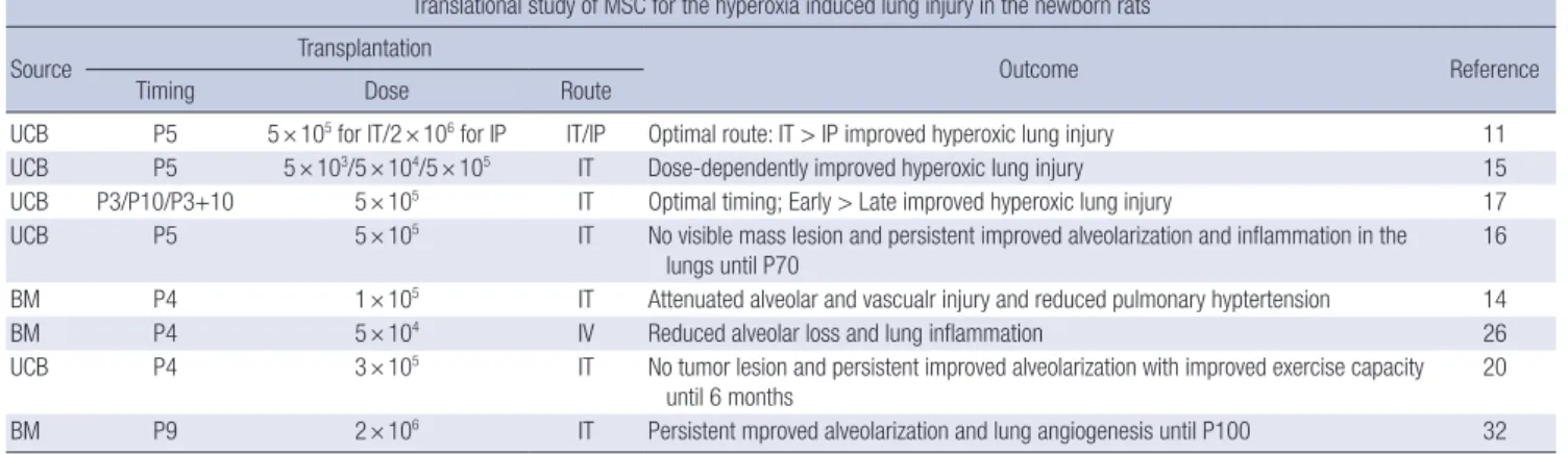
Table 1. Progress of translational research of MSC for neonatal BPD
Translational study of MSC for the hyperoxia induced lung injury in the newborn rats
Source Transplantation
Outcome Reference
Timing Dose Route
UCB P5 5 × 10⁵ for IT/2 × 106 for IP IT/IP Optimal route: IT > IP improved hyperoxic lung injury 11
UCB P5 5 × 10³/5 × 104/5 × 105 IT Dose-dependently improved hyperoxic lung injury 15
UCB P3/P10/P3+10 5 × 10⁵ IT Optimal timing; Early > Late improved hyperoxic lung injury 17
UCB P5 5 × 10⁵ IT No visible mass lesion and persistent improved alveolarization and inflammation in the
lungs until P70 16
BM P4 1 × 10⁵ IT Attenuated alveolar and vascualr injury and reduced pulmonary hyptertension 14
BM P4 5 × 10⁴ IV Reduced alveolar loss and lung inflammation 26
UCB P4 3 × 10⁵ IT No tumor lesion and persistent improved alveolarization with improved exercise capacity
until 6 months 20
BM P9 2 × 10⁶ IT Persistent mproved alveolarization and lung angiogenesis until P100 32
MSC, mesenchymal stem cells; BPD, bronchopulmonary dysplasia; UCB, umbilical cord blood; BM, bone marrow; P, postnatal day; IT, intratracheal; IP, intraperitoneal; IV, intra- venous.
has already been shown (11), the optimal timing of administra
tion is another critical issue that remains to be established. There
fore, we attempted to determine the optimal timing by compar
ing the therapeutic efficacy of early (at P3) versus late (at P10) intratracheal transplantation of MSCs (17). We observed that hyperoxiainduced lung injuries such as impaired alveolariza
tion, increased apoptosis, oxidative stress, inflammation, and fibrosis, as well as reduced VEGF and HGF levels were signifi
cantly attenuated with early but not late transplantation. These findings suggest that the therapeutic time window of MSC trans
plantation for BPD may be narrow during the early but not the late phase of inflammatory responses.
Long-term safety and outcome of MSC transplantation Peirro et al. (20) reported that both human umbilical cordde
rived perivascular cells and MSCs exerted short and longterm (6 months) therapeutic benefits including persistent improve
ment in lung structure and exercise capacity, despite the low engraftment of cells. Moreover, no tumor formation was ob
served, and the beneficial effects of intratracheal transplanta
tion of MSCs in neonatal hyperoxic lung injuries were evident at P5. These beneficial effects, which included improved alveo
lar and vascular growth, were sustained for a prolonged recov
ery period without any longterm adverse effects up to P70 (16).
Overall, these findings support the assumption that transplan
tation of MSCs to prevent or treat BPD in premature infants at a critical early time point might modify and improve the long
term respiratory morbidities of BPD.
PHASE I CLINICAL TRIAL OF MSC FOR BPD
The safety and feasibility of transplanting allogeneic human UCBderived MSCs in preterm infants was assessed. Intratra
cheal transplantation of MSCs was performed in 9 preterm in
fants (3 received 1 × 10⁷ cells/kg and 6 received 2 × 10⁷ cells/kg)
who had a very high risk for developing BPD. The infants in this phase I clinical study had a mean gestational age of 25.3 ± 0.9 weeks, a mean birth weight of 793 ± 127 g and a mean birth age of 10.4 ± 2.6 days (30). The transplantation was well tolerated, without any serious adverse events or doselimiting toxicity.
Tracheal aspirate cytokine levels at day 7 were significantly re
duced compared with the baseline levels. Moreover, BPD se
verity which classified as mild, moderate, and severe according to the consensus of NICHD workshop (31), was significantly lower in the transplant recipients compared with the gestation
al age, body weight, and respiratory severitymatched control group. Overall, these findings suggest that intratracheal trans
plantation of allogeneic human UCBderived MSCs in very pre
term infants at the highest risk for developing BPD is safe and feasible. A longterm followup safety study (NCT01632475) on MSCtreated preterm infants and a phase II doubleblind ran
domized controlled trial to assess the therapeutic efficacy (NCT 01828957) are currently underway.
CONCLUSIONS
In recent years, we have broadened our knowledge and under
standing of stem cell therapy for neonatal lung injury. Contri
butions to this advancement include the various translational research studies supporting the therapeutic potential, safety profile, optimal route, optimal timing, optimal dose, and poten
tial efficacy marker of stem cell therapies for BPD. Moreover, the first phase I clinical trial of MSC transplantation for BPD was conducted successfully, proving its safety and feasibility in the preterm infants. This progress has moved human stem cell therapy for BPD one step closer to clinical translation (Tables 1, 2). We are currently conducting two essential studies to be in
troduced clinically. The first is a phase II clinical trial to assess the therapeutic efficacy (NCT01828957), and the second is a longterm followup safety assessment study of the MSC trans
plant recipients (NCT01897987). Conditional approval of clini
cal use of MSC might be anticipated cautiously after the com
pleteion of the phase II clinical trial with favorable outcome.
DISCLOSURE
Samsung Medical Center and MEDIPOST Co, Ltd have issued or filed patents for “Method of treating lung diseases using cells separated or proliferated from umbilical cord blood” under Yun Sil Chang, Won Soon Park, and Yoon Sun Yang (not affiliated with this article) (application PCT/KR2007/000535).
AUTHOR CONTRIBUTION
All authors participated in writing and revision and agreed to final manuscript.
ORCID
So Yoon Ahn http://orcid.org/0000-0002-1821-3173 Yun Sil Chang http://orcid.org/0000-0001-9201-2938 Won Soon Park http://orcid.org/0000-0002-8245-4692 REFERENCES
1. Walsh MC, Szefler S, Davis J, Allen M, Van Marter L, Abman S, Black
mon L, Jobe A. Summary proceedings from the bronchopulmonary dys- plasia group. Pediatrics 2006; 117: S52-6.
2. Smith LJ, van Asperen PP, McKay KO, Selvadurai H, Fitzgerald DA. Re- duced exercise capacity in children born very preterm. Pediatrics 2008;
122: e287-93.
3. Broström EB, Thunqvist P, Adenfelt G, Borling E, KatzSalamon M. Ob- structive lung disease in children with mild to severe BPD. Respir Med 2010; 104: 362-70.
4. Doyle LW, Faber B, Callanan C, Freezer N, Ford GW, Davis NM. Bron- chopulmonary dysplasia in very low birth weight subjects and lung func- tion in late adolescence. Pediatrics 2006; 118: 108-13.
5. Filippone M, Bonetto G, Corradi M, Frigo AC, Baraldi E. Evidence of unexpected oxidative stress in airways of adolescents born very pre-term.
Eur Respir J 2012; 40: 1253-9.
6. Narang I, Rosenthal M, Cremonesini D, Silverman M, Bush A. Longitu- dinal evaluation of airway function 21 years after preterm birth. Am J Respir Crit Care Med 2008; 178: 74-80.
7. Baraldi E, Filippone M. Chronic lung disease after premature birth. N Engl J Med 2007; 357: 1946-55.
8. Northway WH Jr. Observations on bronchopulmonary dysplasia. J Pedi- atr 1979; 95: 815-8.
9. deLemos RA, Coalson JJ. The contribution of experimental models to our understanding of the pathogenesis and treatment of bronchopulmo- nary dysplasia. Clin Perinatol 1992; 19: 521-39.
10. Yang SE, Ha CW, Jung M, Jin HJ, Lee M, Song H, Choi S, Oh W, Yang YS.
Mesenchymal stem/progenitor cells developed in cultures from UC blood.
Cytotherapy 2004; 6: 476-86.
11. Chang YS, Oh W, Choi SJ, Sung DK, Kim SY, Choi EY, Kang S, Jin HJ, Yang YS, Park WS. Human umbilical cord blood-derived mesenchymal stem cells attenuate hyperoxia-induced lung injury in neonatal rats. Cell Trans- plant 2009; 18: 869-86.
12. Kourembanas S. Stem cell-based therapy for newborn lung and brain injury: feasible, safe, and the next therapeutic breakthrough? J Pediatr 2014; 164: 954-6.
13. Aslam M, Baveja R, Liang OD, FernandezGonzalez A, Lee C, Mitsialis SA, Kourembanas S. Bone marrow stromal cells attenuate lung injury in a murine model of neonatal chronic lung disease. Am J Respir Crit Care Med 2009; 180: 1122-30.
14. van Haaften T, Byrne R, Bonnet S, Rochefort GY, Akabutu J, Bouchen
touf M, ReyParra GJ, Galipeau J, Haromy A, Eaton F, et al. Airway de- livery of mesenchymal stem cells prevents arrested alveolar growth in neonatal lung injury in rats. Am J Respir Crit Care Med 2009; 180: 1131- 42.
15. Chang YS, Choi SJ, Sung DK, Kim SY, Oh W, Yang YS, Park WS. Intratra- cheal transplantation of human umbilical cord blood-derived mesen- chymal stem cells dose-dependently attenuates hyperoxia-induced lung injury in neonatal rats. Cell Transplant 2011; 20: 1843-54.
16. Ahn SY, Chang YS, Kim SY, Sung DK, Kim ES, Rime SY, Yu WJ, Choi SJ, Oh WI, Park WS. Long-term (postnatal day 70) outcome and safety of intratracheal transplantation of human umbilical cord blood-derived mesenchymal stem cells in neonatal hyperoxic lung injury. Yonsei Med J 2013; 54: 416-24.
17. Chang YS, Choi SJ, Ahn SY, Sung DK, Sung SI, Yoo HS, Oh WI, Park WS.
Timing of umbilical cord blood derived mesenchymal stem cells trans- plantation determines therapeutic efficacy in the neonatal hyperoxic lung injury. PLoS One 2013; 8: e52419.
18. Chang YS, Ahn SY, Jeon HB, Sung DK, Kim ES, Sung SI, Yoo HS, Choi SJ, Oh WI, Park WS. Critical role of vascular endothelial growth factor secreted by mesenchymal stem cells in hyperoxic lung injury. Am J Respir Cell Mol Biol 2014; 51: 391-9.
19. Vosdoganes P, Lim R, Moss TJ, Wallace EM. Cell therapy: a novel treat- ment approach for bronchopulmonary dysplasia. Pediatrics 2012; 130:
727-37.
20. Pierro M, Ionescu L, Montemurro T, Vadivel A, Weissmann G, Oudit G, Emery D, Bodiga S, Eaton F, Peault B, et al. Short-term, long-term and paracrine effect of human umbilical cord-derived stem cells in lung inju- ry prevention and repair in experimental bronchopulmonary dysplasia.
Thorax 2013; 68: 475-84.
21. Le Blanc K. Immunomodulatory effects of fetal and adult mesenchymal stem cells. Cytotherapy 2003; 5: 485-9.
22. Amable PR, Teixeira MV, Carias RB, Granjeiro JM, Borojevic R. Protein synthesis and secretion in human mesenchymal cells derived from bone Table 2. Clinical research of MSC for neonatal BPD
Clinical study for the prevention of BPD in the premature infafnts Phase Year ClinicalTrials.gov identifier Status Reference
Phase 1 2011 NCT01297205 Completed 30
Follow-up 2012 NCT01632475, NCT02023788 Ongoing
Phase 2 2013 NCT01828957 Ongoing
Follow-up 2013 NCT01897987 Ongoing
MSC, mesenchymal stem cells; BPD, bronchopulmonary dysplasia.
marrow, adipose tissue and Wharton’s jelly. Stem Cell Res Ther 2014; 5:
53.
23. Choudhery MS, Badowski M, Muise A, Pierce J, Harris DT. Donor age negatively impacts adipose tissue-derived mesenchymal stem cell expan- sion and differentiation. J Transl Med 2014; 12: 8.
24. Kretlow JD, Jin YQ, Liu W, Zhang WJ, Hong TH, Zhou G, Baggett LS, Mi
kos AG, Cao Y. Donor age and cell passage affects differentiation poten- tial of murine bone marrow-derived stem cells. BMC Cell Biol 2008; 9: 60.
25. Berger MJ, Adams SD, Tigges BM, Sprague SL, Wang XJ, Collins DP, Mc
Kenna DH. Differentiation of umbilical cord blood-derived multilineage progenitor cells into respiratory epithelial cells. Cytotherapy 2006; 8: 480-7.
26. Abman SH, Matthay MA. Mesenchymal stem cells for the prevention of bronchopulmonary dysplasia: delivering the secretome. Am J Respir Crit Care Med 2009; 180: 1039-41.
27. Hansmann G, FernandezGonzalez A, Aslam M, Vitali SH, Martin T, Mitsialis SA, Kourembanas S. Mesenchymal stem cell-mediated reversal of bronchopulmonary dysplasia and associated pulmonary hyperten- sion. Pulm Circ 2012; 2: 170-81.
28. Rojas M, Xu J, Woods CR, Mora AL, Spears W, Roman J, Brigham KL.
Bone marrow-derived mesenchymal stem cells in repair of the injured lung. Am J Respir Cell Mol Biol 2005; 33: 145-52.
29. Ortiz LA, Gambelli F, McBride C, Gaupp D, Baddoo M, Kaminski N, Phinney DG. Mesenchymal stem cell engraftment in lung is enhanced in response to bleomycin exposure and ameliorates its fibrotic effects. Proc Natl Acad Sci U S A 2003; 100: 8407-11.
30. Chang YS, Ahn SY, Yoo HS, Sung SI, Choi SJ, Oh WI, Park WS. Mesen- chymal stem cells for bronchopulmonary dysplasia: phase 1 dose-esca- lation clinical trial. J Pediatr 2014; 164: 966-72.e6.
31. Ehrenkranz RA, Walsh MC, Vohr BR, Jobe AH, Wright LL, Fanaroff AA, Wrage LA, Poole K; National Institutes of Child Health and Human De
velopment Neonatal Research Network. Validation of the National In- stitutes of Health consensus definition of bronchopulmonary dysplasia.
Pediatrics 2005; 116: 1353-60.
32. Sutsko RP, Young KC, Ribeiro A, Torres E, Rodriguez M, Hehre D, Devia C, McNiece I, Suguihara C. Long-term reparative effects of mesenchy- mal stem cell therapy following neonatal hyperoxia-induced lung injury.
Pediatr Res 2013; 73: 46-53.