© 2016 The Korean Academy of Medical Sciences.
This is an Open Access article distributed under the terms of the Creative Commons Attribution Non-Commercial License (http://creativecommons.org/licenses/by-nc/4.0) which permits unrestricted non-commercial use, distribution, and reproduction in any medium, provided the original work is properly cited.
pISSN 1011-8934 eISSN 1598-6357
Calculation of the Residual Blood Volume after Acute, Non- Ongoing Hemorrhage Using Serial Hematocrit Measurements and the Volume of Isotonic Fluid Infused: Theoretical Hypothesis Generating Study
Fluid resuscitation, hemostasis, and transfusion is essential in care of hemorrhagic shock.
Although estimation of the residual blood volume is crucial, the standard measuring methods are impractical or unsafe. Vital signs, central venous or pulmonary artery pressures are inaccurate. We hypothesized that the residual blood volume for acute, non- ongoing hemorrhage was calculable using serial hematocrit measurements and the volume of isotonic solution infused. Blood volume is the sum of volumes of red blood cells and plasma. For acute, non-ongoing hemorrhage, red blood cell volume would not change. A certain portion of the isotonic fluid would increase plasma volume. Mathematically, we suggest that the residual blood volume after acute, non-ongoing hemorrhage might be calculated as 0·25N/[(Hct1/Hct2)-1], where Hct1 and Hct2 are the initial and subsequent hematocrits, respectively, and N is the volume of isotonic solution infused. In vivo validation and modification is needed before clinical application of this model.
Keywords: Blood Volume Determination; Hematocrit; Isotonic Solutions; Hemorrhagic Shock; Traumatic Shock
Won Sup Oh1 and Sung-Bin Chon2
1Department of Internal Medicine, Kangwon National University School of Medicine, Chuncheon, Korea; 2Department of Emergency Medicine, Seoul National University College of Medicine, Seoul, Korea
Received: 12 August 2015 Accepted: 16 February 2016 Address for Correspondence:
Sung-Bin Chon, MD
Department of Emergency Medicine, Seoul National University Hospital, 103 Daehak-ro, Jongno-gu, Seoul 03080, Korea E-mail: [email protected]
http://dx.doi.org/10.3346/jkms.2016.31.5.814 • J Korean Med Sci 2016; 31: 814-816
Trauma causes 5 million deaths annually (10% of all-cause mor- tality), and is the most common cause of death (40%) in those aged 10-24 years (1,2). Hemorrhagic shock is the foremost cause of traumatic deaths, and is the main target of resuscitation, es- pecially in the early management stages (3,4). Furthermore, hemorrhagic shock occurs in numerous non-traumatic patients, and optimal treatment is likewise critical in these patients (5).
The cornerstone in the initial management of patients with hemorrhagic shock is damage control resuscitation, character- ized by cautious isotonic fluid resuscitation (permissive hypo- tension) and hemostatic resuscitation with an adequate trans- fusion strategy until definitive control of hemorrhage (6-8). There- fore, the residual blood volume (BV) in hemorrhagic patients at the time of first contact with medical personnel is a reasonable parameter to guide the initial management. In contrast to the management of chronic anemia, however, hematocrit (Hct, %) measurements cannot represent the BV in patients with acute hemorrhagic shock (9,10). The Hct, defined as the red blood cell volume (RBCV) divided by the BV [ = RBCV + plasma vol- ume (PV)], remains unchanged in hyperacute bleeding, and the Hct is not decreased until the PV is increased by shifting the interstitial fluid from the surrounding tissue.
Clinicians usually estimate BV loss according to the guide- lines of the American College of Surgeons. Using vital signs,
urine output, and mental status, the BV loss is categorized as class I-IV (< 15, 15−30, 30−40, > 40% of the total BV, respective- ly) (7). However, this method is semi-quantitative and less ap- plicable in the management of patients who take negative chro- notropes such as beta- or calcium channel blockers, which im- pede compensatory tachycardia, one of the key parameters of the classification. Furthermore, this classification system falls short in management of the elderly and children (7). Central venous and pulmonary artery occlusion pressures are quanti- tative, but cannot accurately reflect the BV (11,12). The refer- ence standards to measure the residual BV using radioisotope- labeled erythrocytes, albumin, indocyanine green, or starch are impractical in the earliest management of hemorrhagic shock due to their complexity and the potential for harm to these pa- tients (13,14). Recently, dynamic indicators to reflect volume responsiveness have been introduced, including the stroke vol- ume variation, pulse pressure variation, systolic pressure varia- tion, and the passive leg raising test (15). However, these meth- ods require specialized equipment and the results are semi-quan- titative.
Given that clinicians generally perform subsequent Hct mea- surements after infusion of certain amounts of isotonic solu- tions during the initial management of patients with hemor- rhagic shock, we hypothesized that the Hct values and isotonic BRIEF COMMUNICATION
Emergency & Critical Care Medicine
Oh WS, et al. • Residual Blood Volume Calculation for Hemorrhagic Shock Patients
http://jkms.org 815
http://dx.doi.org/10.3346/jkms.2016.31.5.814
solution infusion volume could be used to calculate the residu- al BV after acute, non-ongoing hemorrhage.
When patients with hemorrhagic shock present to the emer- gency department (Stage 1: the time of initial contact with med- ical personnel), the standard management consists of obtain- ing blood samples including the initial Hct, rapid infusion of 30 mL/kg of isotonic solutions (usually normal saline) before pos- sible transfusion, and immediate hemostasis as soon as possi- ble. Usually, clinicians check subsequent Hct values (Stage 2:
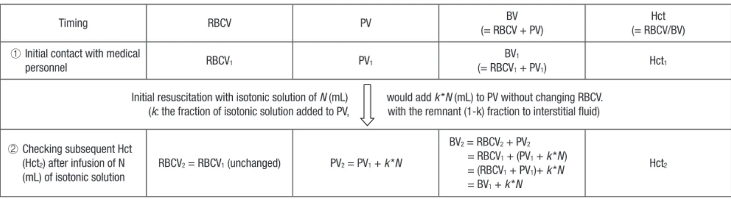
the time to check subsequent Hct), while loading the isotonic solution. Using the designation of subscript of 1 and 2, we can define the BV, PV, and Hct at each time point as described in Fig. 1.
Conceptually, the isotonic solution is distributed to the extra- cellular fluid compartments, which comprise the interstitial fluid compartment and the PV, without entering the intracellu- lar compartments. This distribution of the resuscitation fluid occurs mostly within the first 30 minutes, which would be usu- ally needed for the initial fluid resuscitation in practice from stage 1 to stage 2 (16). If the fraction of isotonic solution distri- bution into the PV is k (where 0 < k < 1), infusion of N (mL) of isotonic solution will add k*N to the PV. Then,
PV2= PV1 + k*N … ①
For a non-ongoing hemorrhage, the RBCV remains unchanged.
RBCV1= RBCV2 … ② Therefore,
BV2= RBCV2 + PV2 [by definition of BV]
= RBCV1 + (PV1 + k*N) [by incorporating ① and ②]
= (RBCV1 + PV1) + k*N
= BV1 + k*N … ③ [by definition of BV]
Meanwhile,
RBCV1= BV1*Hct1 … ④ [by definition]
RBCV2= BV2*Hct2 [by definition]
= (BV1 + k*N)*Hct2 … ⑤ [by incorporating ③]
If we incorporate ④ and ⑤ into ②, which assumes non-sustain- ed hemorrhage, then
BV1*Hct1= (BV1 + k*N)*Hct2 [by incorporating ④ and ⑤]
Then, BV1, the ultimate target of our study can be demonstrated
as follows:
BV1= (k*N)/[(Hct1/Hct2)-1]
Among the components of the equation, N (mL) is the infus ed isotonic solution volume, determined by the clinician, and the Hct1 and Hct2 values are serial measurements.
Additionally, k represents the fraction of the isotonic solution distributed into the PV, which differs among species. If k were to be presumed as 0·25 in human (17), BV1 might be restated more simply as follows:
BV1= 0·25N/[(Hct1/Hct2)-1] (mL)
To simplify the formula for easy use by clinicians, N could be predetermined as 1,000 or 2,000 mL. Then BV1 (mL) might be calculated as follows: 250/[(Hct1/Hct2)-1] (in cases with N = 1,000 mL) and 500/[(Hct1/Hct2)-1] (in cases with N = 2,000 mL) on the condition that following studies support that k is 0·25 even in patients suffering from acute, non-ongoing hemorrhage.
This study demonstrated that the residual BV after control- ling acute hemorrhagic events could be calculated in patients using serial hematocrits and the volume of the isotonic solution infused, by employing the following equation: k*N/[(Hct1/Hct2) -1], where k is the fraction of isotonic solution distribution into the PV, N is the volume of isotonic solution infused between the two hematocrit measurement time points, and Hct1 and Hct2
are the initial and subsequent hematocrits, respectively.
This equation has several merits. Firstly, this simple equation may provide a quantitative measurement of the residual BV, which would guide further intensive management of patients after acute, non-ongoing hemorrhage. Secondly, it utilizes no more resources and does no more harm other than those need- ed for conventional management, unlike other invasive, expen- sive, semi-quantitative, or potentially harmful methods (11-15,18).
This hypothesis-generating study has several limitations. First- ly, this model has been derived mathematically with following assumptions, which may not apply to clinical situations. Accord- ing to the traditional perspectives, the authors assumed that flu- id shift to intravascular compartment after acute hemorrhage would take much time (7,9,10). However, some investigators raised the possibility that this shift might occur faster than ex-
Timing RBCV PV BV
(= RBCV + PV)
Hct (= RBCV/BV) ① Initial contact with medical
personnel RBCV1 PV1 BV1
(= RBCV1 + PV1) Hct1
Initial resuscitation with isotonic solution of N (mL) would add k*N (mL) to PV without changing RBCV.
(k: the fraction of isotonic solution added to PV, with the remnant (1-k) fraction to interstitial fluid)
② Checking subsequent Hct (Hct2) after infusion of N (mL) of isotonic solution
RBCV2 = RBCV1 (unchanged) PV2 = PV1 + k*N
BV2 = RBCV2 + PV2
= RBCV1 + (PV1 + k*N) = (RBCV1 + PV1)+ k*N = BV1 + k*N
Hct2
Fig. 1. The change in red blood cell volume (RBCV), plasma volume (PV), blood volume (BV), and hematocrit (Hct) according to time after acute, non-ongoing hemorrhage.
Oh WS, et al. • Residual Blood Volume Calculation for Hemorrhagic Shock Patients
816 http://jkms.org http://dx.doi.org/10.3346/jkms.2016.31.5.814 pected, which may alter the assumption, although more evi-
dences are needed to support their opinion (19). We neither considered the factors, which may influence the model, such as the integrity of the vasculature, severity of hemorrhage, and re- nal output although the last would be negligible in hemorrhag- ic shock (16,20). All of the limitations arising from the mathe- matical assumptions without these clinical considerations will ultimately alter the k, which we assumed to be 0·25. That is one of the very reasons that we need to validate and modify this mo- del (esp. k) at varying degrees of hemorrhage in vivo before it could be applied to clinical situations.
Another limitation of our model is that it is applicable only to non-ongoing hemorrhage as we assumed so. Although non- ongoing hemorrhage may not result in mortality, the estima- tion of the residual volume would be still valuable in that clini- cians may decide the minimum requisite volume of blood prod- uct by this estimation, which will help lowering the risk of trans- fusion. When the ongoing blood loss is large enough, a more complicated model would be required. To build up this com- plex model, the model suggested by this study might provide the theoretical basis.
In summary, we suggest that the residual BV after acute, non- ongoing hemorrhagic shock might be calculated theoretically using 0·25N/[(Hct1/Hct2)-1], where Hct1 and Hct2 are the initial and subsequent hematocrits, respectively, and N is the volume of isotonic solution infused between these time points. This mo- del needs in vivo validation and modification with successive studies in order to guide the practical management of acute, non-ongoing hemorrhage by estimation of the residual blood volume.
DISCLOSURE
The authors have no potential conflicts of interest to disclose.
AUTHOR CONTRIBUTION
Conception of the theoretical hypothesis: Chon SB. Manuscript preparation, revision, and approval: all authors.
ORCID
Won Sup Oh http://orcid.org/0000-0002-4992-4787 Sung-Bin Chon http://orcid.org/0000-0001-8783-3117 REFERENCES
1. Norton R, Kobusingye O. Injuries. N Engl J Med 2013; 368: 1723-30.
2. Lozano R, Naghavi M, Foreman K, Lim S, Shibuya K, Aboyans V, Abraham J, Adair T, Aggarwal R, Ahn SY, et al. Global and regional mortality from 235 causes of death for 20 age groups in 1990 and 2010: a systematic anal-
ysis for the Global Burden of Disease Study 2010. Lancet 2012; 380: 2095- 128.
3. Sauaia A, Moore FA, Moore EE, Moser KS, Brennan R, Read RA, Pons PT.
Epidemiology of trauma deaths: a reassessment. J Trauma 1995; 38: 185- 93.
4. Teixeira PG, Inaba K, Hadjizacharia P, Brown C, Salim A, Rhee P, Browder T, Noguchi TT, Demetriades D. Preventable or potentially preventable mortality at a mature trauma center. J Trauma 2007; 63: 1338-46.
5. Rose AH, Kotzé A, Doolan D, Norfolk DR, Bellamy MC. Massive transfu- sion--evaluation of current clinical practice and outcome in two large tea- ching hospital trusts in Northern England. Vox Sang 2009; 97: 247-53.
6. Spinella PC, Holcomb JB. Resuscitation and transfusion principles for traumatic hemorrhagic shock. Blood Rev 2009; 23: 231-40.
7. American College of Surgeons, Committee on Trauma. ATLS, Advanced Trauma Life Support for Doctors: Student Course Manual. 8th ed. Chica- go, IL: American College of Surgeons, 2008.
8. Kim Y, Lee K, Kim J, Kim J, Heo Y, Wang H, Lee K, Jung K. Application of damage control resuscitation strategies to patients with severe traumatic hemorrhage: review of plasma to packed red blood cell ratios at a single institution. J Korean Med Sci 2014; 29: 1007-11.
9. Valeri CR, Dennis RC, Ragno G, Macgregor H, Menzoian JO, Khuri SF. Limi- tations of the hematocrit level to assess the need for red blood cell trans- fusion in hypovolemic anemic patients. Transfusion 2006; 46: 365-71.
10. Takanishi DM, Yu M, Lurie F, Biuk-Aghai E, Yamauchi H, Ho HC, Chapital AD. Peripheral blood hematocrit in critically ill surgical patients: an im- precise surrogate of true red blood cell volume. Anesth Analg 2008; 106:
1808-12.
11. Oohashi S, Endoh H. Does central venous pressure or pulmonary capil- lary wedge pressure reflect the status of circulating blood volume in pa- tients after extended transthoracic esophagectomy? J Anesth 2005; 19:
21-5.
12. Shippy CR, Appel PL, Shoemaker WC. Reliability of clinical monitoring to assess blood volume in critically ill patients. Crit Care Med 1984; 12: 107- 12.
13. Iijima T, Iwao Y, Sankawa H. Circulating blood volume measured by pulse dye-densitometry: comparison with (131)I-HSA analysis. Anesthesiology 1998; 89: 1329-35.
14. Jones JG, Wardrop CA. Measurement of blood volume in surgical and in- tensive care practice. Br J Anaesth 2000; 84: 226-35.
15. Marik PE, Monnet X, Teboul JL. Hemodynamic parameters to guide fluid therapy. Ann Intensive Care 2011; 1: 1.
16. Drobin D, Hahn RG. Volume kinetics of Ringer’s solution in hypovolemic volunteers. Anesthesiology 1999; 90: 81-91.
17. Imm A, Carlson RW. Fluid resuscitation in circulatory shock. Crit Care Clin 1993; 9: 313-33.
18. Perel P, Roberts I, Ker K. Colloids versus crystalloids for fluid resuscitation in critically ill patients. Cochrane Database Syst Rev 2013; 2: CD000567.
19. Ryan ML, Thorson CM, Otero CA, Vu T, Schulman CI, Livingstone AS, Proc- tor KG. Initial hematocrit in trauma: a paradigm shift? J Trauma Acute Care Surg 2012; 72: 54-9.
20. Norberg A, Brauer KI, Prough DS, Gabrielsson J, Hahn RG, Uchida T, Tra- ber DL, Svensén CH. Volume turnover kinetics of fluid shifts after hemor- rhage, fluid infusion, and the combination of hemorrhage and fluid infu- sion in sheep. Anesthesiology 2005; 102: 985-94.