137
Immune Network
Apoptosis Protein (c-IAP2) That Contains a Disrupted Ring Domain
Sun-Mi Park, Ji-Su Kim, Ji-Hyun Park, Seung-Goo Kang and Tae Ho Lee
Department of Biology and Protein Network Research Center, Yonsei University, Seoul, Korea
ABSTRACT
Among the members of the inhibitor of apoptosis (IAP) protein family, only Livin and survivin have been reported to have variant forms. We have found a variant form of c-IAP2 through the interaction with the X protein of HBV using the yeast two-hybrid system. In contrast to the wild-type c-IAP2, the variant form has two stretches of sequence in the RING domain that are repeated in the C-terminus that would disrupt the RING domain. We demonstrate that the variant form has an inhibitory effect on TNF-mediated NF-κB activation unlike the wild-type c-IAP2, which increases TNF- mediated NF-κB activation. These results suggest that this variant form has different activities from the wild-type and the RING domain may be involved in the regulation of TNF-induced NF-κB activation. (Immune Network 2002;2(3):137-141)
Key Words: c-IAP2, TNF, NF-κB, TRAF2, TRAF6
Correspondence to: Tae Ho Lee, Department of Biology, Yonsei University, Seoul 120-749, Korea, (Tel) 02-2123-4083, (Fax) 02-312-2242, (E-mail) [email protected]
This work was supported by a grant from the Ministry of Health and Welfare (HMP-00-B-20700-0019).
Introduction
Members of the inhibitor of apoptosis (IAP) protein family have been reported to be involved in signaling that prevent cell death induced by TNFR (1), Fas signaling (2) and etoposide (3). To date, seven human IAP proteins, NAIP (4,5), c-IAP1 (6), c-IAP2 (6), XIAP (7,8), Livin (9), Bruce (10) and survivin (11) have been identified. c-IAP1, c-IAP2 and XIAP all contain two or three copies of the BIR (baculovirus IAP repeat) motif at their N termini and a RING finger at their C termini (12,13). In the case of c-IAP1 and c-IAP2, these molecules contain a CARD domain (14) and interact with TRAF2 (3,15).
Recent reports have shown that NF-κB is required for TNF-mediated induction of human c-IAP2 (16, 17). When c-IAP2 is overexpressed in mammalian cells, c-IAP2 activates NF-κB and suppresses TNF cytotoxicity (1). These c-IAP2 activities are blocked in vivo when a dominant negative form of IκBα that is resistant to TNF-induced degradation is co- transfected (1). A mutant lacking the C-terminal RING domain was shown to inhibit NF-κB activa- tion by TNF and enhances TNF-mediated apoptosis (1). These reports implicate that c-IAP2 is critically
involved in TNF signaling and exerts positive feed- back control on NF-κB via an IκBα targeting mechanism.
There have been reports that have shown variant forms of murine survivin and Livin exist and have different functions (18,19). Three murine survivin cDNA variants are formed through alternative splic- ing and these variants have distinct antiapoptotic functions (18). There has been no previous report that has shown the existence of a variant form of c-IAP2. We have found this variant form of c-IAP2 through the interaction with HBV X protein using yeast two-hybrid system. We have tried to search this variant form through library and genomic screening, however, we failed to find it. In contrast to the wild-type c-IAP2, the variant form has repeated se- quences in the C terminus that would disrupt the RING domain. We demonstrate that c-IAP2V inhib- its TNF-mediated NF-κB, a similar observation that has been reported in a mutant lacking the C-terminal RING domain. However, the variant form increases TRAF2-mediated NF-κB activation whereas the wild- type has no effect. Therefore, these results demonstrate that the variant form has different prop- erties form the wild-type and suggest that it may have distinct functions from the wild-type in vivo.
Materials and Methods
Cell culture and reagents. HEK293, human embryonic kidney cells were grown in Dulbecco's modified
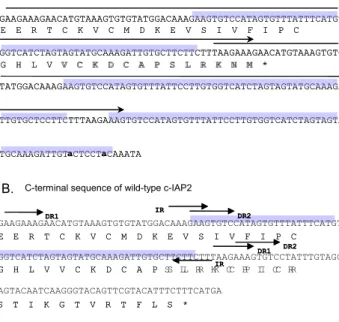
A. C-terminal sequence of variant c- IAP2V
E E R T C K V C M D K E V S I V F I P C
G H L V V C K D C A P S L R K N M *
GAAGAAAGAACATGTAAAGTGTGTATGGACAAAGAAGTGTCCATAGTGTTTATTTCATGT
GGTCATCTAGTAGTATGCAAAGATTGTGCTTCTTCTTTAAGAAAGAACATGTAAAGTGTG
TATGGACAAAGAAGTGTCCATAGTGTTTATTCCTTGTGGTCATCTAGTAGTATGCAAAGA
TTGTGCTCCTTCTTTAAGAAAGTGTCCATAGTGTTTATTCCTTGTGGTCATCTAGTAGTA
TGCAAAGATTGT a CTCCT a CAAATA
GAAGAAAGAACATGTAAAGTGTGTATGGACAAAGAAGTGTCCATAGTGTTTATTTCATGT
GGTCATCTAGTAGTATGCAAAGATTGTGCTTCTTCTTTAAGAAAGTGTCCTATTTGTAGG
AGTACAATCAAGGGTACAGTTCGTACATTTCTTTCATGA
E E R T C K V C M D K E V S I V F I P C
G H L V V C K D C A P S L R K C P I C R
S T I K G T V R T F L S * C-terminal sequence of wild-type c-IAP2
DR1 DR2
DR1 DR2 IR
IR
E E R T C K V C M D K E V S I V F I P C
G H L V V C K D C A P S L R K C P I C R
S T I K G T V R T F L S *
DR1 DR2
DR1 DR2 IR
IR
B.
Figure 1. The C-terminal sequence of variant c-IAP2 encoding the truncated Ring finger motif. A, the DNA and amino acid sequence of the C-terminal of c-IAP2V. The possible gene duplication region is marked by the upper-lined arrow and shadowed. B, the DNA and amino acid sequence of the Ring finger motif of wild-type c-IAP2. The pairs of direct repeats DR1, DR2 and indirect repeat IR are marked.
Eagle's medium (Life Technologies, Inc.) supple- mented with 10% fetal bovine serum in 5% CO2 at 37oC.
Transfection. HEK293 cells were seeded into 12-well dishes the day before transfection and grown to 70%
confluency. Transfection was carried out by the Ca2 PO4-DNA precipitation method using N, N-bis (2- hydroxyethyl)-2-aminoethanesulfonic acid (BES) buffer as described elsewhere (17) with a total of 1μg of DNA containing the indicated constructs. At 16h post-transfection, transfectants were left untreated or treated with TNF at the final concentration of 20 ng/ml for 4 h.
Luciferase assay. Transfectants were lysed in 0.1ml of lysis buffer (Promega) and centrifuged at 10,000 X g for 5 min to remove cell debris. The resulting clear lysates were assayed for luciferase activity according to the manufacturer's instructions and the luciferase activity was measured using the Luciferase Kit (Promega).
Results
A cDNA encoding a variant form of c-IAP2 was found through the interaction with the X protein of HBV using the yeast two-hybrid system and was named c-IAP2V. Sequencing analysis of the wild-type and variant form of c-IAP2 has revealed that the variant form is exactly the same as the wild-type except for the C-terminal portion where insertion of particular stretches of sequence found in the RING domain exist in one or two other sites in the C terminal (Fig. 1).
Pairs of two direct repeat sequence, DR1 (indicated by single arrow) and DR2 (indicated by double line arrow) and an inverted repeat, IR (indicated by thick arrow) exist in the RING domain of the wild-type c-IAP2. The first stretch of sequence that is du- plicated in the c-IAP2V is the 104 bp sequence (indicated by upper arrowed line in Fig. 1A) within the pair of the DR1. The second gene duplication sequence is a 61 bp sequence (indicated by shadowed box in Fig. 1A) between the pair of the DR2 and this sequence is repeated three times whereby the last repeat has only two bp that are different, thus show- ing a nearly perfect repeat. The duplication of these sequences suggests that it may have resulted from somatic gene rearrangement because previous reports have shown that this rearrangement occurs in sites of genes containing direct repeat sequence or inverted repeat sequence. Furthermore, it is unlikely that this variant form is created by incorrect cDNA synthesis because these artifacts usually have a particular se- quence either in the 5'- or 3'- end or have fragments of other cDNAs attached.
The duplication of these sequences causes the dis-
ruption of the RING domain in c-IAP2V that may act as a negative regulator on the activities of c-IAP2.
There are two to three BIR (baculovirus IAP repeat) motifs in the IAP family members and these motifs are essential for the anti-apoptotic function of these proteins. The role of the RING domain in the anti- apoptotic function of the IAPs has not been clearly defined. However, previous studies have shown that c-IAP1 and c-IAP2 without the RING domains ex- hibit anti-apoptotic activities suggesting that the BIR motifs are sufficient for the anti-apoptotic function (3,6). Furthermore, previous report has demonstrated that expression of the RING domain alone increases apoptosis in Drosophila (20). Other studies have shown that full-length c-IAP2 degrades IκBα leading to NF-κB activation, which inhibits TNF- mediated apoptosis (1). Also, truncated form of c-IAP2 without the RING domain blocks c-IAP2 mediated NF-κB activation (1). Thus, the RING finger domain is essential for the role of c-IAP2 in TNF-mediated NF-κB activation.
c-IAP2V with the disrupted RING domain may exhibit different functions from the wild-type. To find out if this variant form has different actions from that of the wild- type, we compared the effect of c-IAP2 and c-IAP2V in TNF-mediated NF-κB activation. We transfected these cDNAs together with the kB-luciferase reporter plasmid in human
0 2000 4000 6000 8000 10000 12000 14000
untreated control c-IAP2 RING c-IAP2V TNF treatment
Luciferase UnitLuciferase Activity
Transfection TNF treatment
pCDM8 pCDM8 c-IAP2 RING c-IAP2V
- + + + +
0 2000 4000 6000 8000 10000 12000 14000
untreated control c-IAP2 RING c-IAP2V TNF treatment
Luciferase UnitLuciferase Activity
Transfection TNF treatment
pCDM8 pCDM8 c-IAP2 RING c-IAP2V
- + + + +
Figure 2. Overexpressed c-IAP2V inhibits TNF-mediated NF- κB activation. HEK293 cells were transiently transfected with 0.1μg of kB-Luc reporter construct together with 0.4μg of c-IAP2, RING and c-IAP2V. After 24 hr of transfection, cells were treated with TNF (20 ng/ml) for 4 hrs. Cell lysates were prepared and assayed for luciferase activity. The data shown represent the result of triplicate transfections in two separate experiments.
0 500 1000 1500 2000 2500
mock control c-IAP2 RING c-IAP2V NIK co-transfection
Luciferase UnitLuciferase Activity
Transfection NIK cotransfection
pCDM8 pCDM8 c-IAP2 RING c-IAP2V
- + + + +
0 500 1000 1500 2000 2500
mock control c-IAP2 RING c-IAP2V NIK co-transfection
Luciferase Unit
0 500 1000 1500 2000 2500
mock control c-IAP2 RING c-IAP2V NIK co-transfection
Luciferase UnitLuciferase Activity
Transfection NIK cotransfection
pCDM8 pCDM8 c-IAP2 RING c-IAP2V
- + + + +
Figure 3. Overexpressed c-IAP2V inhibits NIK-induced NF-κB activation. HEK293 cells were transiently transfected with 0.1μg of kB-Luc reporter construct together with 0.1μg of NIK, 0.4μg of c-IAP2, RING and c-IAP2V. After 24 hr of transfection, cell lysates were prepared and assayed for luciferase activity. PCDM8 indicates cells that were transfected only with the empty vector and not with the NIK construct. The data shown represent the result of triplicate transfections in two separate experiments.
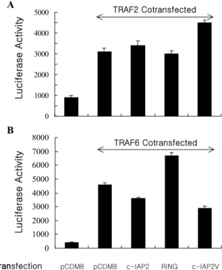
0 500 1000 1500 2000 2500 3000 3500 4000 4500 5000
mock control c-IAP2 RING c-IAP2V TRAF2 co-transfection
Luciferase UnitLuciferase Activity
TRAF2 Cotransfected
0 1000 2000 3000 4000 5000 6000 7000 8000
mock control c-IAP2 RING c-IAP2V
TRAF6 co-transfection
Luciferase UnitLuciferase Activity
Transfection pCDM8 pCDM8 c-IAP2 RING c-IAP2V TRAF6 Cotransfected 0
500 1000 1500 2000 2500 3000 3500 4000 4500 5000
mock control c-IAP2 RING c-IAP2V TRAF2 co-transfection
Luciferase Unit
0 500 1000 1500 2000 2500 3000 3500 4000 4500 5000
mock control c-IAP2 RING c-IAP2V TRAF2 co-transfection
Luciferase UnitLuciferase Activity
TRAF2 Cotransfected
0 1000 2000 3000 4000 5000 6000 7000 8000
mock control c-IAP2 RING c-IAP2V
TRAF6 co-transfection
Luciferase Unit
0 1000 2000 3000 4000 5000 6000 7000 8000
mock control c-IAP2 RING c-IAP2V
TRAF6 co-transfection
Luciferase UnitLuciferase Activity
Transfection pCDM8 pCDM8 c-IAP2 RING c-IAP2V TRAF6 Cotransfected
A
B
Figure 4. Overexpressed c-IAP2 and c-IAP2V show distinct effects on TRAF2 and TRAF6-induced NF-κB activation. Cells were transfected with 0.1μg of kB-Luc reporter construct to- gether with 0.4μg of TRAF2, TRAF6, c-IAP2, RING or c-IAP2V. After 24 hr of transfection, cell lysates were prepared and assayed for luciferase activity. The data shown represent the result of triplicate transfections in two separate experiments.
embryonic kidney cells and treated TNF for 4hrs.
The cells were then processed and the luciferase activity was measured. As shown in Fig. 2 c-IAP2 potentiates TNF-mediated NF-κB activation, show- ing the involvement of c-IAP2 in TNF-mediated NF- κB activation. Transfection of the RING domain alone also increased NF-κB activation by TNF suggesting that the RING domain in c-IAP2 is related to c-IAP2 function in potentiating TNF- mediated NF-κB activation. The result that shows the decrease in NF-κB activation by TNF in c- IAP2V is consistent with the result seen in full- length c-IAP2 and RING domain.
NIK is known to act upstream of the IKKs in TNF-mediated NF-κB activation pathway (21,22).
We investigated the effect of c-IAP2 and c-IAP2V on NIK-induced NF-κB activation. As shown in Fig.
3 transfection of c-IAP2 or the RING domain in- creased NIK-induced NF-κB activation and c-IAP2V decreased the NF-κB activation. This result is similar to that seen in TNF-mediated NF-κB activation.
Members of the TRAF family are known to be involved in the upstream of pathways that lead to NF-κB activation (21,23). Therefore, similar studies were done to investigate the effects of c-IAP2 and c-IAP2V in TRAF2 and TRAF6-mediated NF-κB activation. As shown in Fig. 4, in TRAF2-mediated NF-κB activation, c-IAP2 and the RING domain have no effect, whereas the c-IAP2V shows an in- crease in NF-κB activation, suggesting that the c-IAP2 involvement in NF-κB activation by TRAF2 and TNF is different. In the case of TRAF6-mediated
NF-κB activation, c-IAP2, RING domain and c-IAP2V
show inhibition in NF-κB activation. These results show that c-IAP2 acts differently on NF-κB acti- vation depending on the upstream molecules that
send the NF-κB activation signal.
Discussion
Previous reports have shown that variants of mu- rine survivin and Livin, members of the IAP family, exist and have different functions. Three murine survivin cDNA variants are formed through alter- native splicing and these variants have distinct antia- poptotic functions. These variants either lack the C terminal coiled-coil domain or both the coiled-coil domain and the IAP repeat (18). However, only the variant lacking both the coiled-coil domain and IAP repeat did not suppress caspase-3 activity indicating that the variants have different antiapoptotic prop- erties (18).
The splicing variants of Livin are almost identical proteins that share the amino-terminal BIR and carboxyl-terminal RING domain (19). Livin-β, the shorter variant lacks 18 amino acids in the BIR RING inter-linking region. Both the variant forms showed significant antiapoptotic activity in cell death triggered by TNF and anti-Fas Ab. Livin-α but not Livin-β protected cells from staurosporine-induced apoptosis, however in contrast, apoptosis induced by etoposide was blocked only by the β variant, thus showing differences in biological activities.
This is the first report indicating that a variant form c-IAP2 exists and that it acts differently from the wild-type. Overexpression of c-IAP2 potentiates TNF-mediated and NIK-induced NF-κB activation whereas, c-IAP2V inhibits NF-κB activation in these two cases. However, the opposite effects of c-IAP2 and c-IAP2V were not seen in TRAF2 and TRAF6- induced NF-κB activation. This result suggests that the role of c-IAP2 in the NF-κB activation pathways may be different in TNF-triggered, TRAF2 and TRAF6-induced pathways. The overexpression of RING domain increased NF-κB activation induced by TNF, NIK and TRAF6. This result is in cor- relation with previous reports demonstrating the significance of the RING domain in NF-κB acti- vation.
This work is unique since it suggests for the first time that variants of the IAP family protein can be form through the result of somatic gene rearrange- ment other than alternative splicing and that the variant has distinct activities. These findings indicate that somatic gene rearrangement might play an important role in the fine tuning of c-IAP2 biological function. Further investigation of the mechanism that resulted in the variant form and its biological func- tion may provide further insights into the role of IAP variants in various physiological and pathological conditions.
References
1. Chu ZL, McKinsey TA, Liu L, Gentry JJ, Malim MH, Ballard DW: Suppression of tumor necrosis factor-in- duced cell death by inhibitor of apoptosis c-IAP2 is under NF-κB control. Proc Natl Acad Sci USA 94;10057- 10062, 1997
2. Takahashi R, Deveraux Q, Tamm I, Welsh K, Assa-Munt, N, Salvesen GS, Reed JC: A Single BIR Domain of XIAP Sufficient for Inhibiting Caspases. J Biol Chem 273;7787- 7790, 1998
3. Roy N, Deveraux QL, Takahashi R, Salvesen GS, Reed JC: The c-IAP-1 and c-IAP-2 proteins are direct inhibitors of specific caspases. EMBO J 16;6914-6925, 1997 4. Roy N, Mahadevan MS, McLean M, Shutler G, Yaraghi
Z, Farahani R, Baird S, Besner-Johnston A, Lefebvre C, Kang X, et al: The Gene For Neuronal Apoptosis Inhi- bitory Protein Is Partially Deleted In Individuals with Spinal Muscular Atrophy. Cell 80;167-178, 1995
5. Liston PN, Roy K, Tamai C, Lefevre S, Baird G, Cherton- Horvat R, Farahani M, McLean JE, Ikeda A, Mackenzie Korneluk RG: Suppression of apoptosis in mammalian cells by NAIP and a related family of IAP genes. Nature 379;349-353, 1996
6. Rothe M, Pan MG, Henzel WJ, Ayres TM, Goeddel DV:
The TNFR2-TRAF signaling complex contains two novel proteins related to baculoviral inhibitor of apoptosis pro- teins. Cell 83;1243-1252, 1995
7. Duckett CS, Li F, Wang Y, Tomaselli KJ, Thompson CB, Armstrong RC: Human IAP-like protein regulates pro- grammed cell death downstream of Bcl-xL and cyto- chrome C. Mol Cell Biol 18;608-615, 1998
8. Uren AG, Pakusch M, Hawkins CJ, Puls KL, Vaux DL:
Cloning and expression of apoptosis inhibitory protein homologs that function to inhibit apoptosis and/or bind tumor necrosis factor receptor-associated factors. Proc Natl Acad Sci USA 93;4974-4978, 1996
9. Kasof GM, Gomes BC: Livin, a novel inhibitor of apo- ptosis protein family member. J Biol Chem 276;3238- 3246, 2001
10. Hauser HP, Bardoff M, Pyrowolakis G, Jentsch S: A giant ubiquitin-conjugating enzyme related to IAP apoptosis inhibitors. J Biol Chem 141;1415-1422, 1998
11. Ambrosini G, Adida C, Altieri DC: A novel anti-apoptosis gene, survivin, expressed in cancer and lymphoma. Nat Med 3;917-921, 1997
12. Uren AG, Coulsen EJ, Vaux DL: Conservation of baculo- virus inhibitor of apoptosis repeat proteins (BIRPs) in viruses, nematodes, vertebrates and yeasts. Trends Bio- chem Sci 23;159-162, 1998
13. Sun C, Cai M, Gunasekera AH, Meadows RP, Wang H, Chen J, Zhang H, Wu W, Xu N, Ng SC, Fesik SW: NMR structure and mutagenesis of the inhibitor-of-apoptosis protein XIAP. Nature 401;818-822, 1999
14. Hofmann K, Bucher P, Tschopp J: The CARD domain:
a new apoptotic signalling motif. Trends Biochem Sci 22;155-156, 1997
15. Shu HB, Takeuchi M, Goeddel DV: The tumor necrosis factor receptor 2signal transducers TRAF2 and c-IAP1 are components of the tumor necrosis factor receptor 1signa- lingcomplex. Proc Natl Acad Sci USA 93;13973-13978, 1996
16. Wang CY, Mayo, MW, Korneluk RG, Goeddel DV,
Baldwin AS, Jr: NF-κB Antiapoptosis: Induction of TRAF1 and TRAF2 and c-IAP1 and c-IAP2 to Suppress Caspase-8 Activation. Science 281;1680-1683, 1998 17. Hong SY, Yoon WH, Park JH, Kang SG, Ahn JH, Lee
TH: Involvement of two NF-κB binding elements in TNF-, CD40- and Epstein-Barr virus LMP1-mediated in- duction of the cellular inhibitor of apoptosis protein 2 (c-IAP2/HIAP-1/ MIHC) gene. J Biol Chem 275;18022, 2000
18. Conway EM, Pollefeyt S, Cornelissen J, DeBaere I, Steiner-Mosonyi M, Ong K, Baens M, Collen D, Schuh AC: Three differentially expressed survivin cDNA variants encode proteins with distinct antiapoptotic functions.
Blood 95;1435-1442, 2000
19. Ashhab Y, Alian A, Polliack A, Panet A, Yehuda DB: Two splicing variants of a new inhibitor of apoptosis gene with different biological properties and tissue distribution pat-
tern. FEBS Lett 495;56-60, 2001
20. Hay BA, Wassarman DA, Rubin GM: Drosophila homo- logs of baculovirus inhibitor of apoptosis proteins func- tion to block cell death. Cell 83;1253, 1995
21. Malinin NL, Boldin MP, Kovalenko AV, Wallach D:
MAP3K-related kinase involved in NF-κB induction by TNF, CD95 and IL-1. Nature 385;540-544, 1997 22. Regnier CH, Song HY, Gao X, Goeddel DV, Cao Z,
Rothe M: Identification and characterization of an IkB kinase. Cell 90;373-383, 1997
23. Song HY, Regnier CH, Kirschning CJ, Goeddel DV, Rothe M: Tumor necrosis factor (TNF)-mediated kinase cascades: bifurcation of nuclear factor-kB and c-jun N-terminal kinase (JNK/SAPK) pathways at TNF receptor- associated factor 2. Proc Natl Acad Sci USA 94;9792-979, 1997