INTRODUCTION
Intracranial ependymomas are rare glial cell brain tumors, accounting for 3–9% of all intracranial tumors [1,2]. In patients who have been treated according to best practices, ependy- momas are associated with significant mortality. The 10-year survival rate for adult patients is 70–89% [2-4], and the 10- year overall survival rate is 64% in pediatric patients with ep- endymomas [5-7].
There have been many reports regarding the treatment re-
Ependymomas: Prognostic Factors and Outcome Analysis in a Retrospective Series of 33 Patients
Yong-Hyun Chai1, Shin Jung1, Jung-Kil Lee1, In-Young Kim1, Woo-Youl Jang1, Kyung-Sub Moon1, Jae-Hyoo Kim1, Kyung-Hwa Lee2, Seul-Kee Kim3, Tae-Young Jung1
Departments of 1Neurosurgery, 2Pathology, 3Radiology, Chonnam National University Research Institute of Medical Sciences, Chonnam National University Hwasun Hospital & Medical School, Hwasun, Korea
Received July 4, 2017 Revised September 26, 2017 Accepted September 29, 2017 Correspondence
Tae-Young Jung
Department of Neurosurgery, Chonnam National University Hwasun Hospital, 322 Seoyang-ro, Hwasun-eup, Hwasun 58128, Korea Tel: +82-61-379-7666
Fax: +82-61-379-7673 E-mail: [email protected]
Background The purpose of this study was to evaluate the prognostic factors and outcomes in patients with ependymoma to management plans.
Methods Between 1997 and 2013, 33 patients with 25 ependymomas (WHO grade II) and eight anaplastic ependymomas (WHO grade III) were pathologically diagnosed. Six were pediatric pa- tients (mean age, 6.15 years; range, 1.3-11 years), while 27 were adults (mean age, 47.5 years; range, 19-70 years). Of those, there were 12 adult patients with totally resected ependymomas without ana- plastic pathology and adjuvant treatment. Prognostic factors were assessed in ependymoma patients.
Prognostic factors were studied using Kaplan-Meier estimates in subgroups.
Results For six pediatric patients, the progression-free survival (PFS) was 43.7±13.5 months, and the overall survival (OS) was 58.1±13.7 months. For 27 adult patients, the PFS was 125.6±14.3 months, and the OS was 151.2±12.5 months. Age demonstrated a statistically significant effect on PFS (p=0.03) and OS (p=0.03). In adult ependymomas, the extent of tumor removal significantly af- fected PFS (p=0.03) and trended towards an effect on OS (p=0.06). Out of 12 patients with totally re- sected ependymomas without anaplastic pathology and adjuvant treatment, one patient showed tu- mor recurrence during follow-up (mean, 93.5 months; range, 27.9-162.7 months).
Conclusion Adult patients with ependymomas were found to have better survival rates compared to pediatric patients. We suggest that totally resected adult ependymomas without anaplastic patholo- gy could be observed without any adjuvant treatment, regardless of the tumor location.
Key Words Ependymoma; Surgery; Prognosis; Radiotherapy.
sults of ependymomas. Although some prognostic factors re- main controversial, age, sex, tumor location, pathologic grade, extent of removal, and postoperative adjuvant treatment have all been evaluated as possible prognostic factors [3,8,9]. The treatment options for ependymoma are surgery, radiothera- py, and chemotherapy. Age and the extent of surgery were considered as the major predictors of outcome in patients with ependymomas. Adult patients have better prognosis than chil- dren. Many studies reported that gross total removal was sig- nificantly related to better progression-free survival (PFS) and overall survival (OS) compared to subtotal resection (STR) [2].
Maximal safe resection is the best treatment for ependymo- ma. Surgery and involved-field radiotherapy remain the stan- dard therapy, especially for posterior fossa ependymomas with or without anaplastic pathology. However, in adult patients,
This is an Open Access article distributed under the terms of the Creative Commons Attribution Non-Commercial License (http://creativecommons.org/licenses/by-nc/4.0) which permits unrestricted non-commercial use, distribution, and reproduction in any medium, provided the original work is properly cited.
Copyright © 2017 The Korean Brain Tumor Society, The Korean Society for Neuro- Oncology, and The Korean Society for Pediatric Neuro-Oncology
postoperative radiotherapy could be considered only for ana- plastic ependymomas or suspected residual intracranial tu- mors, because of better prognosis compared to children. In this study, we focused on the prognosis of totally resected adult ependymomas without anaplastic pathology and post- operative adjuvant treatment. This study could be helpful in selecting the subsequent management of adult ependymomas.
MATERIALS AND METHODS Clinical characteristics of patients with ependymomas
Between 1997 and 2013, 33 patients with ependymomas underwent surgery in the Chonnam National University Hospital and Chonnam National University Hwasun Hospi- tal. A retrospective review of medical records was performed in all patients. This study was approved by our Institutional Re- view Board (CNUHH-2017-084). Participants in research con- sented to being involved in this study. Pediatric patients were defined as patients under 18 years old.
Ependymomas were classified as World Health Organiza- tion (WHO) grade II ependymomas or grade III anaplastic ependymomas according to the WHO classification system.
Data regarding patients’ clinical characteristics, such as age, tumor location, extent of removal, pathologic grade, postoper- ative adjuvant treatment, PFS, and OS, were retrospectively analyzed. Postoperative adjuvant treatment was performed for all WHO grade III ependymoma patients. In WHO grade II ependymoma patients, adjuvant treatment was recommended according to the extent of resection. In adult grade II ependy- moma patients with gross total resection (GTR), adjuvant treat- ment was not recommended. One adult grade III ependymo- ma patient with STR did not receive adjuvant treatment due to poor general condition. Radiotherapy was performed 4 weeks after the operation for consideration patient’s general condition and surgical wound. If the patient’s general condi- tion was not good, we considered the patient’s condition when determining treatment timing. Conventional external beam radiotherapy was performed in 13 patients. There was no ra- diation-induced necrosis during follow-up. Adjuvant chemo- therapy was performed for a 15-month-old girl.
The high-risk group was defined as patients with WHO grade III tumors or subtotally resected tumors, and the other patients were assigned to the low-risk group. To evaluate effica- cy of adjuvant treatment, adult patients were divided into three groups: high risk with adjuvant treatment, high risk without adjuvant treatment, and low risk without adjuvant treatment.
We evaluated the extent of removal by evaluating the intra- operative findings and postoperative contrast-enhanced com- puter tomography images. The extent of removal was classi-
fied as total (100%) tumor removal, subtotal (over 50%), or partial (lower than 50%). Tumor recurrence was based on the presence of a tumor on follow-up magnetic resonance images (MRI) or the development of symptoms. We performed fol- low-up brain MRI scans every 6 months. We confirmed tumor recurrence by brain MRI. The related symptoms were absent or present depending on the patients. The pattern of recurrence was classified into local, distant, or cerebrospinal fluid (CSF) dissemination. Local recurrence was defined as a recurrence at the primary tumor site, and distant recurrence was defined as spatially separated recurrence within the brain. CSF dissemi- nation was defined as a recurrence in the ventricles, subarach- noid space, and spine.
Pediatric ependymomas
Table 1 summarizes the patients’ information. Of the six pe- diatric patients with ependymomas receiving treatment, one patient (16.6%) had a supratentorial tumor, and five patients (83.3%) had infratentorial tumors. Three patients (50.0%) had grade II ependymomas, and three patients (50.0%) had grade III. GTR was performed in two patients (33.3%) and STR in four (66.6%). Post-operative adjuvant therapy was adminis- tered to five patients. Radiation treatment was administered to four patients (66.6%), and one patient was treated with che- motherapy (16.6%). In the radiation treatment, three patients received only local brain radiation therapy consisting of 4,000, 4,500, and 5,400 cGy, respectively. One patient received local and craniospinal irradiation (CSI) during the same period. The radiation dose was 3,820 cGy in the local brain and 1,700 cGy in the CSI. Chemotherapy with vincristine, prednisolone, and carmustine was administered to one patient according to the CCG-9445 protocol of the Children’s Cancer Group high- grade glioma study for 5 months. The mean follow-up duration was 48.5 months (range, 16.9–102.8 months). Tumor recur- rence occurred in three patients (50%). Two patients showed local recurrence on follow-up at 32.1 months and 72.0 months, respectively. One patient showed local recurrence and CSF dis- semination at 8.8 months.
Adult ependymomas
A total of 27 adult patients with ependymoma were treated;
22 patients (81.4%) had WHO grade II ependymomas, and five (18.5%) had WHO grade III ependymomas. One patient with WHO grade II ependymoma demonstrated malignant transformation into WHO grade III ependymoma after recur- rence. The tumor was located supratentorially in seven patients (25.9%), infratentorially in 13 (48.1%), and in the spinal cord in seven (25.9%). The tumor was totally resected in 15 patients (55.5%), and an STR was performed in 12 (44.4%). Postopera- tive adjuvant radiation therapy was administered to nine pa-
Statistical analysis
We defined the mean range according to the follow-up du- ration and determined single-variable effects on PFS and OS via univariate analyses. The single variables were age, sex, tu- mor location, extent of removal, pathology, and adjuvant ther- apy. PFS was calculated from the dates of surgery to radiologi- cal progression or the last follow-up visit, and OS was calculated from the dates of surgery to death or final follow-up. Survival probability was calculated using the Kaplan-Meier method by performing comparisons with the log-rank test. All statistical tients (33.3%). Eight patients received 4,500 cGy to 5,400 cGy
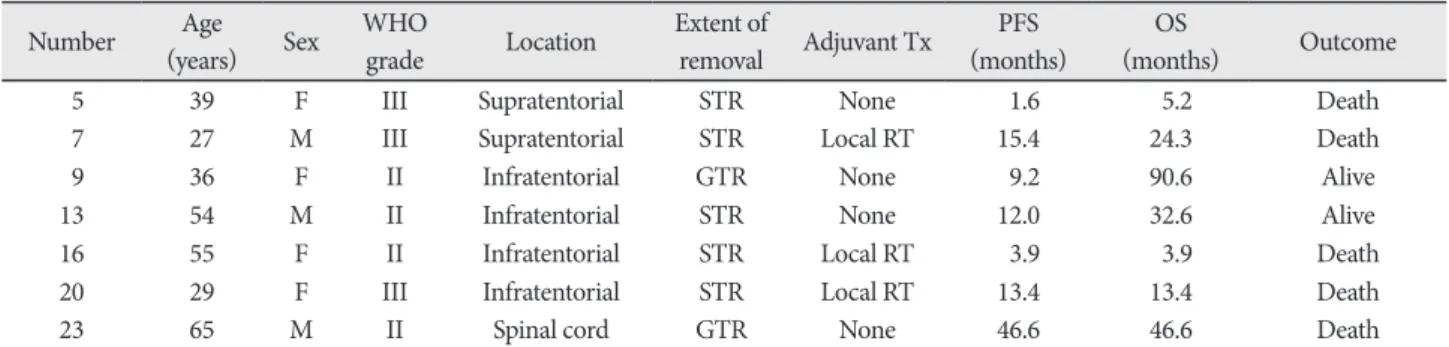
local brain radiotherapy and one patient received 3,240 cGy CSI. The mean follow-up duration was 83.8 months (range, 3.8–182.2 months). Tumor recurrence occurred in seven pa- tients (25.9%). Mean recurrence period of relapsed patients was 12.0 months (range, 1.6–46.6 months). Five patients de- veloped local recurrence, and two patients developed local re- currence combined with CSF dissemination. Table 2 summa- rizes the information of relapsed adult patients.
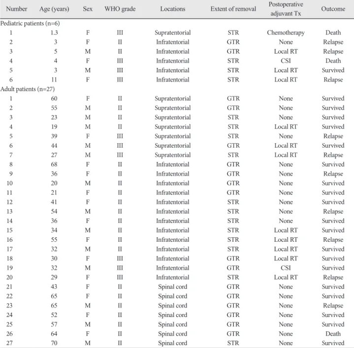
Table 1. Summary of characteristics for all patients
Number Age (years) Sex WHO grade Locations Extent of removal Postoperative
adjuvant Tx Outcome Pediatric patients (n=6)
1 1.3 F III Supratentorial STR Chemotherapy Death
2 3 F II Infratentorial GTR None Relapse
3 5 M II Infratentorial GTR Local RT Relapse
4 4 F III Infratentorial STR CSI Death
5 3 M III Infratentorial STR Local RT Survived
6 11 F III Infratentorial STR Local RT Relapse
Adult patients (n=27)
1 60 F II Supratentorial GTR None Survived
2 55 M II Supratentorial GTR None Survived
3 23 M II Supratentorial STR None Survived
4 19 M II Supratentorial STR Local RT Survived
5 39 F III Supratentorial STR None Relapse
6 44 M III Supratentorial GTR Local RT Survived
7 27 M III Supratentorial STR Local RT Relapse
8 68 F II Infratentorial GTR None Survived
9 36 F II Infratentorial GTR None Relapse
10 20 M II Infratentorial GTR None Survived
11 21 F II Infratentorial GTR None Survived
12 41 F II Infratentorial STR None Survived
13 54 M II Infratentorial STR None Relapse
14 36 F II Infratentorial STR None Survived
15 34 M II Infratentorial STR Local RT Survived
16 55 F II Infratentorial STR Local RT Relapse
17 32 M II Infratentorial STR Local RT Survived
18 30 F III Infratentorial GTR Local RT Survived
19 32 M III Infratentorial GTR CSI Survived
20 29 F III Infratentorial STR Local RT Relapse
21 43 F II Spinal cord GTR None Survived
22 65 F II Spinal cord GTR None Survived
23 65 M II Spinal cord GTR None Relapse
24 52 F II Spinal cord GTR None Survived
25 57 M II Spinal cord GTR None Survived
26 64 F II Spinal cord GTR None Death
27 70 M II Spinal cord STR None Survived
M, male; F, female; WHO, World Health Organization; GTR, gross total removal; STR, subtotal removal; Tx, treatment; WBRT, whole brain radiotherapy; RT, radiotherapy; CSI, craniospinal irradiation
analyses were performed with a significance level of p<0.05 using SPSS 21.0 (IBM Corp., Armonk, NY, USA).
RESULTS
Progression-free survival and overall survival of all ependymomas
Six patients were pediatric patients (mean age, 6.15 years;
range, 1.3–11.0 years) with a male-to-female ratio of 2:4, and 27 were adults (mean age, 47.5 years; range, 19.0–70.0 years) with a male-to-female ratio of 13:14. The mean follow-up duration was 66.7 months (range, 3.9–144.1 months). In all patients, the mean PFS was 114.0±13.8 months and mean OS was 138.0
±12.6 months. There was no statistical significance in the re- lationship between PFS and sex, tumor location, or pathology.
Adult patients were found to be associated with a longer PFS (p=0.03) and OS (p=0.03) (Fig. 1A). Six pediatric patients had
a mean PFS of 43.7±13.5 months and a mean OS of 58.1±13.7 months, while 27 adults had a mean PFS of 127.8±14.8 months and a mean OS of 151.2±12.5 months. Depending on the age and location, we divided the patients into four groups: pediat- ric-infratentorial, adult-supratentorial, adult-infratentorial, and adult-spinal cord. Pediatric-infratentorial group showed a trend of poor prognosis compared to adult-spinal cord group in terms of PFS (p=0.032) (Fig. 1B). PFS was 49.0±15.1 months in pediatric-infratentorial group, 85.5±26.3 months in adult- supratentorial group, 130.7±21.3 months in adult-infratento- rial group, and 113.5±12.2 months in adult-spinal cord group.
Pediatric-infratentroial group was not statistical poor sig- nificance for OS compared adult-group (p=0.358). OS was 66.4±13.7 months in pediatric-infratentorial group, 129.3±27.4 months in adult-supratentorial group, 155.5±17.4 months in adult-infratentorial group, and 113.5±12.2 months in adult- spinal cord group. There was no statistically significant rela- Table 2. Summary of characteristics for seven adult patients with recurrence
Number Age
(years) Sex WHO
grade Location Extent of
removal Adjuvant Tx PFS
(months) OS
(months) Outcome
5 39 F III Supratentorial STR None 1.6 5.2 Death
7 27 M III Supratentorial STR Local RT 15.4 24.3 Death
9 36 F II Infratentorial GTR None 9.2 90.6 Alive
13 54 M II Infratentorial STR None 12.0 32.6 Alive
16 55 F II Infratentorial STR Local RT 3.9 3.9 Death
20 29 F III Infratentorial STR Local RT 13.4 13.4 Death
23 65 M II Spinal cord GTR None 46.6 46.6 Death
M, male; F, female; WHO, World Health Organization; GTR, gross total removal; STR, subtotal removal; Tx, treatment; RT, radiotherapy;
PFS, progression-free survival; OS, overall survival
OS
1.0 0.8 0.6 0.4 0.2
0.0
0 50 100 150 200 Months
AgePediatric Adult
A
PFS
1.0 0.8 0.6 0.4 0.2
0.0
0 50 100 150 200 Months
Age-location Pediatric-infratentorial Adult-supratentorial Adult-infratentorial Adult-spinal cord
B
PFS
1.0 0.8 0.6 0.4 0.2
0.0
0 50 100 150 200 Months
Extent of removal GTRSTR
C
Fig. 1. Kaplan-Meier curves for PFS and OS. A: OS depends on age in all patients. The three-year OS rate was 80.9% for adults, and 66.7%
for pediatric patients (p=0.03). B: PFS depends on age and tumor location in all patients. Pediatric-infratentorial group showed the trend of poor prognosis compared to adult-spinal cord group in terms of PFS. The three-year PFS rate was 40.0% for pediatric-infratentorial group, and 100% for adult-spinal cord group (p=0.03). C: PFS with extent of removal in adult patients. The one-year PFS rate was 100% for GTR, and 75% for STR (p=0.04). PFS, progression-free survival; OS, overall survival; GTR, gross total resection; STR, subtotal resection.
tionship with sex, tumor location, extent of removal, or pathol- ogy. However, there was a statistically significant relationship with age.
Progression-free survival and overall survival of 27 adults ependymomas
These data were summarized in Table 3. Adult ependymo- mas were analyzed for clinical prognostic factors. For PFS, there was no statistically significant relationship with sex (p=0.64), tumor location (p=0.28), pathology (p=0.23), or postoperative adjuvant treatment (p=0.13). However, the extent of removal had a significant effect on PFS (p=0.04) (Fig. 1C); the mean PFS was 154.6±14.0 months for GTR, and 82.7±20.6 months for STR. The one-year PFS rate was 100% for GTR, and 75% for STR. For OS, there was no significant effect of sex (p=0.63), tu- mor location (p=0.78), pathology (p=0.20), or postoperative adjuvant treatment (p=0.31). A trend towards a better OS was found in patients who was underwent GTR (p=0.06); the mean OS was 172.5±9.3 months for GTR and 120.6±22.3 months for STR.
The prognosis of adult patients, especially focused on twelve totally resected ependymomas without anaplastic pathology and postoperative adjuvant treatment
Twelve adult patients underwent total resection of the epen- dymoma without anaplastic pathology and adjuvant treat- ment. The mean age of the patients was 54.3 years (range, 20.0–
68.07 years). The male-to-female ratio was 4:8. The tumor was
located supratentorially in two patients (16.6%), infratentori- ally in four (33.3%), and in the spinal cord in six (50%). The mean follow-up duration was 93.5 months (range, 27.9–162.7 months). One out of the 12 patients developed local recurrence at the primary origin site.
The clinical course of the one case of recurrence is described (Fig. 2). A 36-year-old woman had a 3.7-cm-sized ependy- moma in the fourth ventricle that was totally resected via a telovelar approach. One year later, follow-up MRI showed a 1-cm-sized recurrent lesion associated with enhancement of the fourth ventricle. Local brain radiotherapy (5,400 cGy) and spinal irradiation (2,160 cGy) were performed. Over the course of 92 months of follow-up after radiotherapy, no recurrence was seen.
DISCUSSION
Many reports have published potential prognostic factors for mortality in patients with ependymoma to help plan treat- ments [1,5,10]. Although the results have been controversial, prognostic factors such as age, sex, tumor location, tumor grade, extent of removal, and adjuvant treatment have been evaluat- ed in several studies [1,3,8]. Age and extent of resection can be considered as the major predictors of outcome.
Tumor location was important factor in some studies. In- fratentorial ependymoma was a poorer prognostic factor than supratentorial ependymoma [5]. But some authors maintain that outcome is worse with supratentorial tumor than infraten- torial tumor [4,8]. In our study, pediatric infratentorial epen-
Table 3. Outcomes of adult ependymomas (n=27)
Prognostic factors PFS (months) OS (months) PFS (p value) OS (p value)
Sex
Male (n=13)
Female (n=14) 117.7±20.4
134.5±20.1 152.6±15.3
144.8±19.2
0.64 0.63
Tumor location Supratentorial (n=7) Infratentorial (n=13) Spinal cord (n=7)
85.4±26.3 130.6±21.2 113.5±12.2
129.3±27.4 155.5±17.4 113.5±12.2
0.28 0.78
Extent of removal Total resected (n=15) Subtotal resected (n=12)
154.6±14.0 82.7±20.6
172.5±9.3 120.6±22.3
0.04 0.06
Pathology
WHO grade II (n=21) WHO grade III (n=6)
122.8±13.8 93.0±33.9
152.7±12.0 116.9±35.8
0.23 0.20
Postoperative adjuvant treatment High risk with adjuvant Tx (n=9) High risk without Tx (n=6) Low risk without Tx (n=12)
91.5±27.0 105.6±28.0 151.2±12.5
126.1±26.5 139.4±24.5 152.1±10.1
0.13 0.31
Tx, treatment; PFS, progression-free survival; OS, overall survival; WHO, World Health Organization
dymomas showed a poorer prognosis when compared to adult ependymomas. Location could affect the prognosis differently depending on the patient’s age.
Young age is a major cause of poor outcome [11,12]. Patients in whom ependymomas are detected at an early age are ex- tensively reported as having a very poor prognosis. Some re- ports have suggested that the incidence of high-grade tumors tended to be higher in younger patients and pediatric epen- dymomas were more usually located in the posterior fossa as compared with adult ependymomas. Children aged less than one year were reported to have a 5-year OS of 42.4% [10]. The 5-year OS improved with increasing age to reach 74.7% in pa- tients aged between 5 and 9 years and 81.4% in patients aged between 10 and 14 years [4].
As other prognostic factors, the tumor location and histo- logic grade could be important; however, they remain contro-
versial. Infratentorially located tumors are associated with poorer survival rates than those located supratentorially [5].
Several studies suggested an obvious decline in survival for patients with WHO grade III ependymomas when compared with those with grade II tumors [13]. In this study, there was neither a correlation between location and prognosis nor his- tologic grade. Recently, a molecular grouping study of ependy- momas showed that there is a correlation between two of these groups, ST-EPN-RELA and PF-EPN-A, and a poor prognosis [14]. In the future, genetic characteristics could be considered when formulating the treatment plan with the above clinical prognostic factors.
Surgery is the standard treatment for ependymomas. The extent of resection is a reliable prognostic factor that has been previously reported [1,2,15]. These reports emphasize that the maximal safe resection should be performed in patients with Fig. 2. One case of recurrence out of 12 adult patients with totally resected ependymomas without anaplastic pathology or postoperative adjuvant treatment. A: A 3.7-cm-sized ependymoma in the fourth ventricle (white arrowhead). B: No remnants of the lesion after total resec- tion. C: On first year after surgery, a 1-cm-sized recurrent lesion (white arrowhead). D: No recurrence over the course of 92 months of fol- low-up.
A
C
B
D
ependymomas. One clinical trial showed that GTR was an in- dependent risk factor regardless of the pathologic grade [16].
Maximal surgical resection should be carried out, if possible.
The development of surgical techniques and instruments, such as neuronavigation or intraoperative monitoring, could im- prove the rate of resection and decrease complications. In pedi- atric ependymomas, GTR with adjuvant local radiotherapy has been established as the standard treatment, especially for tu- mors located in the posterior fossa [4,17].
Radiotherapy is an important treatment modality due to the radiosensitivity of ependymomas. CSI has also been ac- cepted in cases of leptomeningeal dissemination. For adult pa- tients, the radiotherapy has postoperatively been performed, regardless of the histologic grade of the tumor and the extent of removal. However, recent studies have suggested that adult patients with WHO grade II ependymoma who received GTR did not immediately need radiotherapy if tumor spreading was not seen in the CSF [18,19]. The optimal management of adult ependymomas remains still controversial.
In this study, postoperative treatment was performed in adult patients with poor prognostic factors, such as subtotal resected or grade III tumor. We divided adult patients into three groups for evaluation of prognosis. Even if there was no statistical sig- nificance, the high-risk group with adjuvant treatment showed the poorer prognosis compared to the low-risk group.
In our study, only one out of the 12 patients who had totally resected ependymomas without anaplastic pathology devel- oped local recurrence. Although recurrence occurred, there was no subsequent recurrence after additional radiotherapy.
This suggests that additional treatment could be needed when recurrence is observed after radiological follow-up. A “wait and see” policy could be recommended in totally resected WHO grade II adult ependymomas, regardless of the tumor location. On the contrary, the prognosis of pediatric ependy- moma is different, and pediatric patients had a poorer progno- sis than adult patients. Similar to previous reports, we also do not recommend a “wait and see” policy especially for infraten- torial pediatric ependymoma.
There were some limitations of this study. The main limita- tion of this study is its retrospective nature and relatively small patient population, which undoubtedly limited the statistical power of some of our analyses. Additionally, we did not per- form a genetic study.
In conclusion, we analyzed 33 ependymoma patients for prognostic factors. Age is an important factor of prognosis. In adult patients, totally resected adult ependymomas without anaplastic pathology could be monitored without any adju- vant treatment, regardless of the tumor location. This study could be helpful in developing the subsequent management plan when treating adult ependymomas.
Conflicts of Interest
The authors have no financial conflicts of interest.
REFERENCES
1. Oya N, Shibamoto Y, Nagata Y, Negoro Y, Hiraoka M. Postoperative ra- diotherapy for intracranial ependymoma: analysis of prognostic factors and patterns of failure. J Neurooncol 2002;56:87-94.
2. Metellus P, Barrie M, Figarella-Branger D, et al. Multicentric French study on adult intracranial ependymomas: prognostic factors analysis and therapeutic considerations from a cohort of 152 patients. Brain 2007;
130(Pt 5):1338-49.
3. Paulino AC, Wen BC, Buatti JM, et al. Intracranial ependymomas: an analysis of prognostic factors and patterns of failure. Am J Clin Oncol 2002;25:117-22.
4. Mansur DB, Perry A, Rajaram V, et al. Postoperative radiation therapy for grade II and III intracranial ependymoma. Int J Radiat Oncol Biol Phys 2005;61:387-91.
5. Tamburrini G, D’Ercole M, Pettorini BL, Caldarelli M, Massimi L, Di Rocco C. Survival following treatment for intracranial ependymoma: a review. Childs Nerv Syst 2009;25:1303-12.
6. Shu HK, Sall WF, Maity A, et al. Childhood intracranial ependymoma:
twenty-year experience from a single institution. Cancer 2007;110:432- 7. van Veelen-Vincent ML, Pierre-Kahn A, Kalifa C, et al. Ependymoma 41.
in childhood: prognostic factors, extent of surgery, and adjuvant thera- py. J Neurosurg 2002;97:827-35.
8. Metellus P, Figarella-Branger D, Guyotat J, et al. Supratentorial epen- dymomas: prognostic factors and outcome analysis in a retrospective series of 46 adult patients. Cancer 2008;113:175-85.
9. Merchant TE, Li C, Xiong X, Kun LE, Boop FA, Sanford RA. Confor- mal radiotherapy after surgery for paediatric ependymoma: a prospec- tive study. Lancet Oncol 2009;10:258-66.
10. Gatta G, Botta L, Rossi S, et al. Childhood cancer survival in Europe 1999-2007: results of EUROCARE-5--a population-based study. Lan- cet Oncol 2014;15:35-47.
11. McGuire CS, Sainani KL, Fisher PG. Both location and age predict survival in ependymoma: a SEER study. Pediatr Blood Cancer 2009;
52:65-9.
12. Ward S, Harding B, Wilkins P, et al. Gain of 1q and loss of 22 are the most common changes detected by comparative genomic hybridisation in paediatric ependymoma. Genes Chromosomes Cancer 2001;32:59- 13. Kurt E, Zheng PP, Hop WC, et al. Identification of relevant prognostic 66.
histopathologic features in 69 intracranial ependymomas, excluding myxopapillary ependymomas and subependymomas. Cancer 2006;
106:388-95.
14. Pajtler KW, Witt H, Sill M, et al. Molecular classification of ependymal tumors across all CNS compartments, histopathological grades, and age groups. Cancer Cell 2015;27:728-43.
15. Korshunov A, Neben K, Wrobel G, et al. Gene expression patterns in ependymomas correlate with tumor location, grade, and patient age.
Am J Pathol 2003;163:1721-7.
16. Gajjar A, Packer RJ, Foreman NK, et al. Children’s Oncology Group’s 2013 blueprint for research: central nervous system tumors. Pediatr Blood Cancer 2013;60:1022-6.
17. Paulino AC. Radiotherapeutic management of intracranial ependy- moma. Pediatr Hematol Oncol 2002;19:295-308.
18. Timmermann B, Kortmann RD, Kühl J, et al. Combined postoperative irradiation and chemotherapy for anaplastic ependymomas in child- hood: results of the German prospective trials HIT 88/89 and HIT 91.
Int J Radiat Oncol Biol Phys 2000;46:287-95.
19. Metellus P, Guyotat J, Chinot O, et al. Adult intracranial WHO grade II ependymomas: long-term outcome and prognostic factor analysis in a series of 114 patients. Neuro Oncol 2010;12:976-84.