pISSN: 0378-6471 eISSN: 2092-9374
DOI : 10.3341/jkos.2011.52.4.454
= 증례보고 =
국소적 망막신경섬유층결손안에서 빛간섭단층촬영의 가음성 결과와 관련된 요인
강성민⋅엄기방 한양대학교 의과대학 안과학교실
목적: 망막신경섬유층촬영에서 국소적 망막신경섬유층결손이 있는 환자가 빛간섭단층촬영(optical coherence tomography, OCT)으로 검출되지 않는 것과 관련된 요인을 알아보고자 하였다.
대상과 방법: 국소적 망막신경섬유층결손이 있는 시야결손전녹내장 24명과 시야결손녹내장 173명을 대상으로 하였고, OCT로 검출 여부에 따라 두 군으로 나누었다. 성별, 연령, 굴절이상, 당뇨병, 고혈압, 중심각막두께, 녹내장 유형, MD, PSD, 평균망막신경섬유층두 께, 유두면적, 사진의 결손과 관련된 변수(위치, 수, 폭)를 비교하였다.
결과: 사진의 망막신경섬유층결손 197안 중 51안(25.9%)이 OCT에서는 검출되지 않았다. 사진의 결손과 OCT결손의 폭과 위치는 유의 한 상관관계가 있었다(상관계수 각각 0.98, 0.64). 로지스틱 회귀분석에서 OCT의 가음성과 관련된 요인은 평균망막신경섬유층두께(odds ratio=1.106, 95% 신뢰구간=1.057-1.156, p<0.001), 결손폭(odds ratio=0.929, 95% 신뢰구간=0.884-0.977, p=0.004)이었다.
결론: 망막신경섬유층의 두께가 두꺼울수록, 결손폭이 좁을수록 OCT로 검출되지 않을 확률이 높았다.
<대한안과학회지 2011;52(4):454-461>
■ 접 수 일: 2010년 9월 27일 ■ 심사통과일: 2010년 11월 9일
■ 게재허가일: 2011년 2월 15일
■ 책 임 저 자: 엄 기 방
서울시 성동구 행당동 17 한양대학교병원 안과
Tel: 02-2290-8570, Fax: 02-2291-8517 E-mail: [email protected]
* 본 논문의 요지는 2010년 대한안과학회 제104회 학술대회에서 포스터로 발표되었음.
망막신경섬유층결손은 시신경유두의 변화와 시야결손보 다 더 초기에 나타나며, 망막신경절세포 축삭이 30-50%
소실되어야 시야결손이 나타난다고 알려져 있다.1-5따라서 망막신경섬유층검사는 시야검사보다 녹내장 조기 진단에 유용하다.
망막신경섬유층사진은 비용이 적게 들고, 안저를 관찰할 수 있어 임상에서 널리 사용되나 단점으로는 정성적이거나 밝기, 질감, 혈관이 비춰 보이는 정도를 grading system으 로 분류하므로 반 정량적이다.6 또한 안저의 색소침착이 적 으면 망막신경섬유층이 잘 보이지 않는다. 매체가 투명해야 잘 보이고, 양질의 상을 얻으려면 동공을 최대로 산동시켜야 한다. 반면에 빛간섭단층촬영(optical coherence tomography, OCT)은 동공이 5 mm 정도면 가능하나, 고가의 장비이고 시신경유두주위 망막신경섬유층의 스캔 자료만을 제공한다.
망막신경섬유층을 정량적으로 평가하기 위하여 여러 장 비가 개발되어 사용하고 있으며, 그중 하나가 OCT이다.
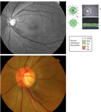
OCT는 망막신경섬유층을 포함한 여러 망막층의 횡단면을 측정하여 객관적인 자료를 바로 제공한다. 또한 정상안과 녹내장안을 구별해 낼 수 있다고 보고되어 있다.7-11그러나 망막신경섬유층촬영에서 국소적 망막신경섬유층결손이 있 음에도 불구하고 OCT의 12개 시구역의 sector average 분 석에서 정상으로 나타나는 경우를 종종 볼 수 있었다(Fig.
1). 따라서 본 연구에서는 사진에서 나타난 국소적 망막신 경섬유층결손이 있는 환자들을 대상으로 OCT의 가음성과 관련된 요인을 조사하였다. 더불어 OCT의 유용성을 알아 보고자 사진과 OCT에서 검출된 국소적 망막신경섬유층결 손의 위치와 폭의 상관관계를 분석하였다.
대상과 방법
2007년 8월부터 2010년 4월까지 한양대학교병원 안과 를 방문하여 최소한 한쪽 눈에서 망막신경섬유층결손이 관 찰된 197명의 환자를 후향적으로 분석하였다. 시야결손전 녹내장 24명은 국소적 망막신경섬유층결손이 있고 시야결 손이 없는 경우, 시야결손녹내장 173명은 국소적 망막신경 섬유층결손이 있고 녹내장성 시야결손을 보이는 경우였다.
병력조사(고혈압, 당뇨병 등), 시력측정, 골드만 안압측 정, 세극등(Haag-Streit 900, Bern, Switzerland)을 이용 한 전안부검사, 앞방각경검사, 초음파를 이용한 중심각막 두께측정(Sp-300, Tomey Co., Japan), 검안경을 이용한
Figure 1. Example of false negative finding. Inferior localized
retinal nerve fiber layer defect was observed on the red-free photograph, but optical coherence tomography result was nor- mal at the corresponding location on the sector average analysis.Figure 2. The angular locations of localized retinal nerve fiber
layer (RNFL) defects on red-free fundus photography were measured to evaluate the angular width and location of lo- calized RNFL defects. Reference line was from the optic disc center to the temporal margin of the optic disc (0°). The angles between reference line and the line from the center of the disc to the points at which the upper and lower margins of the localized RNFL defect met with the optic disc margin were measured.The directional angle was assessed in a clockwise direction in right eyes and a counter-clockwise direction in left eyes.
시신경유두검사, Humphrey C 30-2 시야검사(Humphrey Instruments, San Leandro, CA), 망막신경섬유층 및 시신 경유두사진촬영(TRC-50 IA, Topcon Inc., Tokyo, Japan), Stratus OCT (version 4.0.2, Optical coherence tomog- raphy, Humphrey systems Inc., Dublin, CA, USA), HRT II (version 2.01, Heidelberg Engineering, Heidelberg, Germany) 검사를 시행하였다.
경미한 백내장 이외의 당뇨망막병증 등 다른 안질환이 있는 경우, 경사유두, 시신경유두드루젠, 시신경결손이 있 거나 시야결손의 원인이 되는 신경학적 질환이 있는 경우 는 대상에서 제외하였다. 최대교정시력이 0.5 이상, 구면렌 즈 대응치가 -6.0D에서 +3.0D 사이인 경우가 연구대상에 포함되었다.
자동시야검사
녹내장성 시야결손은 Humphrey C 30-2 threshold test 에서 최소한 2회 이상 glaucoma hemifield test가 outside normal limit이거나 corrected pattern standard deviation 이 5% 미만이거나 pattern deviation plot에서 가장자리를 제외한 부위에서 인접한 3개 이상 점의 역치가 정상의 5%
미만으로 나타나고 그중 한 개 이상은 1% 미만인 경우로 정하였다. 주시상실 20% 미만, 가양성과 가음성 반응이
30% 미만으로 신뢰할 만한 결과와 재현성 있는 경우를 대 상으로 하였다.
망막신경섬유층사진 촬영
충분히 산동시킨 후 주사레이저검안경(F-10, Nidek Inc., Japan) 또는 안저카메라와 내장된 무적색광 필터(TRC- 50IA, Topcon Inc., Tokyo, Japan)를 이용하였다. 양안이 대상인 경우 망막신경섬유층결손이 잘 보이는 쪽의 눈을 택하였다. 촬영된 사진은 명암과 대비를 조절하여 결손의 경계가 잘 보이도록 하였다. 국소적 망막신경섬유층결손은 시신경유두연에서 1유두직경 떨어진 지점에서 망막의 가장 큰 혈관의 직경보다는 넓고, 시신경유두에서 멀어질수록 폭 이 넓어지며, 시신경유두연에 닿는 틈새모양 또는 쐐기모양 이 있는 경우로 정의하였다. 선모양이나 방추형(slit-like or spindle-like)인 경우, 미만성 결손인 경우, 결손의 경계 가 불분명한 경우는 대상에서 제외하였다.
망막신경섬유층결손의 위치는 우안을 기준으로 표시하 였다. Adobe photoshopTM 7.0 (Adobe System Inc., CA, USA)을 이용하여 컴퓨터 모니터에서 시신경유두의 수평직 경과 수직직경의 중앙에서 그은 교차점이 만나는 곳을 시 신경유두의 중앙으로 하였고, 이 곳에서 이측으로 수평선 을 그은 선(기준선)이 시신경유두연과 만나는 곳을 0도로 정하였다. 시신경유두의 상측은 90도, 비측은 180도, 하측
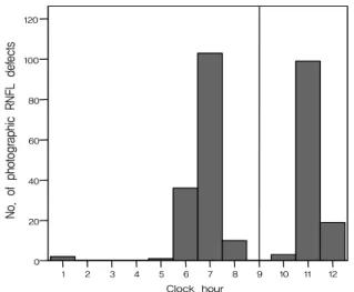
Figure 3. The number of eyes (total = 197) and retinal nerve fiber
layer (RNFL) defects (total = 273) by location on red-free RNFL photographs.Figure 4. Frequency distribution of photographic retinal nerve
fiber layer (RNFL) defects by clock-hour location on red-free RNFL photographs. Defects were most commonly observed at 7 o’clock and 11 o’clock.은 270도가 된다(시계 방향). 좌안 결손 위치는 Adobe photoshopTM의 캔바스 수평 뒤집기를 이용하여 좌안 망막 신경섬유층 사진을 뒤집어 위치가 우안과 같이 되도록 하 여 기록하였다. 시신경유두 중앙에서 망막신경섬유층결손 이 시신경유두연에 닿는 곳의 양측 경계에 선을 그어 두 선 이 이루는 각을 사진의 망막신경섬유층결손 폭으로 하여 측정하였고, 시신경유두연에 위치한 결손폭의 중앙을 망막 신경섬유층결손 위치로 정하였고 각도와 시간으로 기록하 였다(Fig. 2).
빛간섭단층촬영
소프트웨어 버전 4.0.1인 Stratus OCT를 이용하여 측정 하였다. 시신경유두주위 망막신경섬유층에 반지름 1.7 mm 의 동심원을 따라 256개의 지점에서 망막신경섬유층두께 를 측정하는 fast retinal nerve fiber layer thickness scan 을 이용하였다. 30도 간격으로 12등분한 시구역, 90도 간격으 로 4등분한 사분면, 360도의 평균망막신경섬유층두께(average retinal nerve fiber layer thickness)를 구하였다. 망막 상 이 잘 맺히고, 시신경유두를 중심으로 한 동심원, 6 이상의 signal strength를 보이는 경우에만 대상에 포함시켰다.
OCT 결손은 사진의 망막신경섬유층결손에 해당되는 위 치에 있는 정상인의 5% 미만인 비정상 시구역으로 정하였 다. OCT 결손폭은 비정상 구역들의 합으로 각도로 기록하 였다. 우안의 OCT 결손 위치는 결손의 중앙으로 사진의 결 손에서와 같이 각도와 시간으로 표시하였다. 좌안의 OCT 결손 위치는 Adobe photoshopTM의 캔바스 수평 뒤집기를 이용하여 기록하였다.
사진의 결손 위치를 기준으로 하여 OCT 결손 위치가 사
진 결손 위치의 30도 이내에 있거나, 사진 결손이 2개 이상 인 경우 OCT 결손에서 1개라도 사진의 결손과 일치하면 사진의 국소 망막신경섬유층결손이 OCT에서 검출된 것으 로 간주하였다.
통계학적 분석
사진의 국소적 망막신경섬유층결손이 OCT로 검출되지 않는 것과 관련된 인자들을 알아보고자 OCT로 결손이 검 출되지 않은 군과 검출된 군으로 나누어 연속변수는 in- dependent t-test, Mann-Whitney U-test로, 범주형 변수 는 Chi-square test로 두 군을 비교하였다. 그 결과 p<0.1 인 변수들을 로지스틱 회귀분석에서 공변량(covariate)으 로 하고, 사진의 망막신경섬유층결손이 OCT로 검출된 여 부를 종속변수로 하여 독립변수를 일시에 투입하여 logit모 형을 구하는 enter 방식의 회귀분석으로 위험인자를 알아 보았다. 사진의 망막신경섬유층결손과 OCT결손의 위치와 폭의 상관관계는 상관분석을 하였다. p-value가 0.05 미만 인 경우 통계적으로 유의한 것으로 간주하였다. SPSS version 13.0 (SPSS Inc., Chicago, Illinois, USA)을 이용 하여 분석하였다.
결 과
대상의 평균 연령은 56.6 ± 12.4세(평균 ± 표준편차)였 고, 남자 113명, 여자 84명이었다. 구면렌즈대응치는 평균 -0.86 ± 2.52디옵터였고, 안압은 평균14.1 ± 2.2 mmHg였 다(범위: 9-20 mmHg). 시야의 mean deviation은 평균
Figure 5. Scatterplot of the angular locations of the retinal
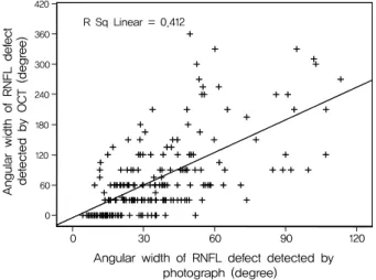
nerve fiber layer (RNFL) defects detected by red-free RNFL photograph and those of RNFL defects detected by optical co- herence tomography (OCT) (R = 0.980).Figure 6. Scatterplot of the angular widths of the retinal nerve
fiber layer (RNFL) defects detected by red-free RNFL photo- graph and those of RNFL defects detected by optical coher- ence tomography (OCT) (R = 0.642).-6.85 ± 6.04 dB이었고, corrected pattern standard de- viation은 6.19 ± 4.11 dB이었다. 시야결손이 있는 173안 을 Hodapp-Parrish-Anderson 분류에 따라 시야결손의 정도를 나누면 초기 시야결손 88안, 중기 시야결손 42안, 심한 시야결손은 43안이었다.
사진의 결손 위치와 수는 하측은 78안에서 1개, 상측 45 안에서 1개 2안에서 2개, 상하 양측은 70안에서 2개 2안에 서 3개 있었으며, 총 결손 수는 273개였다(Fig. 3). 시간별 로 사진의 결손 수를 보았을 때 7시에 103개(37.7%), 11 시에 99개(36.3%)로 많았다(Fig. 4). 사진의 결손폭은 평 균 36.3 ± 23.1도였다.
사진의 결손 위치와 OCT결손 위치와는 유의한 상관관계 가 있었다(R=0.98, p<0.001) (Fig. 5). 사진의 결손폭과 OCT 결손폭과도 유의한 상관관계가 있었다(R=0.64, p< 0.001) (Fig. 6).
사진의 망막신경섬유층결손은 197안 중 51안(25.9%)에 서 OCT로 검출되지 않았다. OCT로 결손이 검출되지 않 은 군과 검출된 군으로 나누어 단변량분석으로 비교한 결 과 고혈압, 녹내장 유형, 시야 MD, 시야 PSD, 국소적 망막 신경섬유층결손의 위치, 결손 수, 결손폭의 합계, 평균망막 신경섬유층두께, 시신경유두면적은 p값이 0.1 미만이었다 (Table 1). 이를 로지스틱 회귀분석에서 공변량(covariate) 으로 하여 위험인자를 알아 본 결과, OCT의 가음성과 관련 된요인은 평균망막신경섬유층두께(odds ratio=1.106, 95%
신뢰구간=1.057-1.156, p<0.001)와 결손폭(odds ratio=
0.929, 95% 신뢰구간=0.884-0.977, p=0.004)이었다(Table 2).
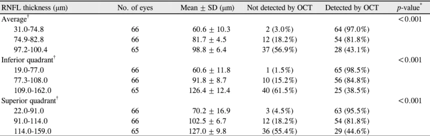
평균망막신경섬유층두께, 하사분면 망막신경섬유층두께, 상사분면 망막신경섬유층두께를 각각 두께순으로 3등분하
여 망막신경섬유층두께에 따라 검출률의 차이가 있는지 비교 하였을 때 모두 망막신경섬유층두께가 두꺼울수록 유의하게 OCT로 검출되지 않는 비율이 높았다(Pearson Chi-Square test, 각각 p<0.001) (Table 3). 사진의 총결손폭(망막신 경섬유층결손들의 폭을 모두 합한 값)을 결손폭에 따라 다 섯 군으로 세분하여 분석한 결과, 총결손폭이 감소할수록 OCT로 검출되지 않는 비율이 유의하게 높았다(Pearson Chi-square test, p<0.001) (Table 4).
고 찰
Stratus OCT는 조직의 여러 층에서 반사되는 빛의 시간 차이를 간섭계로 측정하여 망막과 시신경유두의 고해상도 의 횡단면 영상을 제공하는 비접촉, 비침습적인 영상 장비 이며, 재현성이 높고, 정상안과 녹내장안을 구별해 낼 수 있 다고 하였다.12,13
국소적 망막신경섬유층결손은 7시(하이측)와 11시(상이 측)에 흔히 나타난다고 하였으며, 본 연구에서도 273개의 결손 중 7시에서 103개(37.7%), 11시에서 99개(36.3%) 로 많이 나타나 같은 결과를 보였다.14,15시신경유두의 하이 측과 상이측은 손상을 받기 쉬운 가장 취약한 부위로 알려 져 있다.16
본 연구에서 사진의 국소적 망막신경섬유층결손에 대한 OCT의 검출률은 74.1%였고, OCT결손의 위치와 폭은 사 진의 망막신경섬유층결손의 위치와 폭과 상당하거나 강한 지형적 관련이 있었다. 이전의 연구에서도 OCT의 검출률 이 85.9%이고, 두 검사가 지형적으로 강한 관련이 있다고 하였다.17 사진의 국소적 망막신경섬유층결손의 위치와 폭
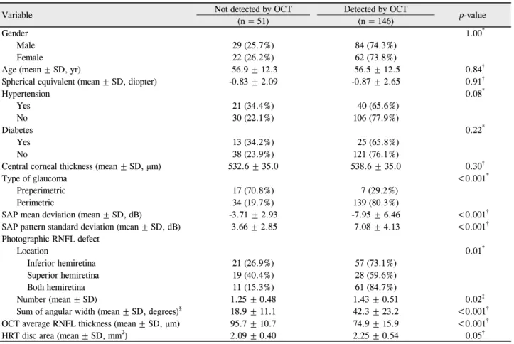
Table 1. Comparison between photographic retinal nerve fiber layer (RNFL) defects not detected by optical coherence tomography
(OCT) group and detected by OCT groupVariable Not detected by OCT Detected by OCT
p-value
(n = 51) (n = 146)
Gender 1.00*
Male 29 (25.7%) 84 (74.3%)
Female 22 (26.2%) 62 (73.8%)
Age (mean ± SD, yr) 56.9 ± 12.3 56.5 ± 12.5 0.84†
Spherical equivalent (mean ± SD, diopter) -0.83 ± 2.09 -0.87 ± 2.65 0.91†
Hypertension 0.08*
Yes 21 (34.4%) 40 (65.6%)
No 30 (22.1%) 106 (77.9%)
Diabetes 0.22*
Yes 13 (34.2%) 25 (65.8%)
No 38 (23.9%) 121 (76.1%)
Central corneal thickness (mean ± SD, μm) 532.6 ± 35.0 538.6 ± 35.0 0.30†
Type of glaucoma <0.001*
Preperimetric 17 (70.8%) 7 (29.2%)
Perimetric 34 (19.7%) 139 (80.3%)
SAP mean deviation (mean ± SD, dB) -3.71 ± 2.93 -7.95 ± 6.46 <0.001†
SAP pattern standard deviation (mean ± SD, dB) 3.66 ± 2.85 7.08 ± 4.13 <0.001†
Photographic RNFL defect
Location 0.01*
Inferior hemiretina 21 (26.9%) 57 (73.1%)
Superior hemiretina 19 (40.4%) 28 (59.6%)
Both hemiretina 11 (15.3%) 61 (84.7%)
Number (mean ± SD) 1.25 ± 0.48 1.43 ± 0.51 0.02‡
Sum of angular width (mean ± SD, degrees)§ 18.9 ± 11.1 42.3 ± 23.2 <0.001†
OCT average RNFL thickness (mean ± SD, μm) 95.7 ± 10.7 74.9 ± 15.9 <0.001†
HRT disc area (mean ± SD, mm2) 2.09 ± 0.40 2.25 ± 0.54 0.05†
Data are expressed as mean ± standard deviation.
*By Chi-square test; †By unpaired t-test; ‡By Mann-Whitney U test; §sum of angular widths of defects: the summated width of the angle of RNFL defects around the optic disc.
HRT = heidelberg retina tomograph.
Table 2. Factors associated with false negative results of optical coherence tomography in eyes with localized retinal nerve fiber lay-
er (RNFL) defectsFactors Logistic regression p-value
Odds ratio 95% confidence interval
Average RNFL thickness (μm) 1.106 1.057-1.156 <0.001
Angular width of defect (degree) 0.929 0.884-0.977 0.004
을 측정하기 위하여 본 연구에서는 망막신경섬유층결손이 시신경유두연에 닿는 곳에서, 이전의 연구에서는 OCT 비 디오모드의 원과 비슷한 위치에 그린 사진의 원에서 결손 의 폭과 위치를 측정하였다.17OCT 결손의 폭과 위치는 저 자들은 정상치의 5% 미만인 시구역을, 다른 연구는 TSNIT (temporal, superior, inferior, nasal) 그래프를 이용하였 다.18본 연구와 다른 연구와는 측정 방법에 다소간 차이가 있고, 대상 및 검출기준이 다르나 유사한 결과를 보였다.
그러나 사진에서 망막신경섬유층결손이 뚜렷이 보임에 도 불구하고 197안 중 51안(25.9%)은 OCT로 검출되지 않았다. 로지스틱 회귀분석으로 위험인자를 알아 본 결과,
OCT의 가음성과 관련된 요인은 평균망막신경섬유층두께 (odds ratio=1.106), 결손폭(odds ratio=0.929)이었다. 이 는 평균망막신경섬유층두께가 1 μm 증가하면 결손이 검출 되지 않을 확률이 검출될 확률보다 1.106배로 높아지고, 결 손폭이 1도 증가하면 결손이 검출되지 않을 확률이 검출될 확률보다 0.929배로 낮아짐을 의미한다. 즉 국소적 평균망 막신경섬유층결손의 두께가 두꺼울수록, 결손폭이 좁을수 록 OCT로 검출되지 않을 확률이 높았다. 평균망막신경섬 유층두께, 하사분면 망막신경섬유층두께, 상사분면 망막신 경섬유층두께를 각각 두께순으로 3등분하여 망막신경섬유 층두께에 따라 검출률의 차이가 있는지 비교하였을 때 모
Table 3. Retinal nerve fiber layer (RNFL) thickness and detection of photographic defects by optical coherence tomography (OCT)
RNFL thickness (μm) No. of eyes Mean ± SD (μm) Not detected by OCT Detected by OCT p-value*Average† <0.001
31.0-74.8 66 60.6 ± 10.3 2 (3.0%) 64 (97.0%)
74.9-82.8 66 81.7 ± 4.5 12 (18.2%) 54 (81.8%)
97.2-100.4 65 98.8 ± 6.4 37 (56.9%) 28 (43.1%)
Inferior quadrant† <0.001
19.0-77.0 66 60.6 ± 11.8 1 (1.5%) 65 (98.5%)
77.3-108.0 66 91.8 ± 8.7 10 (15.2%) 56 (84.8%)
109.0-162.0 65 126.4 ± 12.4 40 (61.5%) 25 (38.5%)
Superior quadrant† <0.001
22.0-91.0 66 70.2 ± 16.9 3 (4.5%) 63 (95.5%)
91.0-114.0 66 102.5 ± 6.7 12 (18.2%) 54 (81.8%)
114.0-159.0 65 127.0 ± 9.8 36 (55.4%) 29 (44.6%)
*by Pearson Chi-Square test; †Average, inferior quadrant, and superior quadrant retinal nerve fiber layer thickness were divided into tertile.
Table 4. Sum of angular width of photographic retinal nerve fiber layer (RNFL) defect (the summated width of the angle of RNFL
defects around the disc) and detection of photographic defects by optical coherence tomography (OCT)Sum of angular width of photographic RNFL defect (degree) Not detected by OCT Detected by OCT p-value*
<0.001
≤10 11 (91.7%) 1 (8.3%)
11-20 24 (58.5%) 17 (41.5%)
21-30 6 (13.3%) 39 (86.7%)
31-40 8 (24.2%) 25 (75.8%)
>40 2 (3.0%) 64 (97.0%)
Total 51 (25.9%) 146 (74.1%)
*by Pearson Chi-Square test.
두 망막신경섬유층두께가 두꺼울수록 OCT로 검출되지 않 는 비율이 높아짐을 알 수 있었다. 또한 총결손폭이 감소할 수록 OCT로 검출되지 않는 비율이 높아짐을 확인할 수 있 었다.
Hwang et al18은 사진에서 국소적 망막신경섬유층결손이 있으나 OCT로 검출되지 않은 시야결손녹내장 2안을 관찰 한 결과 OCT의 TSNIT 그래프에서 사진의 국소결손과 상 응하는 위치에서 그래프 선이 급격히 꺾였다고 하였다. 이 경우 그래프를 보면 정상치 5% 미만에 도달하지는 못했으 나 5% 비정상에 근접한 상태로 사진의 결손 부위에 해당하 는 OCT 망막신경섬유층두께가 국소적으로 얇아져 있다.
Jeoung et al17은 Stratus OCT의 민감도는 국소 망막신경 섬유층결손의 폭에 비례한다고 하였다. 하지만 이 연구들 은 망막신경섬유층에 영향을 미칠 수 있는 인자들을 보정 하지 않은 결과였다. 망막신경섬유층두께는 나이, 인종, 안 구길이, 시신경유두크기 등에 영향을 받는다고 알려져 있
다.19-22다변량분석으로 이를 보정한 본 연구의 결과는 망
막신경섬유층두께가 두꺼울수록, 결손폭이 좁을수록 OCT 로 검출되지 않을 확률이 높다는 것으로 위의 결과들과 부 합된다.
Kim et al14은 국소적 망막신경섬유층결손이 있고 시야
는 정상인 시야결손전녹내장 49명을 대상으로 Stratus OCT 여러 측정치들의 진단능력을 알아본 연구에서 민감도 는 2.0-40.8%라고 하였다. 가장 민감도가 높았던 Stratus OCT 측정치는 정상치 5% 미만의 1시간 이상 비정상인 망 막신경섬유층두께라고 하였다. 본 연구에서는 시야결손전 녹내장 24안 중 7안(29.2%)에서 OCT로 검출되었고, 녹내 장성 시야결손이 있는 시야결손녹내장 173안 중 139안 (80.3%)에서 OCT로 검출되어 위의 결과와 같이 시야결손 전녹내장에서는 진단능력이 부족하였다. Stratus OCT의 정상치 자료는 대부분 서양인으로 구성된 328명의 자료를 수집한 것으로 연구결과에 영향을 미칠 수 있다. 아시아인 이 백인보다 망막신경섬유층두께가 두껍기 때문에 아시아 인에서는 가음성이 생길 가능성이 높다고 하였다.23한국인 정상치를 이용한 기준이 Stratus OCT 정상치를 이용한 기 준보다 민감도가 높았고 특이도는 차이가 없다는 이전의 연구도 있다.24
본 연구에서는 사진의 국소적 망막신경섬유층결손의 위 치와 폭을 망막신경섬유층결손이 시신경유두연에 닿는 곳 에서 측정하였으므로 OCT에서 측정한 위치와 약간 다를 것이다. 반면에 OCT 비디오모드의 원과 비슷한 위치에 그 린 사진의 원에서 결손의 폭과 위치를 측정하는 방법은 위
치적으로 OCT과 비슷하겠으나 재현성은 저자들의 방법보 다 높지 않을 것으로 생각된다.18
결론적으로 망막신경섬유층결손의 두께가 두꺼울수록, 결손폭이 좁을수록 OCT로 검출되지 않을 확률이 높았다.
사진의 결손의 위치와 폭과 OCT 결손의 위치와 폭은 지형 적 관련성이 있어서 OCT가 녹내장 진단장비로 유용하였 다. OCT는 망막신경섬유층을 포함한 여러 망막층의 횡단 면을 측정하여 객관적인 자료를 제공하고 내장된 정상치와 비교하여 정상 비정상 여부를 바로 알 수 있으며, 안저의 색소침착이 적은 경우, 미만성 망막신경섬유층결손이 의심 되는 경우 OCT가 망막신경섬유층사진보다 나아 보인다.
또한 OCT는 정신적 육체적 장애로 시야검사를 할 수 없는 경우 도움이 되며, 큰 시신경유두에 유두함몰이 큰 경우 모 든 측정치들이 정상범위 내이면 의미 있는 신경섬유의 소 실이 없다고 보아도 될 것이다.
그러나 사진에서 망막신경섬유층결손이 잘 보임에도 불 구하고 51안(25.9%)에서 OCT로 검출되지 않았다는 점에 서 OCT의 결과에 너무 의존하기 보다는 녹내장 진단에 보 조적으로 이용되어야 하며, 망막신경섬유층사진 등 여러 소 견들을 종합하여 진단하는 것이 필요하다고 본다. OCT의 진단능력을 높이기 위해서는 scan density의 증가와 새로 운 분석방법 등 현재보다 더 나은 하드웨어와 소프트웨어 의 개발이 있어야 하겠다.
참고문헌
1) Quigley HA, Addicks EM, Green WR. Optic nerve damage in hu- man glaucoma. III. Quantitative correlation of nerve fiber loss and visual field defect in glaucoma, ischemic neuropathy, papilledema, and toxic neuropathy. Arch Ophthalmol 1982;100:135-46.
2) Sommer A, Katz J, Quigley HA, et al. Clinically detectable nerve fiber atrophy precedes the onset of glaucomatous field loss. Arch Ophthalmol 1991;109:77-83.
3) Airaksinen PJ, Alanko HI. Effect of retinal never fiber loss on the optic nerve head configuration in early glaucoma. Graefes Arch Clin Exp Ophthalmol 1983;220:193-6.
4) Tuulonen A, Lehtola J, Airaksinen PJ. Nerve fiber layer defects with normal visual fields. Do normal optic disc and normal visual field indicate absence of glaucomatous abnormality? Ophthalmology 1993;100:587-97.
5) Tuulonen A, Airaksinen PJ. Initial glaucomatous optic disk and retinal nerve fiber layer abnormalities and their progression. Am J Ophthalmol 1991;111:485-90.
6) Quigley HA, Reacher M, Katz J, et al. Quantitative grading of nerve fiber layer photographs. Ophthalmology 1993;100:1800-7.
7) Chen HY, Huang ML. Discrimination between normal and glau- comatous eyes using Stratus optical coherence tomography in Taiwan Chinese subjects. Graefes Arch Clin Exp Ophthalmol 2005;243:894-902.
8) Medeiros FA, Zangwill LM, Bowd C, et al. Evaluation of retinal
nerve fiber layer, optic nerve head, and macular thickness meas- urements for glaucoma detection using optical coherence tomography. Am J Ophthalmol 2005;139:44-55.
9) Wollstein G, Ishikawa H, Wang J, et al. Comparison of three opti- cal coherence tomography scanning areas for detection of glau- comatous damage. Am J Ophthalmol 2005;139:39-43.
10) Manassakorn A, Nouri-Mahdavi K, Caprioli J. Comparison of reti- nal nerve fiber layer thickness and optic disk algorithms with opti- cal coherence tomography to detect glaucoma. Am J Ophthalmol 2006;141:105-15.
11) Song YM, Uhm KB. Discrimination between normal and early stage of glaucomatous eyes using the stratus optical coherence tomography. J Korean Ophthalmol Soc 2007;48:1675-85.
12) Paunescu LA, Schuman JS, Price LL, et al. Reproducibility of nerve fiber thickness, macular thickness, and optic nerve head measurements using StratusOCT. Invest Ophthalmol Vis Sci 2004;
45:1716-24.
13) Budenz DL, Michael A, Chang RT, et al. Sensitivity and specificity of the StratusOCT for perimetric glaucoma. Ophthalmology 2005;
112:3-9.
14) Kim TW, Park UC, Park KH, Kim DM. Ability of Stratus OCT to identify localized retinal nerve fiber layer defects in patients with normal standard automated perimetry results. Invest Ophthalmol Vis Sci 2007;48:1635-41.
15) Jonas JB, Schiro D. Localised wedge shaped defects of the retinal nerve fibre layer in glaucoma. Br J Ophthalmol 1994;78:285-90.
16) Airaksinen PJ, Mustonen E, Alanko HI. Optic disc haemorrhages precede retinal nerve fibre layer defects in ocular hypertension.
Acta Ophthalmol (Copenh) 1981;59:627-41.
17) Jeoung JW, Park KH, Kim TW, et al. Diagnostic ability of optical coherence tomography with a normative database to detect lo- calized retinal nerve fiber layer defects. Ophthalmology 2005;
112:2157-63.
18) Hwang JM, Kim TW, Park KH, et al. Correlation between topo- graphic profiles of localized retinal nerve fiber layer defects as de- termined by optical coherence tomography and red-free fundus photography. J Glaucoma 2006;15:223-8.
19) Kanamori A, Escano MF, Eno A, et al. Evaluation of the effect of aging on retinal nerve fiber layer thickness measured by optical co- herence tomography. Ophthalmologica 2003;217:273-8.
20) Budenz DL, Anderson DR, Varma R, et al. Determinants of normal retinal nerve fiber layer thickness measured by Stratus OCT.
Ophthalmology 2007;114:1046-52.
21) Nagai-Kusuhara A, Nakamura M, Fujioka M, et al. Association of retinal nerve fibre layer thickness measured by confocal scanning laser ophthalmoscopy and optical coherence tomography with disc size and axial length. Br J Ophthalmol 2008;92:186-90.
22) Han JI, Lim HW, Song YM, Uhm KB. Factors influencing optic disc and retinal nerve fiber layer parameters measured by optical coherence tomography. J Korean Ophthalmol Soc 2007;48:1073- 81.
23) Da Pozzo S, Iacono P, Marchesan R, et al. The effect of ageing on retinal nerve fibre layer thickness: an evaluation by scanning laser polarimetry with variable corneal compensation. Acta Ophthalmol Scand 2006;84:375-9.
24) Kang SM, Lee SB, Uhm KB. Diagnostic ability of stratus OCT us- ing Korean normative database for early detection of normal-tension glaucoma. J Korean Ophthalmol Soc 2008;49:798-810.
=ABSTRACT=
False Negative Findings of Optical Coherence Tomography in Eyes with Localized Nerve Fiber Layer Defects
Sung Min Kang, MD, Ki Bang Uhm, MD, PhD
Department of Ophthalmology, Hanyang University College of Medicine, Seoul, Korea
Purpose: To identify the risk factors associated with false negative findings of optical coherence tomography (Stratus OCT) in patients with photographic localized retinal nerve fiber layer (RNFL) defects.
Methods: Twenty-four patients with preperimetric glaucoma and 173 patients with perimetric glaucoma, all with localized RNFL defects were included in the present study. The patients were divided into 2 groups according to the presence or ab- sence of detection of photographic defects by OCT. Gender, age, refractive error, diabetes, hypertension, central corneal thickness, type of glaucoma, mean deviation, pattern standard deviation, average RNFL thickness, disc area, and photo- graphic RNFL defect related variables (location, number, and angular width) were compared between the 2 groups. Each variable was initially evaluated by univariate analysis and significant variables (p < 0.1) were included in the logistic re- gression analysis.
Results: Photographic RNFL defects were not detected by OCT in 51 (25.9%) of the 197 eyes. The angular locations and widths of RNFL defects by OCT were significantly correlated with those of RNFL defects by red-free RNFL photographs (Pearson correlation coefficient R = 0.98 and 0.64, respectively). Logistic regression analysis revealed the risk factors for false negative findings of OCT included average RNFL thickness (odds ratio = 1.106, 95% confidence interval [CI] = 1.057-1.156, p < 0.001) and angular width of defect (odds ratio = 0.929, 95% CI = 0.884-0.977, p = 0.004).
Conclusions: This present study suggests that false negative findings of OCT in patients with photographic localized RNFL defects were associated with thicker RNFL thickness and smaller angular width of RNFL defect.
J Korean Ophthalmol Soc 2011;52(4):454-461
Key Words: Glaucoma, Optical coherence tomography, Retinal nerve fiber layer defect, Risk factor
Address reprint requests to Ki Bang Uhm, MD, PhD
Department of Ophthalmology, Hanyang University Seoul Hospital
#17 Haengdang-dong, Seongdong-gu, Seoul 133-792, Korea
Tel: 82-2-2290-8570, Fax: 82-2-2291-8517, E-mail: [email protected]