■Received: 2013. 6. 15. ■ Revised: 2013. 11. 13.
■Accepted: 2014. 3. 15.
■Address reprint requests to Kyung Seek Choi, MD Department of Ophthalmology, Soonchunhyang University Seoul Hospital, #59 Daesagwan-ro, Yongsan-gu, Seoul 140-743, Korea
Tel: 82-2-709-9354, Fax: 82-2-709-9083 E-mail: [email protected]
J Korean Ophthalmol Soc 2014;55(4):534-540 pISSN: 0378-6471⋅eISSN: 2092-9374
http://dx.doi.org/10.3341/jkos.2014.55.4.534
Original Article
내경계막 제거술 후 발생한 활꼴모양의 망막신경섬유층의 부종
Arcuate Retinal Nerve Fiber Swelling after Internal Limiting Membrane Peeling
이시형⋅이성진⋅최경식
Si Hyung Lee, MD, Sung Jin Lee, MD, Kyung Seek Choi, MD
순천향대학교 의과대학 안과학교실
Department of Ophthalmology, Soonchunhyang University College of Medicine, Seoul, Korea
Purpose: To evaluate the cause of dark arcuate striae observed in infrared photographs in idiopathic epiretinal membrane (ERM) and macular hole patients after internal limiting membrane (ILM) peeling.
Methods: Forty patients (41 eyes) of idiopathic ERM and macular hole who underwent trans pars plana vitrectomy with ILM peel- ing and gas tamponade were included in the present study. The best corrected visual acuity (BCVA) was recorded at pre- operative and postoperative 6 months. Infrared (IR) photography with spectral domain optical coherence tomography (SD-OCT) were obtained at preoperative and postoperative 1 week, 1 month, 3 months, and 6 months. When abnormal findings were visi- ble on IR photographs, additional SD-OCT was performed at the corresponding sites.
Results: Of 40 patients, 4 patients demonstrated dark striae extending from the optic nerve to near macular area in IR photo- graphs at 1 week postoperatively. SD-OCT images of the dark striae region revealed the swelling of retinal nerve fiber layer (RNFL). At postoperative 6 months, however, RNFL swelling previously observed subsided in all 4 cases, while temporal retinal thinning and dimples were observed in 3 cases. The preoperative and postoperative BCVA did not show any statistical difference between the patients with the RNFL swelling and the patients without the swelling.
Conclusions: Swelling of RNFL may occur in idiopathic ERM and macular hole patients after pars plana vitrectomy and ILM peel- ing, which may present as dark striae in IR photographs.
J Korean Ophthalmol Soc 2014;55(4):534-540
Key Words: Epiretinal membrane, Internal limiting membrane, Macular hole, Retinal nerve fiber layer
ⓒ2014 The Korean Ophthalmological Society
This is an Open Access article distributed under the terms of the Creative Commons Attribution Non-Commercial License (http://creativecommons.org/licenses/by-nc/3.0/) which permits unrestricted non-commercial use, distribution, and reproduction in any medium, provided the original work is properly cited.
내경계막은 망막과 유리체 사이의 해부학적 경계부에 위 치한 망막의 가장 내측 단층의 구조층이다.1 이러한 내경계 막은 세포증식의 골격으로 작용하여 망막전막의 발생과 연 관이 있다고 알려져 있으며,2,3 후유리체피질과 함께 황반부
의 전후 및 접선방향의 견인을 형성하여 황반원공의 발생 에도 관여한다고 알려져 있다.4,5 또한 망막전막 및 황반원 공 환자에서 유리체절제술 시행 시 내경계막 제거의 유용 성에 대한 많은 연구가 보고되었다.6-10
하지만 내경계막 제거술은 망막신경섬유층에 기계적인 손상을 유발할 수 있다.11-13 Tadayoni et al14은 최초로 망막 전막 환자에서 유리체 절제술 및 내경계막 제거술 후 활꼴 모양의 망막 변화를 blue filter 안저촬영상 관찰하였고 망막 신경섬유층의 해리에 의한 변화임을 보고하였다. 망막신경 섬유층의 해리는 내경계막 제거술을 시행한 경우에 술 후 1개월에서 3개월 후 발생하며 빛간섭단층촬영상 망막신경
Table 1. Demographic characteristics of subjects in this study
SRNFL No SRNFL Total
Sex (male/female) 3/1 12/24 15/25
Retinal pathology (ERM/MH) 3/1 23/14 26/15
Age* (years) 68.75 ± 11.15 61.81 ± 9.42 62.49 ± 9.67
Range (years) 53-78 43-83 43-83
Values are presented as mean ± SD.
SRNFL = swelling of retinal nerve fiber layer; ERM = epiretinal membrane; MH = macular hole.
*Standard deviation.
Figure 1. Pre- and post-operative infrared (IR) images of the left eye of a 78-year-old man who underwent vitrectomy and ILM peel-
ing for epiretinal membrane. (A) Preoperative IR image with signs of macular pucker due to epiretinal membrane. (B) IR image of postoperative 7 days demonst rating dark arcuate striae radiating from the optic nerve head toward the macula. Several round dark spots are visible at temporal ends of the striae, which are considered as signs of trauma from the ILM forceps. (C) IR image of post- operative 6 months revealing subsided swelling of the retinal nerve fiber layer.섬유층의 패임으로 관찰됨을 보고하였다.15-18 이외에도 내 경계막 제거술 후 microperimetry상 중심부근암점이 관찰된 증례들도 보고된 바 있다.12,19 이러한 손상은 술 후 드물지 않게 발생하고 있으나,14,15 시력검사 혹은 자동 시야 검사상 변화를 가져올 정도로 황반 기능에 영향을 미치지 않았다.14
최근의 발표된 연구에서는 내경계막 제거술 후 촬영한 적외선 안저사진 상 망막신경섬유층의 해리와 다른 형태의 활꼴모양의 망막변화를 관찰하였으며, 해당부위의 빛간섭 단층촬영 결과 망막신경섬유층의 부종이 관찰된 증례들을 보고한 바 있다.20 하지만 아직까지 국내에서 이와 같은 증 례는 보고된 바 없어, 이에 저자들은 망막전막 및 황반원공 에서 유리체절제술 및 내경계막 제거술 시행 후 촬영한 적 외선 안저촬영 상 활꼴모양의 망막변화 및 빛간섭단층촬영 상 해당 부위의 망막신경섬유층의 부종이 발생한 증례들에 대해 보고하는 바이다.
대상과 방법
2011년 9월부터 2012년 9월까지 본원 안과에서 특발성 망막전막 또는 황반원공으로 진단받고 유리체 절제술 및
내경계막 제거술을 시행 받은 40명의 환자(41안)를 대상으 로 후향적 분석을 하였으며, 41안 중 망막전막은 26안, 황반 원공은 3기 6안, 4기 9안으로 모두 15안이었다. 연구 대상은 특발성 망막전막 및 황반원공 환자만을 포함시키기 위해 수 술 전 다른 황반부 질환이 함께 동반되어 있는 경우와 속발 성 망막전막 및 황반원공을 일으킬 수 있는 다른 안내 병증 의 기왕력이 있는 경우는 제외하였으며, 술 후 최대교정시력 및 망막신경섬유층의 시간에 따른 변화를 관찰하기 위해 최 소 12개월 이상 추적 관찰이 가능한 경우만을 포함시켰다.
모든 수술은 두 술자에 의해 시행되었다. 수술방법은 3개 의 모양체 평면부 공막창을 통한 23게이지 평면부 유리체절 제술을 시행하여 후유리체박리를 일으킨 후, triamcinolone acetonide를 도포하거나 indocyanine green (ICG) 염색하여 망 막전막 및 내경계막을 잘 보이게 하였으며, 유리체 절제침과 눈속집게를 이용하여 망막전막 및 내경계막을 벗겨내고 이 를 제거하였다. 황반원공인 경우에 액체공기 교환술을 공기 압 30 mmHg로 유지하며 시행한 후, 황반부 상태에 따라 SF6
18% 혹은 C3F8 14% 가스 주입술을 시행하였다. 백내장 수술 을 함께 시행한 경우, 백내장 초음파 유화술 및 인공수정체 삽입술을 먼저 시행한 후 유리체절제술을 시행하였다.
A B C
Figure 3. Color and infrared (IR) images of postoperative day 7 in a 69-year-old woman who underwent vitrectomy
and ILM peeling for a epiretinal membrane. (A) The color photograph demonstrates the gas bubble above and endo- diathermy site inferior to the macula, but no visible striae. (B) The IR image demonstrates one thick dark striae, radi- ating from optic nerve head to below the papillomacular bundle (arrowhead), which was not visible in the color fun- dus photograph. A gas bubble is seen above, and endodiathermy site is visible at the end of the dark striae.Figure 2. Postoperative infrared (IR) photographs and spectral
domain optical coherent tomography (SD-OCT) images of the patient who demonstrated swelling of retinal nerve fiber layer (RNFL). (A) SD-OCT image at 1-week postoperatively dem- onstrates swelling of the RNFL (arrowheads), which corre- sponds to the dark striae on the IR image (arrow). (B) Postoperative 6 months follow-up IR and SD-OCT image at the same plane reveals resolution of the dark striae (arrow) and the corresponding swelling of RNFL (arrowheads).수술 전 및 술 후 외래 내원 시 세극등검사 및 안저촬영 기(KOWA Nonmyd-7; KOWA, Tokyo, Japan)를 사용하여 안 저사진을 촬영하였고, 술 후 1주, 1개월, 3개월, 6개월에 적외선 안저촬영 및 빛간섭단층촬영(optical coherent tomography; OCT) 을 빛간섭단층촬영기(Spectralis HRA+OCT; Heidelberg en-
gineering, CA, USA)를 사용하여 시행하였으며, 적외선 안저 사진 상 망막 이상 소견이 관찰되면 해당 부위의 빛간섭단층 촬영을 추가로 시행한 후 망막 각 층의 두께를 Spectralis OCT의 안구추적기능(active eye tracking)을 이용하여 추적관 찰기간 동안 관찰하였다. 최대교정시력은 술 전, 술 후 6개 월 시점에 측정하였으며, logMAR 시력으로 변환 후 시간에 따른 변화를 알아보았다. 통계학적 분석은 SPSS 20.0 프로그 램을 사용하였고, Wilcoxon signed rank test 및 Mann-Whitney test를 이용하여 결과를 분석하였다. 이때 p값이 0.05 미만인 경우에 통계적으로 유의하다고 판단하였다.
결 과
연구 대상의 성별 분포는 남자가 15명, 여자가 25명이었고, 평균 연령은 62.49 ± 9.67세 (43-83세)였다(Table 1). 술 전 검 사한 적외선 안저촬영에서 어두운 선 등의 이상 소견은 관 찰되지 않았으며, 빛간섭단층촬영상 망막전막 혹은 황반원 공 외에 망막신경섬유층의 이상 소견은 관찰되지 않았다 (Fig. 1). 내경계막 제거 중 1안에서 망막 열공이 발생하여 열공 주변으로 경계 레이저 광응고술을 시행하였으며, 그 외에 다른 합병증은 발생하지 않았다. 술 후 1주일째 9.7%
(41안 중 4안)에서 적외선 안저사진상 시신경유두에서 시작 되어 황반부에서 끝나는 활꼴모양의 어두운 선이 관찰되었 으며, 그에 해당하는 망막신경섬유층의 부종이 빛간섭단층 촬영상 관찰되었다(Fig. 2).
술 후 1주일째 발견된 망막신경섬유층의 부종 후 새로이
A
B
A B
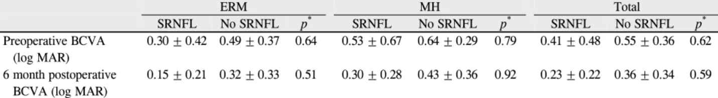
Table 2. Pre- and postoperative visual acuities in patients with and without swelling of retinal nerve fiber layer
ERM MH Total
SRNFL No SRNFL p*** SRNFL No SRNFL p*** SRNFL No SRNFL p*** Preoperative BCVA
(log MAR)
0.30 ± 0.42 0.49 ± 0.37 0.64 0.53 ± 0.67 0.64 ± 0.29 0.79 0.41 ± 0.48 0.55 ± 0.36 0.62 6 month postoperative
BCVA (log MAR)
0.15 ± 0.21 0.32 ± 0.33 0.51 0.30 ± 0.28 0.43 ± 0.36 0.92 0.23 ± 0.22 0.36 ± 0.34 0.59
Values are presented as mean ± SD.
SRNFL = swelling of retinal nerve fiber layer; ERM = epiretinal membrane; MH = macular hole; BCVA = best corrected visual acuity.
*Mann-Whitney test.
A
B
C
Figure 4. Postoperative images of infrared (IR) photographs
and spectral domain optical coherent tomography (SD-OCT) at 1 week, 3 months, and 6 months postvitrectomy for macular hole. (A) One week postoperative IR image illustrates swel- ling of the retinal nerve fiber layer (RNFL) radiating above the macular area (arrow), and the SD-OCT image demonstrates a relatively normal temporal macula after surgery. (B) Three-month postoperative IR image illustrates resolution of RNFL swelling, and SD-OCT image reveals slight thinning of the temporal reti- na and the development of “dimples” similar to that described in dissociated optic nerve fiber layer (arrowhead). (C) Six-month postoperative images demonstrate further thinning and deep- ening of “dimples” in the temporal macula (arrowhead).발생한 어두운 선이나 망막신경섬유층의 부종은 없었다. 망 막신경섬유층의 부종은 술 후 수개월에 걸쳐 서서히 호전 되었으며, 6개월 후 촬영한 빛간섭단층촬영에서 모든 안에 서 부종이 호전되었다(Fig. 2). 도상검안경을 이용한 안저 검사 및 칼라 안저 촬영에서는 망막신경섬유층의 부종을 관찰할 수 없었으며, 적외선 안저촬영상에서만 관찰 가능하 였다(Fig. 3). 본 연구에 포함된 모든 안에서 술 후 최대교정 시력은 술 전에 비해 호전되었다. 이 때 술 전 및 술 후 평 균 logMAR 시력은 0.53 ± 0.36과 0.35 ± 0.33였으며, 이는 통계학적으로 유의한 차이를 보였다(Wilcoxon signed rank test, p=0.002).
술 후 망막신경섬유층의 부종이 발생한 군의 술 전 및 술 후 6개월째의 최대교정시력은 각각 0.41 ± 0.48과 0.23 ±
0.22였으며, 부종이 발생하지 않은 군의 술 전 및 술 후 6개 월 최대교정시력은 각각 0.55 ± 0.36과 0.36 ± 0.34였다. 두 군간의 술 전 및 술 후 최대교정시력은 통계학적으로 유의한 차이가 없었다. 또한 부종이 발생한 환자와 발생하지 않은 환 자에서 망막전막과 황반원공 군으로 나누어 비교하였을 경 우에도 통계학적으로 유의한 차이가 없었다(Mann-Whitney test, p>0.05) (Table 2).
망막신경섬유층의 부종이 관찰된 4 증례 중 3 증례에서 술 후 경과 관찰 중 촬영한 적외선 안저촬영 및 빛간섭단층 촬영상 황반 이측부 망막이 점차적으로 얇아지는 양상을 관찰할 수 있었다. 또한 망막신경섬유층 해리에서 관찰되는 신경섬유층 패임 소견이 황반부 이측의 망막 내층에서 관 찰되었다(Fig. 4).
고 찰
망막전막 및 황반원공에서 내경계막 제거술에 대한 효과 는 여러 연구에서 보고된 바 있으며, 현재 망막전막 및 황반 원공의 수술적 치료에 있어 기본적인 술기로 알려져 있다. 하지만 유리체절제술 및 내경계막 제거술 시 합병증으로 망막신경섬유층의 손상이 발생할 수 있음을 여러 연구에서 보고하였으며, 그 원인으로는 술 후 안압 상승,21,22 눈속 가 스 및 조명 독성,21,23 내경계막 제거술 중 일어날 수 있는 신 경섬유층 손상, 액체-공기 교환술 시 발생할 수 있는 신경섬 유층의 탈수, ICG염색 시 망막 독성 등이 있다. 이러한 경우 망막신경섬유층의 손상으로 인해 중심부 혹은 주변부에 시 야 결손이 흔히 동반된다. 그 외에 내경계막 제거술 후 발생 한 망막신경섬유층의 해리에 대한 증례들도 보고된 바 있
다.14-16,18 Clark et al20은 내경계막 제거술 후 발생한 활꼴모
양의 망막신경섬유층의 부종에 대해 보고하였으며, 본 연구 보다는 다소 높은 31.6%의 환자에서 활꼴모양의 망막신경 섬유층의 부종이 관찰되었다고 보고하였다.
본 논문에 제시된 증례들에서는 유리체절제술 및 내경계 막 제거술 후 적외선 안저 사진 상 망막신경섬유층 해리와 비슷한 양상의 활꼴모양의 어두운 선이 관찰되었으나, 빛간
섭단층촬영상 기존의 망막신경섬유층 해리 소견으로 알려 진 신경섬유층의 패임 소견 대신에 망막신경섬유층의 부종 이 관찰되었다. 이러한 망막신경섬유층의 변화는 적외선 안 저촬영에서만 관찰 가능하였으며, 세극등 검사 및 안저 촬 영에서는 관찰되지 않았다. 따라서 내경계막 제거술을 시행 한 경우 술 후 적외선 안저촬영을 통해 망막신경섬유층의 변화를 확인해 볼 필요성이 있다. 본 연구에서 사용한 Spectralis OCT는 적외선안저촬영과 빛간섭단층촬영을 동 시에 촬영할 수 있어 술 후 발생한 망막신경섬유층의 이상 소견을 발견할 수 있었으며, 동시에 이상소견 부위의 빛간 섭단층촬영을 시행하여 신경섬유층의 부종을 확인할 수 있 었다. 적외선 안저촬영 상 관찰된 활꼴모양의 어두운 선은 망막신경섬유층의 부종으로 인한 국소적인 적외선 파장의 침투 저하 때문으로 생각할 수 있다.
망막신경섬유층의 부종의 발생 원인에 대해서는 크게 두 가지를 고려해 볼 수 있다. 첫 번째 원인으로 내경계막 제거 술 시행 시 안내집게에 의한 직접적인 망막 내층의 손상을 생각해 볼 수 있다. Fig. 1에서 활꼴모양의 어두운 선이 끝 나는 부위에 수개의 원형모양의 암 점들을 관찰할 수 있다.
이러한 암 점들은 안내집게로 내경계막을 잡은 후 내경계 막 제거술이 시작되는 부위라고 생각할 수 있으며, 그 부근 에서 망막 내층의 직접적인 손상이 발생했을 가능성을 고 려해 볼 수 있다. 망막 내층의 손상은 신경섬유층의 ax- oplasmic flow을 방해할 수 있으며, 이로 인해 시신경유두부 위까지 활꼴모양 형태로 진행하는 망막신경섬유층의 부종 이 발생했을 가능성이 있다.
두 번째 원인으로 내경계막 제거술 시 내경계막과 함께 뮐러세포의 일부가 제거되면서 망막내층에 손상이 생길 수 있다. 내경계막은 뮐러세포의 end feet과 연결된 기저막으
로,13,24 망막신경섬유층을 구조적으로 지지해주는 역할을
한다.25 여러 기존 연구들에서 내경계막 제거술 후 투과전자 현미경을 통해 제거된 내경계막의 미세구조를 관찰한 결과 내경계막뿐 아니라 뮐러세포의 end feet이 내경계막의 망막 면에서 발견되었다고 보고하였으며, 내경계막 제거 시 뮐러 세포의 손상을 통해 망막신경섬유층을 비롯한 망막내층에 손상이 생길 수 있음을 보고하였다.11,13 또한 이는 내경계막 제거술을 시행한 안에서 망막신경섬유층의 손상 중의 하나 인 망막신경섬유층 해리의 높은 빈도의 발생률과도 일치한
다.15,18 따라서 내경계막 제거술 시 발생하는 뮐러세포의 손
상은 본 연구에서 관찰된 망막신경섬유층의 부종의 형성에 관여하였다고 추정해 볼 수 있다.
망막신경섬유층의 해리와 비교하였을 때 본 연구에서 관 찰된 신경섬유층의 부종은 다른 임상 양상을 보였다. 망막 신경섬유층의 해리는 일반적으로 술 중 뮐러세포의 손상
발생 후 뮐러세포의 점진적인 형태학적 변화에 의해 술 후 기간이 어느 정도 지나야 발생하는 것으로 알려져 있다.26 이에 반해 망막신경섬유층의 부종은 술 후 비교적 일찍(술 후 1주일) 발생하였으며, 망막신경섬유층의 부종이 나타난 4 증례 중 3 증례에서는 시간이 지남에 따라 부종이 호전되 면서 망막신경섬유층의 해리가 관찰되었다. 이러한 소견을 고려하였을 때 망막신경섬유층의 부종은 뮐러세포의 손상 과 연관이 있으나, 망막신경섬유층의 해리에 비해 보다 급 성 손상에 의해 발생한 것으로 고려할 수 있다.
그 외에 다른 원인으로는 기존의 연구들에서 제시되었던 광 독성, ICG와 같은 염색약 독성, 혹은 술 중 삽입되는 액 체 및 가스에 의한 손상을 고려해 볼 수 있다. 하지만 이러 한 요인들은 본 연구에서 관찰된 부분적인 손상보다는 전 반적인 손상을 유발하기 때문에 가능성이 낮다고 할 수 있 다. 또한 신경섬유층의 부종은 술 후 수개월 후 망막 손상을 남기지 않은 채 호전되었기 때문에, 이는 기존에 알려진 광 독성에 의한 망막 손상과 다르다 할 수 있다.
망막전막 및 황반원공 환자에서 활꼴모양의 망막신경섬 유층의 부종은 술 후 1개월, 6개월 시점의 최대교정시력에 영향을 미치지 않았으며, 이는 기존의 망막신경섬유층의 해 리에 대한 연구들과 같은 결과이다(Table 2).14,18 하지만 본 연구에서 술 후 부종이 발생한 증례 수가 적어 망막신경섬 유층의 부종이 최대교정시력에 미치는 영향에 대해서는 추 가적인 연구가 필요할 것으로 사료된다. 또한 3 증례에서 망 막신경섬유층의 부종이 호전되면서 망막신경섬유층의 해리 소견이 나타나는 것을 고려하였을 때 신경섬유층의 부종과 해리는 내경계막 제거술 후 일시적으로 발생하는 망막신경 섬유층의 형태학적 변화 단계 중 하나로 생각할 수 있다.
결론적으로 저자들은 국내에서 이전에 보고된 바 없는 황반원공 및 망막전막 환자에서 내경계막 제거술 후 발생 한 망막신경섬유층 부종을 여러 증례 경험하였으며, 기존의 알려진 망막신경섬유층의 해리와는 다른 소견임을 알 수 있었다. 특발성 망막전막 및 황반원공 환자에서 유리체 절제 술 및 내경계막 제거술 후 적외선 안저 사진 상 시신경유두에 서부터 시작되는 활꼴 모양의 어두운 선이 관찰될 수 있으며, 이는 직접적 혹은 간접적으로 망막신경섬유층의 손상이 가해 져 부종이 발생한 것으로 생각해 볼 수 있다. 추후 이러한 망 막신경섬유층의 변화와 내경계막 제거술 간의 직접적인 연관 성에 대해서는 추가적인 연구가 필요할 것으로 생각한다.
REFERENCES
1) Fine BS. Limiting membranes of the sensory retina and pigment epithelium. An electron microscopic study. Arch Ophthalmol 1961;66:847-60.
2) Poliner LS, Olk RJ, Grand MG, et al. Surgical management of pre- macular fibroplasia. Arch Ophthalmol 1988;106:761-4.
3) de Bustros S, Thompson JT, Michels RG, et al. Vitrectomy for idio- pathic epiretinal membranes causing macular pucker. Br J Ophthalmol 1988;72:692-5.
4) Gass JD. Idiopathic senile macular hole. Its early stages and pathogenesis. Arch Ophthalmol 1988;106:629-39.
5) Johnson RN, Gass JD. Idiopathic macular holes. Observations, stages of formation, and implications for surgical intervention.
Ophthalmology 1988;95:917-24.
6) Tognetto D, Grandin R, Sanguinetti G, et al. Internal limiting mem- brane removal during macular hole surgery: results of a multicenter retrospective study. Ophthalmology 2006;113:1401-10.
7) Kwok AKh, Lai TY, Yuen KS. Epiretinal membrane surgery with or without internal limiting membrane peeling. Clin Experiment Ophthalmol 2005;33:379-85.
8) Park DW, Dugel PU, Garda J, et al. Macular pucker removal with and without internal limiting membrane peeling: pilot study.
Ophthalmology 2003;110:62-4.
9) Kwok AK, Lai TY, Li WW, et al. Indocyanine green-assisted in- ternal limiting membrane removal in epiretinal membrane surgery:
a clinical and histologic study. Am J Ophthalmol 2004;138:194-9.
10) Bainbridge J, Herbert E, Gregor Z. Macular holes: vitreoretinal re- lationships and surgical approaches. Eye (Lond) 2008;22:1301-9.
11) Gandorfer A, Haritoglou C, Gass CA, et al. Indocyanine green-as- sisted peeling of the internal limiting membrane may cause retinal damage. Am J Ophthalmol 2001;132:431-3.
12) Haritoglou C, Gass CA, Schaumberger M, et al. Macular changes after peeling of the internal limiting membrane in macular hole surgery. Am J Ophthalmol 2001;132:363-8.
13) Wolf S, Schnurbusch U, Wiedemann P, et al. Peeling of the basal mem- brane in the human retina: ultrastructural effects. Ophthalmology 2004;111:238-43.
14) Tadayoni R, Paques M, Massin P, et al. Dissociated optic nerve fi- ber layer appearance of the fundus after idiopathic epiretinal mem- brane removal. Ophthalmology 2001;108:2279-83.
15) Mitamura Y, Ohtsuka K. Relationship of dissociated optic nerve fi- ber layer appearance to internal limiting membrane peeling.
Ophthalmology 2005;112:1766-70.
16) Miura M, Elsner AE, Osako M, et al. Dissociated optic nerve fiber layer appearance after internal limiting membrane peeling for idio- pathic macular hole. Retina 2003;23:561-3.
17) Mitamura Y, Suzuki T, Kinoshita T, et al. Optical coherence tomo- graphic findings of dissociated optic nerve fiber layer appearance.
Am J Ophthalmol 2004;137:1155-6.
18) Ito Y, Terasaki H, Takahashi A, et al. Dissociated optic nerve fiber layer appearance after internal limiting membrane peeling for idio- pathic macular holes. Ophthalmology 2005;112:1415-20.
19) Imai H, Ohta K. Microperimetric determination of retinal sensi- tivity in areas of dissociated optic nerve fiber layer following in- ternal limiting membrane peeling. Jpn J Ophthalmol 2010;54:435- 40.
20) Clark A, Balducci N, Pichi F, et al. Swelling of the arcuate nerve fi- ber layer after internal limiting membrane peeling. Retina 2012;
32:1608-13.
21) Pendergast SD, McCuen BW 2nd. Visual field loss after macular hole surgery. Ophthalmology 1996;103:1069-77.
22) Paques M, Massin P, Santiago PY, et al. Visual field loss after vi- trectomy for full-thickness macular holes. Am J Ophthalmol 1997;124:88-94.
23) Hutton WL, Fuller DG, Snyder WB, et al. Visual field defects after macular hole surgery. A new finding. Ophthalmology 1996;103:
2152-8.
24) Terasaki H, Miyake Y, Nomura R, et al. Focal macular ERGs in eyes after removal of macular ILM during macular hole surgery.
Invest Ophthalmol Vis Sci 2001;42:229-34.
25) Hollander H, Makarov F, Dreher Z, et al. Structure of the macroglia of the retina: sharing and division of labour between astrocytes and Muller cells. J Comp Neurol 1991;313:587-603.
26) Haritoglou C, Gandorfer A, Kampik A. NFL appearance after peeling. Ophthalmology 2006;113:1690.
= 국문초록 =
내경계막 제거술 후 발생한 활꼴모양의 망막신경섬유층의 부종
목적: 특발성 망막전막 및 황반원공 환자에서 내경계막 제거술 후 적외선 안저촬영상 관찰된 활꼴모양의 어두운 선과 이에 해당하는 부위의 망막신경섬유층의 부종에 대해 보고하고자 한다.
대상과 방법: 특발성 망막전막 혹은 황반원공으로 진단받은 환자 40명의 41안을 대상으로 적외선 안저촬영과 빛간섭단층촬영은 술 전 및 술 후 1주일, 1개월, 3개월, 6개월째에 시행하였다. 술 후 적외선 안저촬영상 활꼴모양의 어두운 선이 관찰된 경우, 해당 부위의 빛간섭단층촬영을 추가로 시행하여 망막 내층의 변화를 관찰하였다.
결과: 내경계막 제거술 후 9.7% (41안 중 4안)에서 적외선 안저촬영상 활꼴모양의 어두운 선이 관찰되었으며, 해당 부위의 빛간섭단층 촬영상 망막신경섬유층의 부종이 관찰되었다. 술 후 6개월째에 망막신경섬유층의 부종은 모든 증례에서 호전되었으나, 4 증례 중 3 증례에서 이측 망막내층의 얇아짐 및 망막신경섬유층의 패임이 관찰되었다. 술 전후의 최대교정시력은 망막신경섬유층의 부종이 발생한 환자군과 부종이 발생하지 않은 환자 군간에 통계학적으로 유의한 차이가 없었다.
결론: 망막전막 혹은 황반원공에서 유리체 절제술 및 내경계막 제거술 후에 일시적인 활꼴모양의 망막신경섬유층의 부종이 발생할 수 있다.
<대한안과학회지 2014;55(4):534-540>