© 2012 Korean Breast Cancer Society. All rights reserved. http://ejbc.kr | pISSN 1738-6756
INTRODUCTION
Ultrasound-guided core needle biopsy (CNB) has become an alternative to surgical biopsy as a primary diagnostic modality because of its accuracy and cost-effectiveness [1]. Its other advantages include that there is no requirement for ionizing radiation, non-dedicated equipment can be used, and real-time visualization of the biopsy needle is possible [2]. However, CNB has inherent limitations that can result in malignancy being missed.
Atypical ductal hyperplasia (ADH) belongs to the patholog- ical category of ‘proliferative disease with atypia.’ The reported rate of underestimation of carcinoma in CNB-diagnosed ADH is as high as 48% [3-8]. Therefore, complete surgical excision is generally recommended for ADH lesions diagnosed using CNB. However, it may be possible to avoid surgical excision if clinicians are able to identify lesions with a low probability of harboring malignant foci. Therefore, we previously developed a scoring system for predicting malignancy in CNB-diagnosed ADH [1,5]. The score takes into account patient age, palpabil- ity, microcalcification, lesion size, and multiplicity. We have termed this score the U score, with U standing for underesti- mation.
The present study was designed to validate our scoring system. The study involved 85 women with CNB-diagnosed ADH who underwent subsequent surgical excision. We iden- tified factors associated with underestimation, and determined whether the U score was a predictor of malignancy, mainly focusing on whether a 100% negative predictive value (NPV)
Validation of a Scoring System for Predicting Malignancy in Patients
Diagnosed with Atypical Ductal Hyperplasia Using an Ultrasound-Guided Core Needle Biopsy
Jisun Kim1, Wonshik Han1,2, Eun-Young Go3, Hyeong-Gon Moon1, Soo Kyung Ahn1, Hee-Chul Shin4, Jee-Man You5, Jung Min Chang6, Nariya Cho6, Woo Kyung Moon6, In Ae Park7, Dong-Young Noh1,2
1Department of Surgery and 2Cancer Research Institute, Seoul National University College of Medicine, Seoul; 3Department of General Surgery, Boramae Medical Center, Seoul; 4Department of Surgery, Chung-Ang University College of Medicine, Seoul; 5Department of Surgery, Sun General Hospital, Daejeon;
Departments of 6Radiology and 7Pathology, Seoul National University College of Medicine, Seoul, Korea ORIGINAL ARTICLE
Purpose: The need for surgical excision in patients with ultra- sound-guided core needle biopsy (CNB)-diagnosed atypical ductal hyperplasia (ADH) remains an issue of debate. The present study sought to validate a scoring system (the U score, for under- estimation) that we have previously developed for predicting malignancy in CNB-diagnosed ADH. Methods: The study pro- spectively enrolled 85 female patients with CNB-diagnosed ADH who underwent subsequent surgical excision. Underestimation was defined as a surgical specimen having malignant foci.
Results: The overall underestimation rate was 37% (31/85).
Multivariate analysis showed that a clinically palpable mass, microcalcification on imaging, size >15 mm and a patient age of
≥50 years were independently associated with underestimation.
When applied to the scoring system, the validation score was significant (p<0.001; area under the curve, 0.852). No patient with a U score <3.5 had an underestimated lesion. Conclusion:
The present study successfully validated the efficacy of our scoring system for predicting malignancy in CNB-diagnosed ADH. A U score of ≤3.5 indicates that surgical excision may not be necessary.
Key Words: Breast hyperplasia, Breast neoplasms, Diagnostic errors, Needle biopsy
Correspondence to: Wonshik Han
Department of Surgery, Seoul National University College of Medicine, 101 Daehak-ro, Jongno-gu, Seoul 110-744, Korea
Tel: +82-2-2072-1958, Fax: +82-2-3673-4250 E-mail: [email protected]
This work was supported by National Research Foundation of Korea (NRF) grant funded by the Korean government (20110031417 and 20110032148).
Received: March 13, 2012 Accepted: November 30, 2012
Cancer
(of cut-off 3.5) shows consistency as in the previous datasets.
METHODS
The study prospectively enrolled 85 women who consecu- tively underwent CNB with subsequent surgical excision between 2007 and 2011 in the Seoul National University Hospital. Patients who underwent CNB in other institutions were excluded due to the absence of information such as in- itial image findings and the gauge of the needle used. We have previously reported on our scoring system for predicting malignancy in CNB-diagnosed ADH [1], which is based on values given to five clinicopathological factors of age, palpability, microcalcification, size of lesion, and multiplicity [U score=
3.5×age (age ≤50=0, age >50 =1)+2.0×palpability (non- palpable=0, palpable=1)+2.0×microcalcification (no=0, yes=1)+3.5×sonographic size (≤1.5 cm=0, >1.5 cm=1)+
3.5×multiplicity (focal=0, multiple=1)]. The constant of each variable was derived from the logistic regression model as previously described [1]. Histological underestimation was defined as upstaging of the CNB-based diagnosis to invasive carcinoma or carcinoma in situ after surgical excision. This study was approved by the Institutional Review Board (No.
H-1106-069-366) of the Seoul National University Hospital.
All patients underwent CNB using the 14-gauge automated gun method (Bard Peripheral Technologies, Covington, USA) or the 8-, 11-gauge vacuum-assisted device method (Mammo- tome®; Ethicon-Endosurgery, Cincinnati, USA). The choice of method was made by the surgeon and the radiologist based on the pre-procedural condition of the lesion. For both meth- ods, high resolution ultrasound guidance was carried out with 10 or 12 MHz linear transducers (Voluson 730, Kretz-Medison, Zipf, Austria; HDI 5000, Advanced Technology Laboratories, Bothell, USA).
Statistical analysis was performed using a statistical software package (SPSS version 17.0; SPSS Inc., Chicago, USA) and a two-sided p-value ≤0.05 was considered as indicating signifi- cance. The differences in the distribution of categorical vari- ables were analyzed using the Pearson chi-square test and Student’s t-test was used for continuous variables. The stepwise logistic regression model was used for multivariate analysis.
RESULTS
Predictors of underestimation
The mean patient age was 48.6 years (range, 24-70 years) and the mean lesion size was 15.5 mm (range, 3-67 mm). The over- all underestimation rate was 37% (31/85). The excision speci- mens from the 85 patients showed that four (5%) had invasive
carcinomas and 27 (32%) had in situ carcinomas (Table 1).
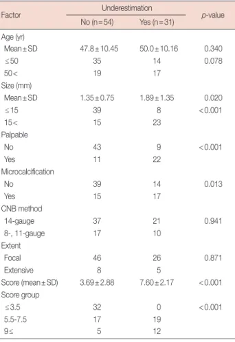
Univariate analysis showed that palpability, microcalcifica- tion, and the sonographic size of the lesion were all associated with underestimation (p<0.001, p=0.013, and p=0.020, respectively) (Table 2). The CNB method and the pathological extent of ADH in the CNB specimen were not found to be factors. Multivariate analysis showed that palpability, micro- calcification, a sonographic size >15 mm and an age >50
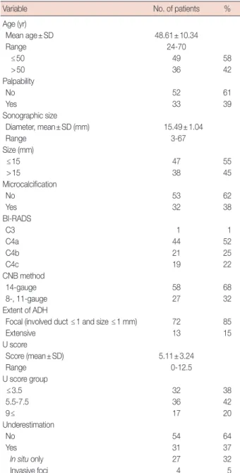
Table 1. Patient demographics (n=85)
Variable No. of patients %
Age (yr)
Mean age±SD 48.61±10.34
Range 24-70
≤50 49 58
>50 36 42
Palpability
No 52 61
Yes 33 39
Sonographic size
Diameter, mean±SD (mm) 15.49±1.04
Range 3-67
Size (mm)
≤15 47 55
>15 38 45
Microcalcification
No 53 62
Yes 32 38
BI-RADS
C3 1 1
C4a 44 52
C4b 21 25
C4c 19 22
CNB method
14-gauge 58 68
8-, 11-gauge 27 32
Extent of ADH
Focal (involved duct ≤1 and size ≤1 mm) 72 85
Extensive 13 15
U score
Score (mean±SD) 5.11±3.24
Range 0-12.5
U score group
≤3.5 32 38
5.5-7.5 36 42
9≤ 17 20
Underestimation
No 54 64
Yes 31 37
In situ only 27 32
Invasive foci 4 5
The demographic characteristics of the study patients are shown. The overall underestimation rate was 37% (32% with in situ carcinoma and 5% with inva- sive foci).
BI-RADS=Breast Imaging Reporting and Data System; CNB=core needle biopsy; ADH=atypical ductal hyperplasia.
years were independent factors associated with underestima- tion (p=0.001, p=0.007, p=0.022, and p=0.017, respectively) (Table 3).
Validation
The mean U score was 5.11 (range, 0-12.5). For analysis, U scores were divided into three groups: ≤3.5, 5.5 to 7.5, and
>9 as in the previous study [1]. The U score was found to be associated with underestimation (p<0.001). The area under the curve (AUC) of the receiver operating characteristic (ROC) Table 2. Univariate analysis
Factor Underestimation
p-value No (n=54) Yes (n=31)
Age (yr)
Mean±SD 47.8±10.45 50.0±10.16 0.340
≤50 35 14 0.078
50< 19 17
Size (mm)
Mean±SD 1.35±0.75 1.89±1.35 0.020
≤15 39 8 <0.001
15< 15 23
Palpable
No 43 9 <0.001
Yes 11 22
Microcalcification
No 39 14 0.013
Yes 15 17
CNB method
14-gauge 37 21 0.941
8-, 11-gauge 17 10
Extent
Focal 46 26 0.871
Extensive 8 5
Score (mean±SD) 3.69±2.88 7.60±2.17 <0.001 Score group
≤3.5 32 0 <0.001
5.5-7.5 17 19
9≤ 5 12
Univariate analysis was performed to identify clinicopathological factors asso- ciated with underestimation.
CNB=core needle biopsy.
Table 3. Multivariate analysis Factor
Underestimation
β-coefficient OR 95% CI p-value
No (n=54)
Yes (n=31) Age (yr)
≤50 35 14 1.94 6.93 1.40-34.23 0.017
50< 19 17
Size (mm)
≤15 39 8 1.61 4.99 1.26-19.86 0.022
15< 15 23
Palpable
No 43 9 4.21 67.53 6.06-753.10 0.001
Yes 11 22
Microcalcification
No 39 14 3.12 22.64 2.34-219.49 0.007
Yes 15 17
Extent
Focal 46 26 1.02 2.78 0.51-15.09 0.236
Extensive 8 5
Multivariate analysis was performed. Palpability, microcalcification, sonographic size >1.5 cm and age >50 years were found to be independent factors (p-values of 0.001, 0.007, 0.022, and 0.017, respectively).
OR=odds ratio; CI=confidence interval.
Figure 1. The receiver operating characteristic curve for the U scoring system. U score=3.5×age (age ≤50=0, age >50=1)+2.0×palpability (non-palpable=0, palpable=1)+2.0×microcalcification (no=0, yes=1) +3.5×sonographic size (≤1.5 cm=0, >1.5 cm=1)+3.5×multiplicity (focal=0, multiple=1). Area under the curve=0.852 (p<0.001; 95%
confidence interval, 0.729-0.907).
Sensitivity
0.0 0.2 0.4 0.6 0.8 1.0
1-Specificity 1.0
0.8
0.6
0.4
0.2
0.0
curve of the U score was analyzed to evaluate the performance of the scoring system. The curve was articulated using SPSS by calculating the probability of being underestimated (no underestimation=0, underestimation=1) and the likelihood of being predicted with the scoring system. The AUC was 0.852 (p<0.001; 95% CI, 0.73-0.91) (Figure 1). No patient with a U score ≤3.5 was found to have a lesion that was underdiag- nosed, which indicates a NPV of 100%.
DISCUSSION
The overall underestimation rate was 37%, which was lower than that in the previous study (46%) from an analysis of 74 patients between 2000 and 2007. Palpability, microcalcifica- tion, sonographic size and age >50 years were independent factors associated with underestimation. The extent didn’t reach statistical significance compared to the previous study.
The U score was validated with an AUROC of 0.852. Chae et al. [9] have reported a 22.2% (10/45) underestimation rate with age >50 as an independent risk factor, which is consis- tent with our data. Nonetheless, ‘size >1 cm’ and ‘microcalci- fication combined with mass finding’ lost their statistical power in multivariate analysis, which is thought to be due to the small number of analyzed data.
ADH is a borderline proliferative intraductal breast lesion that involves a four to five times greater risk of developing a subsequent breast carcinoma. There are two histological types:
one with the cytological features of low-grade ductal carcino- ma in situ (DCIS) with insufficient architectural atypia, and the other called ‘mini-DCIS’ with similar cytology and archi- tectural atypia but of a smaller size (<2 mm) [3]. It is difficult to histologically differentiate between ADH and DCIS [4].
Moreover, it is much more difficult to differentiate ADH from DCIS in a CNB specimen, which explains the high upgrade rate of ADH to DCIS after complete excision following CNB.
Some studies have shown a lower underestimation rate when using the 11-gauge vacuum-assisted device compared to the 14-gauge automated gun device, suggesting that a greater amount of biopsy tissue reduces the risk of underesti- mation [1,5,6]. However, the present study found that the rate of underestimation was similar for both the 11- and 14-gauge methods. We compared the demographics of 11- and 14- gauge methods to identify any potential selection bias. In uni- variate comparison, non-palpable lesion and lesions with mi- crocalcification were selected for the 11-gauge method (32/58 vs. 21/27, p=0.045, and 13/58 vs. 18/27, p<0.001, respective- ly). When stepwise logistic regression analysis was performed adjusting for the confounding factors, microcalcification was the only independent different factor between two groups
(odds ratio [OR], 6.54; 95% confidence interval [CI], 2.37- 18.07; p<0.001). From these analyses, it is quite acceptable that no significant difference of underestimation was observed between two groups, considering that microcalcification is a risk factor of underestimation. The use of the 11-gauge vacu- um-assisted device or excision has also become more popular for benign breast lesions especially in younger women [7,8,10].
But since there is not enough data to confirm the significant benefit of vacuum-assisted device excision in terms of under- estimation, patient selection should be done with caution considering major issues such as whether adequate margin evaluation is possible, and that later surgery may be difficult due to the associated tissue destruction [11].
Wagoner et al. [12] have reported an underestimation rate of 17.9% after excision of core needle biopsy diagnosed ADH.
Ninety-four point three percent cases were diagnosed using a stereotactic vacuum-assisted device with 11-gauge needle. In this study, the extent of the ADH lesion and histologic type were associated with underestimation. Lesions with 1 to 2 ADH foci (n=82), three foci (n=23), and four or more foci (n=18) were upstaged to DCIS in 7%, 13%, and 72% of cases, respectively. They also demonstrated that lesions with micro- papillary feature had a higher number of ADH foci compared to the cribriform type, resulting in a significantly higher rate of underestimation. In the same context, Koo et al. [13] found that the presence of stromal alterations around the ADH lesion is an independent risk factor of underestimation, after analyz- ing 50 cases of ADH with subsequent surgical excision. These findings indicate the necessity of more discrete and categorized pathologic review followed by detailed description of CNB diagnosed ADH lesions, in terms of predicting the underesti- mation risk so as to enable surgeons to determine the treatment strategy.
Sixty-eight cases of mammographic findings in ADH were reviewed to identify the difference which discriminates DCIS from pure ADH [14]. It was found that 14 out of 29 granular calcification were underestimated and 6 out of 8 cases show- ing segmental/linear branching distribution of microcalcifica- tion were upstaged to DCIS. No underestimation was found in lesions with fine, rounded microcalcification. More detailed assessment of the mammographic finding of microcalcifica- tion would provide more accurate predictive information about underestimation when added to the U score.
The main issue when predicting underestimation is decid- ing whether to surgically excise the lesion or not when core needle biopsy diagnoses atypical ductal hyperplasia. McGhan et al. [15] have reviewed 114 cases of CNB-diagnosed ADH with subsequent excisional biopsy. Young age (<50 years) with focal atypia and no residual calcifications after post-biopsy
may represent a low risk of being underestimated, and thus the potential avoidance of surgical excision. There are also similar data showing that when post-biopsy mammography reveals no residual lesion, surgical excision may be unneces- sary in small sized ADH with less than three foci [12,16]. In this study, after initial CNB-diagnosis, no post-biopsy imag- ing follow-up was performed as this is not a routine practice in our center. However, these data support the potential role of post-biopsy images in decision-making about whether to perform surgical excision or close observation. Linda et al.
[17] analyzed the role of post-biopsy MRI in CNB diagnosed ADH and reported a low negative predictive value, i.e., that magnetic resonance imaging is not helpful in predicting residu- al malignancies. Mammography would be beneficial as post- biopsy imaging. When added to the U score, post-biopsy findings may have more predictive value.
The present study successfully validated the efficacy of our scoring system for predicting malignancy in CNB-diagnosed ADH. The study found that a score of ≤3.5 indicates that sur- gical excision may not be necessary. This scoring system can be applied in clinical settings.
CONFLICT OF INTEREST
The authors declare that they have no competing interests.
REFERENCES
1. Ko E, Han W, Lee JW, Cho J, Kim EK, Jung SY, et al. Scoring system for predicting malignancy in patients diagnosed with atypical ductal hy- perplasia at ultrasound-guided core needle biopsy. Breast Cancer Res Treat 2008;112:189-95.
2. Cho N, Moon WK, Cha JH, Kim SM, Kim SJ, Lee SH, et al. Sonograph- ically guided core biopsy of the breast: comparison of 14-gauge auto- mated gun and 11-gauge directional vacuum-assisted biopsy methods.
Korean J Radiol 2005;6:102-9.
3. Ingegnoli A, d’Aloia C, Frattaruolo A, Pallavera L, Martella E, Crisi G, et al. Flat epithelial atypia and atypical ductal hyperplasia: carcinoma un- derestimation rate. Breast J 2010;16:55-9.
4. Masood S, Rosa M. Borderline breast lesions: diagnostic challenges and
clinical implications. Adv Anat Pathol 2011;18:190-8.
5. Jang M, Cho N, Moon WK, Park JS, Seong MH, Park IA. Underestima- tion of atypical ductal hyperplasia at sonographically guided core biop- sy of the breast. AJR Am J Roentgenol 2008;191:1347-51.
6. Lee JW, Han W, Ko E, Cho J, Kim EK, Jung SY, et al. Sonographic lesion size of ductal carcinoma in situ as a preoperative predictor for the pres- ence of an invasive focus. J Surg Oncol 2008;98:15-20.
7. Londero V, Zuiani C, Linda A, Battigelli L, Brondani G, Bazzocchi M.
Borderline breast lesions: comparison of malignancy underestimation rates with 14-gauge core needle biopsy versus 11-gauge vacuum-assist- ed device. Eur Radiol 2011;21:1200-6.
8. Ancona A, Capodieci M, Galiano A, Mangieri F, Lorusso V, Gatta G.
Vacuum-assisted biopsy diagnosis of atypical ductal hyperplasia and patient management. Radiol Med 2011;116:276-91.
9. Chae BJ, Lee A, Song BJ, Jung SS. Predictive factors for breast cancer in patients diagnosed atypical ductal hyperplasia at core needle biopsy.
World J Surg Oncol 2009;7:77.
10. Park HL, Kim LS. The current role of vacuum assisted breast biopsy system in breast disease. J Breast Cancer 2011;14:1-7.
11. Forgeard C, Benchaib M, Guerin N, Thiesse P, Mignotte H, Faure C, et al. Is surgical biopsy mandatory in case of atypical ductal hyperplasia on 11-gauge core needle biopsy? A retrospective study of 300 patients. Am J Surg 2008;196:339-45.
12. Wagoner MJ, Laronga C, Acs G. Extent and histologic pattern of atypi- cal ductal hyperplasia present on core needle biopsy specimens of the breast can predict ductal carcinoma in situ in subsequent excision. Am J Clin Pathol 2009;131:112-21.
13. Koo JS, Kim MJ, Kim EK, Jung W. Factors in the breast core needle bi- opsies of atypical ductal hyperplasia that can predict carcinoma in the subsequent surgical excision specimens. J Breast Cancer 2010;13:132-7.
14. Hoang JK, Hill P, Cawson JN. Can mammographic findings help dis- criminate between atypical ductal hyperplasia and ductal carcinoma in situ after needle core biopsy? Breast 2008;17:282-8.
15. McGhan LJ, Pockaj BA, Wasif N, Giurescu ME, McCullough AE, Gray RJ. Atypical ductal hyperplasia on core biopsy: an automatic trigger for excisional biopsy? Ann Surg Oncol 2012;19:3264-9.
16. Sneige N, Lim SC, Whitman GJ, Krishnamurthy S, Sahin AA, Smith TL, et al. Atypical ductal hyperplasia diagnosis by directional vacuum- assisted stereotactic biopsy of breast microcalcifications. Considerations for surgical excision. Am J Clin Pathol 2003;119:248-53.
17. Linda A, Zuiani C, Furlan A, Lorenzon M, Londero V, Girometti R, et al. Nonsurgical management of high-risk lesions diagnosed at core nee- dle biopsy: can malignancy be ruled out safely with breast MRI? AJR Am J Roentgenol 2012;198:272-80.