© 2018 Korean Breast Cancer Society. All rights reserved. http://ejbc.kr | pISSN 1738-6756
INTRODUCTION
Neoadjuvant chemotherapy (NAC) is being increasingly used for patients with operable breast cancer to allow for more
minimal surgery [1]. Moreover, NAC has been shown to ef- fectively downstage axillary lymph nodes (ALNs) [2]. To avoid complications associated with ALN dissection (ALND), it is preferable to identify nodal disease with a less invasive sentinel lymph node (SLN) surgical procedure, which results in less morbidity [3].
Because identification of the SLNs could be interrupted by blocked lymph passages, there is a risk of a high false negative rate (FNR) of SLN biopsy (SLNB) [4]. There have been several studies examining whether SLNB can precisely predict ALN metastasis after NAC in breast cancer patients with cytology proven node metastasis. Recently, a study reported a SLN iden- tification rate of 89.6% and a FNR of 14.2% after NAC [5].
Although about 30% of patients had pathologic complete response (pCR) in the axilla after NAC, ALND has been sug- gested for breast cancer patients with biopsy-determined node metastasis breast cancer regardless of response to NAC treat- ment [6,7]. Some evidence has suggested that the nodal stage after NAC reflects the prognosis more accurately than the ini- tial axillary status [8]. Therefore, removal of lymph nodes dur-
Use of Sentinel Lymph Node Biopsy after Neoadjuvant Chemotherapy in Patients with Axillary Node-Positive Breast Cancer in Diagnosis
Hee Jun Choi, Isaac Kim, Emad Alsharif, Sungmin Park, Jae-Myung Kim, Jai Min Ryu, Seok Jin Nam, Seok Won Kim, Jonghan Yu, Se Kyung Lee, Jeong Eon Lee
Division of Breast Surgery, Department of Surgery, Samsung Medical Center, Sungkyunkwan University School of Medicine, Seoul, Korea ORIGINAL ARTICLE
Purpose: This study aimed to evaluate the effects of sentinel lymph node biopsy (SLNB) on recurrence and survival after neoadjuvant chemotherapy (NAC) in breast cancer patients with cytology-proven axillary node metastasis. Methods: We selected patients who were diagnosed with invasive breast cancer and axillary lymph node metastasis and were treated with NAC fol- lowed by curative surgery between January 2007 and December 2014. We classified patients into three groups: group A, negative sentinel lymph node (SLN) status and no further dissection;
group B, negative SLN status with backup axillary lymph node dissection (ALND); and group C, no residual axillary metastasis on pathology with standard ALND. Results: The median follow- up time was 51 months (range, 3–122 months) and the median number of retrieved SLNs was 5 (range, 2–9). The SLN identifi- cation rate was 98.3% (234/238 patients), and the false negative
rate of SLNB after NAC was 7.5%. There was no significant dif- ference in axillary recurrence-free survival (p=0.118), disease- free survival (DFS; p=0.578) or overall survival (OS; p=0.149) among groups A, B, and C. In the subgroup analysis of breast pathologic complete response (pCR) status, there was no signifi- cant difference in DFS (p=0.271, p=0.892) or OS (p=0.207, p=0.300) in the breast pCR and non-pCR patients. Conclusion:
These results suggest that SLNB can be feasible and oncologi- cally safe after NAC for cytology-determined axillary node me- tastasis patients and could help reduce arm morbidity and lymphedema by avoiding ALND in SLN-negative patients.
Key Words: Breast neoplasms, Lymph node excision, Neoadjuvant therapy, Sentinel lymph node biopsy
Correspondence to: Jeong Eon Lee https://orcid.org/0000-0002-3148-9166
Division of Breast and Endocrine Surgery, Department of Surgery, Samsung Medical Center, Sungkyunkwan University School of Medicine, 81 Irwon-ro, Gangnam-gu, Seoul 06351, Korea
Tel: +82-2-3410-3479, Fax: +82-2-3410-6982 E-mail: [email protected]
This research was supported by the Basic Science Research Program through the National Research Foundation of Korea funded by the Ministry of Education (2015R1D1A1A01057585); by a grant from the Korea Health Technology R&D Project through the Korea Health Industry Development Institute, funded by the Ministry of Health & Welfare, Republic of Korea (HI14C3418); and by a National Research Foundation of Korea grant funded by the Korean government (Ministry of Science, ICT and Future Planning) (2016R1A5A2945889).
This study was presented in part as a poster at the 2018 ASCO Annual Meeting, Chicago, IL, USA on June 2018.
Received: August 22, 2018 Accepted: October 15, 2018
Cancer
ing ALND may not be needed for patients with a complete re- sponse (CR). Specifically, there have been several studies of SLNB feasibility in clinical ALN metastasis but not in cytol- ogy-determined axillary node metastasis [3,9,10]. In our study, we evaluated the feasibility of SLNB in cytology-determined node-positive breast cancer and evaluated disease-free surviv- al (DFS) and overall survival (OS) in patients who underwent SLNB alone versus those who underwent SLNB with ALND.
METHODS
This study is a registered medical record review based on a prospectively collected database. We evaluated 506 patients who were diagnosed with invasive breast cancer and ALN metastasis by ultrasound of the axilla and have undergone surgical treatment after NAC treatment at Samsung Medical Center between January 2007 and December 2014. Inclusion criteria were as follows: (1) diagnosis of enlarged ALN by breast ultrasonography and by fine needle aspiration cytology upon initial examination, (2) presence of cytology-deter- mined, positive ALN status, (3) completion of the planned- regimen NAC, and (4) completion of excision of a breast can- cer and SLNB or ALND. Exclusion criteria were as follows: (1) presence of bilateral breast cancer, (2) inflammatory breast cancer, (3) previous ipsilateral axillary surgery, and (4) distant metastasis.
Radiotherapy was administered in all patients treated by breast-conserving surgery and in some patients treated by to- tal mastectomy. Most patients (95.1%) received NAC with an- thracycline- or taxane-based regimens. These regimens in- cluded anthracycline plus cyclophosphamide, followed by an- thracycline-based, taxane-based, or trastuzumab regimens.
Clinical response to treatment was evaluated by breast ultra- sonography and breast magnetic resonance imaging (MRI).
Clinical CR was explained as disappearance of residual tumor on breast ultrasonography or breast MRI.
Sentinel node biopsy was performed with technetium-99m sulfur-colloid diluted in normal saline solution and/or vital blue dye (0.8% indigo carmine). The site and timing of agent administration were at the physician’s discretion. Radiolabeled colloid was injected 2 to 6 hours before surgery and/or 5 mL of 0.8% indigo carmine was injected periareolarly and the breast was massaged for 5 minutes. For the sulfur-colloid in- jection, a handheld gamma detection probe was used to scan the axilla transcutaneously and identify the most radioactive area. All radioactive and/or blue lymph nodes and palpable lymph nodes were excised and submitted as SLNs. The clini- cal CR of the breast was defined as a disappearance of all of the tumor deposits on MRI scan or on breast ultrasonogram.
The quantification of response by using the categories of CR, partial response, stable disease/progressive disease served as a gross estimate of tumor chemosensitivity.
We sectioned the removed SLNs transversely into 24 sec- tions. After pathological evaluation of the sections, we fixed the tissue with 10% formalin and embedded it in paraffin; the embedded tissue was processed further to prepare hematoxy- lin and eosin (H&E)-stained sections. We designated meta- static foci of 0.2 to 2 mm as micrometastases, and metastatic clusters smaller than 0.2 mm as isolated tumor cells, whether detected by H&E or by immunohistochemistry. We used anti- estrogen receptor and anti-progesterone receptor monoclonal antibodies to perform immunohistochemical staining of the formalin-fixed paraffin-embedded tissue. Only nuclear stain- ing was scored. A positive test was defined as positive staining of greater than or equal to 1% of tumor cells, while a negative test was defined as staining of less than 1% of tumor cells. We also performed immunohistochemical staining of the forma- lin-fixed paraffin-embedded tissue using anti-human epider- mal growth factor receptor 2 (HER2) monoclonal antibodies.
A positive test was defined as staining with a score of 3+. A score of 2+ was interpreted as equivocal. A negative test was defined as staining with a score of 0/1+.
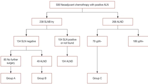
Three of five breast surgeons at our center have started try- ing SLNB procedures since 2011 in breast cancer patients un- der NAC treatment, except in cases of disease progression in follow-up breast MRI or breast ultrasonography. Among pa- tients with negative SLN, patients with suspicious enlarged nodes at the time of surgery underwent further ALND. We classified patients into three groups: group A, negative SLN metastasis and no further dissection; group B, negative SLN metastasis however further ALND; and group C, axillary CR with ALND regardless of clinical response (Figure 1). We col- lected oncological data on axillary recurrence, distant metas- tasis, and survival outcome. We evaluated and compared on- cology outcomes among all groups.
We were aided by the statistical team at the Samsung Medical Center. We categorized patients in whom SLNs were identi- fied as true positive (TP) or false negative (FN). The FNR was calculated as follows: FN/(FN+TP)×100%. The independent t-test was used to compare continuous variables, and the chi- square test and the Fisher exact test were used to compare dis- crete variables. We used the Kaplan-Meier method with the log-rank test to construct survival curves. Differences were assumed to be significant when the p-value was less than 0.05.
All statistical analyses, including the logistic regression and the chi-square tests were performed using the IBM SPSS Sta- tistics for Window, version 23 software (IBM Corp., Armonk, USA). This study was approved by the Institutional Review
Board of Samsung Medical Center, Seoul, Korea (IRB file no.
2017-09-051).
RESULTS
In this study, we included 506 breast cancer patients with cytology proven node metastasis who underwent NAC treat- ment followed by curative surgery. The mean age at surgery was 44.4 ±9.3 years. The median follow-up time was 51 months (range, 3–122 months). Of these patients, 134 were SLN metastasis-negative on frozen SLNB. Eighty-five patients with negative SLNs metastasis had no further surgery (group A), while 49 patients with negative SLN metastasis had back- up ALND (group B). One hundred and four patients with positive SLN metastasis or undetected SLNs by the radioactive and/or vital blue method had further ALND. Of the patients who did not undergo SLNB, 79 had ALND with no residual axillary metastasis (group C), and 189 patients with patholog- ical node-positive disease underwent ALND (Figure 1).
The clinicopathological and treatment characteristics of pa- tients included in this study are showed in Table 1. Regarding the breast cancer subtype, 37.5% of patients had hormone re- ceptor (HR)-positive/HER2-negative breast cancer, 16.2% had HR-positive/HER2-positive breast cancer, 17.0% had HR- negative/HER2-positive breast cancer, and 29.2% had triple- negative breast cancer (TNBC). For NAC, 65.6% of the pa- tients received anthracycline- and taxane-based regimens and
506 Neoadjuvant chemothrapy with positive ALN
238 SLNB try
134 SLN negative
85 No further surgery
Group A
79 ypN−
268 ALND
104 SLN positive or not found
49 ALND
Group B
104 ALND
Group C
189 ypN+
Figure 1. Algorithm of patient selection and grouping of patients with initial cytology-determined nodal disease.
ALN=axillary lymph node; SLNB=sentinel lymph node biopsy; SLN=sentinel lymph node; ALND=axillary lymph node dissection.
Table 1. Clinicopathological and treatment characteristics of 506 pa- tients
Characteristic No. (%)
Age at surgery (yr)* 44.4±9.3
Clinical T stage
cT1 35 (6.9)
cT2 270 (53.4)
cT3 159 (31.4)
cT4 42 (8.3)
Clinical N stage
cN1 205 (40.5)
cN2 169 (33.5)
cN3 132 (26.0)
Clinical tumor subtype
HR+/HER2– 190 (37.5)
HR+/HER2+ 82 (16.2)
HR–/HER2+ 86 (17.0)
Triple-negative 148 (29.2)
Neoadjuvant chemotherapy regimen
Anthracycline+taxane 332 (65.6)
Anthracycline, no taxane 49 (9.7)
Taxane, no anthracycline 6 (1.2)
H-containing regimen 94 (18.6)
Other 25 (4.9)
Type of surgery
Mastectomy 212 (41.9)
Breast-conserving surgery 294 (58.1)
HR=hormone receptor; HER2=human epidermal growth factor receptor 2;
H=trastuzumab.
*Mean±SD.
18.6% of the patients received trastuzumab-containing regi- men. Clinical nodal stage data showed an incidence of 40.5%
for clinical N1 stage, 33.5% for clinical N2 stage, and 26.0%
for clinical N3 stage. Clinical tumor stage data showed an in- cidence of 6.9% for clinical T1 stage, 53.4% for clinical T2 stage, 31.4% for clinical T3 stage, and 8.3% for clinical T4
stage.
The SLN identification rate was 98.3% (234/238 patients).
The median number of retrieved SLNs was 5 (range, 2–9), and 104 of the 238 patients who underwent SLNB had positive SLNs. The FNR of SLNB after NAC was 7.5% (8/106 patients).
The characteristics of the SLN biopsy group and pathologi- Table 2. Characteristics of the sentinel lymph node biopsy-negative group and pathologic node-negative ALND group
Variable Group A (n=85)
No. (%) Group B (n=49)
No. (%) Group C (n=79)
No. (%) Total (n=213)
No. (%) p-value
Age (yr)* 43.3±9.1 46.4±8.4 45.7±10.5 44.5±9.7 0.128
No. of SLNs* 4.6±1.8 5.3±2.3 - 4.9±2.1 0.154
No. of nodes after ALND* - 15.4±4.6 16.5±5.9 16.1±5.3 0.217
Type of surgery 0.001
Conserving surgery 70 (82.4) 33 (67.3) 43 (54.4) 146 (68.5)
Mastectomy 15 (17.6) 16 (32.7) 36 (45.6) 67 (31.5)
Clinical tumor subtype 0.154
HR+/HER2– 21 (24.7) 11 (22.4) 21 (26.6) 53 (24.9)
HR+/HER2+ 15 (17.6) 12 (24.5) 15 (19.0) 42 (19.7)
HR–/HER2+ 12 (14.2) 10 (20.4) 23 (29.1) 45 (21.1)
Triple-negative 37 (43.5) 16 (32.7) 20 (25.3) 73 (34.3)
Chemotherapy regimen 0.163
Anthracycline+taxane 59 (69.4) 31 (63.3) 40 (50.6) 130 (61.0)
Anthracycline, no taxane 2 (2.4) 2 (4.1) 8 (10.1) 12 (5.6)
Taxane, no anthracycline 1 (1.2) 0 1 (1.3) 2 (0.9)
H-containing regimen 17 (20.0) 14 (28.6) 27 (34.2) 58 (27.2)
Others 6 (7.1) 2 (4.1) 3 (3.8) 11 (5.2)
Clinical tumor stage 0.001
cT1 12 (14.1) 3 (6.1) 3 (3.8) 18 (8.5)
cT2 55 (64.7) 32 (65.3) 35 (44.3) 122 (57.3)
cT3 14 (16.5) 11 (22.4) 27 (34.2) 52 (24.4)
cT4 4 (4.7) 3 (6.1) 14 (17.7) 21 (9.9)
Clinical node stage 0.022
cN1 50 (58.8) 18 (36.7) 29 (36.7) 97 (45.5)
cN2 21 (24.7) 16 (32.7) 32 (40.5) 69 (32.4)
cN3 14 (16.5) 15 (30.6) 18 (22.8) 47 (22.1)
Pathologic tumor stage 0.993
ypT0-is 40 (47.1) 24 (49.0) 37 (46.8) 101 (47.4)
ypT1 30 (35.3) 17 (34.7) 26 (32.9) 73 (34.3)
ypT2 13 (15.3) 6 (12.2) 11 (13.9) 30 (14.1)
ypT3 2 (2.4) 2 (4.1) 5 (6.3) 9 (4.2)
Pathologic node stage <0.001
ypN0 84 (98.8) 41 (83.7) 79 (100) 204 (95.8)
ypN1 1 (1.2)† 8 (16.3) 0 9 (4.2)
ypN2 0 0 0 0
Clinical response after NAC 0.387
CR 43 (50.6) 28 (57.2) 43 (54.4) 114 (53.5)
PR 37 (43.5) 18 (36.7) 26 (32.9) 81 (38.0)
SD or PD 5 (5.9) 3 (6.1) 10 (12.7) 18 (8.5)
Lymphovascular invasion 0.872
Absent 72 (84.7) 43 (87.8) 67 (64.8) 182 (85.4)
Present 13 (15.3) 6 (12.2) 12 (15.2) 31 (14.6)
ALND=axillary lymph node dissection; SLN=sentinel lymph node; HR=hormone receptor; HER2=human epidermal growth factor receptor 2; H=trastuzumab;
NAC=neoadjuvant chemotherapy; CR=complete response; PR=partial response; SD=stable disease; PD=progressive disease.
*Mean±SD; †Negative in frozen biopsy but microinvasive in permanent biopsy.
cal node negative ALND group are shown in Table 2. Group C showed a higher mastectomy rate (p=0.001), clinical tumor stage (p=0.001), clinical node stage (p=0.022), and pathol- ogic nodal stage (p<0.001) than did groups A and B. However there was no significant difference in age, clinical tumor subtype, chemotherapy regimen, pathologic tumor stage, clinical response after NAC and lymphovascular invasion among the three groups. Sixteen of the 85 patients in the SLNB alone group (group A) experienced recurrences. Six patients were pathologic T0 stage, six were pathologic T1 stage, and four were pathologic T2 stage. Six patients were HR-positive/HER2-negative, six were HR-negative/HER2-
positive, and four had TNBC. Systemic metastasis occurred in the bone, lungs, and brain in eight patients, and axillary me- tastasis occurred in two patients (Table 3).
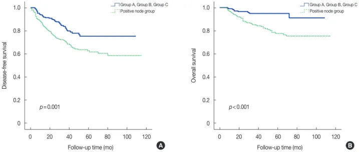
We compared survival outcomes between the node-nega- tive group (combined groups A, B, and C) and the node-posi- tive group. There was a significant difference in DFS and in OS between the node-negative (combined groups A–C) and the node-positive groups (5-year DFS: 78.3% vs. 62.7%, p=0.001; 5-year OS: 94.9% vs. 77.8%, p<0.001) (Figure 2).
Figure 3 compares the survival outcomes in groups A, B, and C. Axillary recurrence-free survival was not significantly dif- ferent among groups (5-year axillary recurrence-free survival:
Table 3. Types of recurrence in the sentinel lymph node biopsy alone group after neoadjuvant chemotherapy
Case cT cN ypT ypN Subtype Recurrence site Disease-free time (mo) ALN recurrence No. of SLN
1 2 1 1 0 HR+/HER2− Breast 25 No 5
2 1 1 0 0 HR+/HER2− Skin 22 No 4
3 3 2 2 0 HR+/HER2− Lung 9 No 8
4 2 1 2 0 HR+/HER2− Bone 32 No 3
5 3 3 2 0 HR+/HER2− Bone 12 No 8
6 3 2 0 0 HR+/HER2− Brain 6 No 3
7 2 1 1 0 HR–/HER2+ Breast 7 No 8
8 3 2 1 0 HR–/HER2+ Axilla 13 Yes 4
9 2 1 1 0 HR–/HER2+ SCN 37 No 7
10 4 1 1 0 HR–/HER2+ Lung 29 No 5
11 2 1 0 0 HR–/HER2+ Bone 5 No 5
12 4 3 0 0 HR–/HER2+ Brain 5 No 3
13 3 2 0 0 TNBC Breast 43 No 4
14 2 3 1 0 TNBC Axilla, SCN 6 Yes 5
15 3 2 2 0 TNBC Chest wall 7 No 6
16 3 2 0 0 TNBC Brain 26 No 5
ALN=axillary lymph node; SLN=sentinel lymph node; HR=hormone receptor; HER2=human epidermal growth factor receptor 2; SCN=supraclavicular lymph node; TNBC=triple-negative breast cancer.
Figure 2. Kaplan-Meier curves for disease-free survival (A) and overall survival (B) between combined groups A–C and node-positive groups.
1.0
0.8
0.6
0.4
0.2
0
1.0
0.8
0.6
0.4
0.2
0
0 20 40 60 80 100 120 0 20 40 60 80 100 120
p=0.001 p<0.001
Follow-up time (mo) Follow-up time (mo)
Disease-free survival Overall survival
A B
Group A, Group B, Group C Group A, Group B, Group C
Positive node group Positive node group
Figure 4. Kaplan-Meier curves for dis- ease-free survival and overall survival among groups A, B, and C in hormone receptor (HR)-positive (A) and HR-neg- ative (B) patients.
1.0 0.8 0.6 0.4 0.2 0
1.0 0.8 0.6 0.4 0.2 0
1.0 0.8 0.6 0.4 0.2 0
1.0 0.8 0.6 0.4 0.2 0 0 20 40 60 80 100 120
0 20 40 60 80 100 120
0 20 40 60 80 100 120
0 20 40 60 80 100 120 Follow-up time (mo)
Follow-up time (mo)
Follow-up time (mo)
Follow-up time (mo)
Disease-free survivalDisease-free survival Overall survivalOverall survival
A
B
A Group
B C
A Group
B C
p=0.767
p=0.581
Figure 3. Kaplan-Meier curves for axillary recurrence-free survival (A), disease-free survival (B), and overall survival (C) among groups A, B, and C.
1.0 0.8 0.6 0.4 0.2 0
1.0 0.8 0.6 0.4 0.2 0
1.0 0.8 0.6 0.4 0.2 0
0 20 40 60 80 100 120 0 20 40 60 80 100 120 0 20 40 60 80 100 120
Follow-up time (mo) Follow-up time (mo) Follow-up time (mo)
Axillary recurrence-free survival Disease-free survival Overall survival
A B C
A Group
B C
A Group
B C
A Group
B
p=0.118 p=0.578 p=0.149 C
p=0.428
p=0.333
group A, 97.4% vs. group B, 90.9% vs. group C, 98.7%, p=0.118). There was also no difference among groups A, B, and C in DFS (81.2% vs. 85.7% vs. 77.2%, p=0.578) or in OS (92.9% vs. 100% vs. 93.7%, p=0.149).
In the subgroup analysis by HR status, there was no signifi- cant difference in DFS (p=0.767) or OS (p=0.428) among groups A, B, and C in the HR-positive subgroup. There was also no difference in DFS (p=0.581) or OS (p=0.333) in the HR-negative subgroup (Figure 4). Similarly, there was no sig-
nificant difference in DFS (p=0.052) or OS (p=0.410) among groups A, B, and C in the HER2-positive subgroup and no significant difference in DFS (p=0.811) or OS (p=0.358) in the HER2-negative subgroup (Figure 5). In the subgroup analysis of breast pCR status, there was no significant differ- ence in DFS (85.0% vs. 95.8% vs. 86.5%, p=0.271) or OS (95.0% vs. 100% vs. 97.3%, p=0.207) among groups A, B, and C in the breast pCR subgroup and no significant difference in DFS (77.8% vs. 76.0% vs. 69.0%, p=0.892) or OS (91.1% vs.
Figure 5. Kaplan-Meier curves for dis- ease-free survival and overall survival among groups A, B, and C in human epidermal growth factor receptor 2 (HER2)-positive (A) and HER2-negative (B) patients.
Figure 6. Kaplan-Meier curves for dis- ease-free survival and overall survival among groups A, B, and C in breast pathologic complete response (pCR) (A) and non-pCR (B) patients.
1.0 0.8 0.6 0.4 0.2 0
1.0 0.8 0.6 0.4 0.2 0 1.0 0.8 0.6 0.4 0.2 0
1.0 0.8 0.6 0.4 0.2 0
1.0 0.8 0.6 0.4 0.2 0
1.0 0.8 0.6 0.4 0.2 0 1.0 0.8 0.6 0.4 0.2 0
1.0 0.8 0.6 0.4 0.2 0 0 20 40 60 80 100 120
0 20 40 60 80 100 120 0 20 40 60 80 100 120
0 20 40 60 80 100 120
0 20 40 60 80 100 120
0 20 40 60 80 100 120 0 20 40 60 80 100 120
0 20 40 60 80 100 120 Follow-up time (mo)
Follow-up time (mo) Follow-up time (mo)
Follow-up time (mo)
Follow-up time (mo)
Follow-up time (mo) Follow-up time (mo)
Follow-up time (mo)
Disease-free survivalDisease-free survivalDisease-free survivalDisease-free survival Overall survivalOverall survivalOverall survivalOverall survival
A
A B
B
A Group
B C
A Group
B C A Group
B C
A Group
B C
p=0.052
p=0.271 p=0.811
p=0.892
p=0.410
p=0.207 p=0.358
p=0.300
100% vs. 90.5%, p=0.300) in the breast non-pCR subgroup (Figure 6).
DISCUSSION
Several studies have been published on the effectiveness and role of SLNB after NAC. The results of these studies showed that SLNB with NAC may be feasible and acceptable option [11-14]. ALN metastases can be extirpated by NAC in some patients, and consequently, ALND may be avoided in these. However, SLNB after NAC might not be accurate be- cause anatomical alterations of the lymphatic drainage by che- motherapy can destroy lymphatic vessels or lymphatic chan- nels due to inflammation or fibrosis. Previous studies showed that for patients with clinically positive nodes, the identifica- tion rate for the SLNB after NAC is 89.0% to 98.0% [9,10,15, 16]. Our study showed an identification rate of 98.3% for SLNB, which falls within the published range [17]. Because this study defined suspicious palpable lymph nodes as SLNs, the high SLN number could have affected the identification rate. We have previously studied the feasibility and prognostic effect of SLNB after NAC [4]. However, the median follow-up time was as short at 37 months, and only 329 patients were surveyed. Therefore a study with a longer follow-up and a larger sample size was needed.
In several studies, the FNR of SLNB was 14% in node-posi- tive patients treated with NAC. This was more than about 4%
FNR of SLNB for node-negative patients who did not receive NAC treatment [18,19]. Also, this study did not use targeted axillary dissection (TAD), which ensures resection of the clipped lymph node at the time of SLNB. TAD could poten- tially lead to an even lower FNR of 4.2% and allow for a more precise way of performing SLNB [20]. We did not use TAD at SLNB after NAC, however, in our study, the median number of retrieved SLNs was five and the overall FNR was 7.5%. The overall FNR of this study was a rate less than the pre-specified rate of ≤10%, although we performed SLNB in breast cancer patients with cytologically-determined node metastasis.
Many studies have demonstrated the use of SLNB with NAC in breast cancer patients with node metastasis [3,21,22]. In our study, the patient population included biopsy-determined, node metastasis patients. In contrast, in the Sentinel Neoadjuvant (SENTINA) study, the node metastasis was clinically diagnosed;
in that study, the diagnosis of 592 patients with clinically node- metastasis cancer was changed to negative SLN after NAC, with a 14.2% FNR. The overall FNR was 12.6% for the American College of Surgeons Oncology Group (ACOSOG) Z1071 trial and 8.4% for the Sentinel Node Biopsy Following Neoadjuvant Chemotherapy in Biopsy Proven Node-Positive Breast Cancer
(SN-FNAC) trial. However, the FNR was 7.3% in the SENTINA trial and 9.1% in the ACOSOG Z1071 trial when three or more SLNs were retrieved. Similarly, in this study, the FNR was 7.5%
and the median number of removed SLNs was five.
A recent study showed no statistical difference in DFS among subgroups according to HR status [15]. Similarly, we also found that the OS and DFS rates did not differ signifi- cantly between subgroups according to HR, HER2, or pCR status. Furthermore, our study included a longer follow-up than the previous studies. A meta-analysis suggested that more than 20% of patients treated with breast surgery would develop lymphedema [23,24]. Lymphedema following breast cancer treatment can be an irreversible condition with nega- tive effects on quality of life. The data from several studies have shown that patients with NAC treatment had more lymphedema [24]. In our study, lymphedema and arm mo- tion morbidity were observed in 7.1% (6/85) of the SLNB- alone group and in 27.3% (35/128) of the ALND group. Radi- ation therapy was administered to 82.4% (70/84) in the SLNB- alone group and to 80.5% (103/128) in the ALND group.
Therefore, SLNB may be associated with less lymphedema and arm motion morbidity than is ALND after NAC.
Although this study was performed in a single comprehen- sive cancer institution in Korea and the number of patients was relatively small, it did not investigate clinically suspected node-positive patients but instead investigated node-positive patients confirmed by cytology. Therefore, the results obtained from the analysis of the small sample population are still meaningful. Additionally, this study was not a prospective randomized clinical trial; group C was higher than group A and B in clinical tumor stage and node stage. Thus, the distri- bution of patients may have had some effect on the results of regional control. Also, we began performing SLNB alone after NAC since 2011, so the median follow-up period for group A was less than that for group B and group C. The longest fol- low-up time was 73 months in group A and 113 months in group B. However, this study had a median follow-up time of 51 months, and the results reveal clinical effect and provide important insights regarding feasibility of SLNB after NAC.
In conclusion, our study of breast cancer patients who had ALN conversion from cytology-determined positive to nega- tive following NAC indicated that SLNB and ALND without SLNB had similar rates of axillary recurrence, DFS and OS.
Therefore, SLNB may be acceptable after NAC for patients with cytology-determined, node-metastasis-positive breast cancer with a reasonable identification rate. Also, in some pa- tients, SLNB can help identify possible downstaging to a nega- tive nodal stage, and since ALND is avoided, this may reduce arm motion morbidity and lymphedema. We suggest the use
of SLN surgery as an alternative to ALND after NAC in pa- tients with cytology-determined axillary node metastasis- positive breast cancer.
CONFLICT OF INTEREST
The authors declare that they have no competing interests.
REFERENCES
1. Fisher B, Brown A, Mamounas E, Wieand S, Robidoux A, Margolese RG, et al. Effect of preoperative chemotherapy on local-regional disease in women with operable breast cancer: findings from National Surgical Adjuvant Breast and Bowel Project B-18. J Clin Oncol 1997;15:2483- 93.
2. Vlastos G, Mirza NQ, Lenert JT, Hunt KK, Ames FC, Feig BW, et al. The feasibility of minimally invasive surgery for stage IIA, IIB, and IIIA breast carcinoma patients after tumor downstaging with induction che- motherapy. Cancer 2000;88:1417-24.
3. Boughey JC, Suman VJ, Mittendorf EA, Ahrendt GM, Wilke LG, Taback B, et al. Sentinel lymph node surgery after neoadjuvant chemo- therapy in patients with node-positive breast cancer: the ACOSOG Z1071 (Alliance) clinical trial. JAMA 2013;310:1455-61.
4. Park S, Lee JE, Paik HJ, Ryu JM, Bae SY, Lee SK, et al. Feasibility and prognostic effect of sentinel lymph node biopsy after neoadjuvant che- motherapy in cytology-proven, node-positive breast cancer. Clin Breast Cancer 2017;17:e19-29.
5. Enokido K, Watanabe C, Nakamura S, Ogiya A, Osako T, Akiyama F, et al. Sentinel lymph node biopsy after neoadjuvant chemotherapy in patients with an initial diagnosis of cytology-proven lymph node-posi- tive breast cancer. Clin Breast Cancer 2016;16:299-304.
6. Shen J, Gilcrease MZ, Babiera GV, Ross MI, Meric-Bernstam F, Feig BW, et al. Feasibility and accuracy of sentinel lymph node biopsy after preoperative chemotherapy in breast cancer patients with documented axillary metastases. Cancer 2007;109:1255-63.
7. Hennessy BT, Hortobagyi GN, Rouzier R, Kuerer H, Sneige N, Buzdar AU, et al. Outcome after pathologic complete eradication of cytologi- cally proven breast cancer axillary node metastases following primary chemotherapy. J Clin Oncol 2005;23:9304-11.
8. Rouzier R, Extra JM, Klijanienko J, Falcou MC, Asselain B, Vincent- Salomon A, et al. Incidence and prognostic significance of complete axillary downstaging after primary chemotherapy in breast cancer patients with T1 to T3 tumors and cytologically proven axillary meta- static lymph nodes. J Clin Oncol 2002;20:1304-10.
9. Fu JF, Chen HL, Yang J, Yi CH, Zheng S. Feasibility and accuracy of sentinel lymph node biopsy in clinically node-positive breast cancer after neoadjuvant chemotherapy: a meta-analysis. PLoS One 2014;9:
e105316.
10. Kang YJ, Han W, Park S, You JY, Yi HW, Park S, et al. Outcome following sentinel lymph node biopsy-guided decisions in breast cancer patients with conversion from positive to negative axillary lymph nodes after neoadjuvant chemotherapy. Breast Cancer Res Treat 2017;166:473-80.
11. Mamounas EP, Brown A, Anderson S, Smith R, Julian T, Miller B, et al.
Sentinel node biopsy after neoadjuvant chemotherapy in breast cancer:
results from National Surgical Adjuvant Breast and Bowel Project Protocol B-27. J Clin Oncol 2005;23:2694-702.
12. van Deurzen CH, Vriens BE, Tjan-Heijnen VC, van der Wall E, Albregts M, van Hilligersberg R, et al. Accuracy of sentinel node biopsy after neoadjuvant chemotherapy in breast cancer patients: a systematic review. Eur J Cancer 2009;45:3124-30.
13. Mocellin S, Goldin E, Marchet A, Nitti D. Sentinel node biopsy perfor- mance after neoadjuvant chemotherapy in locally advanced breast cancer: a systematic review and meta-analysis. Int J Cancer 2016;138:
472-80.
14. Lee S, Kim EY, Kang SH, Kim SW, Kim SK, Kang KW, et al. Sentinel node identification rate, but not accuracy, is significantly decreased after pre-operative chemotherapy in axillary node-positive breast cancer patients. Breast Cancer Res Treat 2007;102:283-8.
15. Kim JY, Kim MK, Lee JE, Jung Y, Bae SY, Lee SK, et al. Sentinel lymph node biopsy alone after neoadjuvant chemotherapy in patients with initial cytology-proven axillary node metastasis. J Breast Cancer 2015;
18:22-8.
16. Newman EA, Sabel MS, Nees AV, Schott A, Diehl KM, Cimmino VM, et al. Sentinel lymph node biopsy performed after neoadjuvant chemo- therapy is accurate in patients with documented node-positive breast cancer at presentation. Ann Surg Oncol 2007;14:2946-52.
17. Lyman GH, Giuliano AE, Somerfield MR, Benson AB 3rd, Bodurka DC, Burstein HJ, et al. American Society of Clinical Oncology guide- line recommendations for sentinel lymph node biopsy in early-stage breast cancer. J Clin Oncol 2005;23:7703-20.
18. Miltenburg DM, Miller C, Karamlou TB, Brunicardi FC. Meta-analysis of sentinel lymph node biopsy in breast cancer. J Surg Res 1999;84:138- 42.
19. Fraile M, Rull M, Julián FJ, Fusté F, Barnadas A, Llatjós M, et al. Sentinel node biopsy as a practical alternative to axillary lymph node dissection in breast cancer patients: an approach to its validity. Ann Oncol 2000;
11:701-5.
20. Caudle AS, Yang WT, Krishnamurthy S, Mittendorf EA, Black DM, Gilcrease MZ, et al. Improved axillary evaluation following neoadju- vant therapy for patients with node-positive breast cancer using selec- tive evaluation of clipped nodes: implementation of targeted axillary dissection. J Clin Oncol 2016;34:1072-8.
21. Boileau JF, Poirier B, Basik M, Holloway CM, Gaboury L, Sideris L, et al. Sentinel node biopsy after neoadjuvant chemotherapy in biopsy- proven node-positive breast cancer: the SN FNAC study. J Clin Oncol 2015;33:258-64.
22. Kuehn T, Bauerfeind I, Fehm T, Fleige B, Hausschild M, Helms G, et al.
Sentinel-lymph-node biopsy in patients with breast cancer before and after neoadjuvant chemotherapy (SENTINA): a prospective, multicen- tre cohort study. Lancet Oncol 2013;14:609-18.
23. DiSipio T, Rye S, Newman B, Hayes S. Incidence of unilateral arm lymphoedema after breast cancer: a systematic review and meta-analy- sis. Lancet Oncol 2013;14:500-15.
24. Warren LE, Miller CL, Horick N, Skolny MN, Jammallo LS, Sadek BT, et al. The impact of radiation therapy on the risk of lymphedema after treatment for breast cancer: a prospective cohort study. Int J Radiat Oncol Biol Phys 2014;88:565-71.