Jailing Technique Using a Catheter-based Open-cell Stent System in Internal Carotid Artery Sidewall Aneurysms Unfeasible to Simple Coiling
Sun Geon Yoon1, Sung-Chul Jin2, Seung Hwan Kim2, Kyoung Dong Jeon2, Doo Young Kim2, Sun-Il Lee2, Hae Woong Jeong3
1Department of Neurosurgery, Busan Paik Hospital, Inje University, Busan, Korea
2Department of Neurosurgery, Haeundae Paik Hospital, Inje University, Busan, Korea
3Department of Diagnostic Radiology, Busan Paik Hospital, Inje University, Busan, Korea
Objective : An open cell stent system may offer better apposition of cell struts to vessel wall than a closed cell stent system in acute vasculature.
The purpose of this study was to evaluate the feasibility of coiling using the jailing technique with the Neuroform EZ stent system.
Methods : The jailing technique using the open-cell stent system of the Neuroform EZ stent was planned in 22 consecutive patients with 22 cer- ebral aneurysms. We retrospectively evaluated the technical success of the jailing technique and the occurrence of interference between two micro- catheters as well as the factors influencing this interference.
Results : The jailing technique was successful in 19/22 patients (86.4%), and interference between two microcatheters occurred in 6/21 (28.6%).
The jailing technique failed in 3/22 patients, with problems that included failure of the stent delivery system to advance into the positioned micro- catheter in one, interference between the microcatheters during the ad- vancement of the stent delivery system in one, and failure of micro- catheter insertion into the aneurysm sac in the remaining patient.
Interference between the two microcatheters developed during the ad- vance of the stent delivery system into the positioned microcatheter in all cases. One factor that influences interference between two microcatheters more than expected by chance is the carotid siphon angle (p = 0.019).
Conclusion : The acuteness of the carotid siphon angle influences the in- terference between two microcatheters. Therefore, the jailing technique using the Neuroform EZ stent should be performed carefully in cerebral aneurysms with an acute carotid siphon angle because the procedure may possibly fail.
J Cerebrovasc Endovasc Neurosurg.
2013 December;15(4):293-298 Received : 27 June 2013
Revised : 7 August 2013 Accepted : 23 October 2013 Correspondence to Sung-Chul Jin
Department of Neurosurgery, Inje University Haeundae Paik Hospital, 1435 Jwa-dong, Haeundae-gu, Busan 612-043, Korea Tel : 82-51-797-0607
Fax : 82-51-703-0434 E-mail : [email protected]
This is an Open Access article distributed under the terms of the Creative Commons Attribution Non- Commercial License (http://creativecommons.org/li- censes/by-nc/3.0) which permits unrestricted non- commercial use, distribution, and reproduction in any medium, provided the original work is properly cited.
Keywords Carotid siphon, Microcatheter, Neuroform, Jailing
INTRODUCTION
The jailing technique is one of the stent-assisted coil- ing modalities in wide-necked cerebral aneurysms that
are recalcitrant to simple coiling.1)4)5) A major advant- age of the jailing technique is strengthening the stabil- ity of the microcatheter in cerebral aneurysms into which insertion of the microcatheter might be difficult.
Patient
No. Sex/Age Aneurysm
location Carotid siphon
angle (degree) Aneurysm size
(height × width) Aneurysm
neck size Initial treatment
design Success of Jailing
Neuroform stent deployment
1 M/52 Cave 60 4.5×4 mm 4 mm Stent-assisted Success Success
2 F/52 Ophthalmic 89 6.2×4.6 mm 4.1 mm Stent-assisted Success Success
3 M/47 Pcoma 104 2.7×2 mm 2 mm Simple coiling Success Success
4 M/56 Cave 109 1.9×3 mm 1.6 mm Stent-assisted Success Success
5 F/50 Ophthalmic 35 3.6×2.3 mm 2.2 mm Stent-assisted Failure Success
6 F/51 Cave 95 3.4×3 mm 3 mm Stent-assisted Success Success
7 F/66 Ventral 96 3.4×3.1 mm 3.2 mm Stent-assisted Success Success
8 F/69 Cave 64 4.5×4 mm 4 mm Stent-assisted Success Success
9 F/77 Pcom 68 3.4×2.1 mm 2.5 mm Stent-assisted Success Success
10 F/65 Sup. hypophyseal 53 3×3 mm 3 mm Stent assisted Success Success
11 M/66 Ventral 39 2×3 mm 3.1 mm Stent-assisted Failure Failure
12 F/48 Pcoma 84 4×3.5 mm 3.5 mm Stent-assisted Success Success
13 F/47 Ventral 76 2.1×3.3 mm 3.7 mm Stent-assisted Success Success
14 F/53 Sup. hypophyseal 133 7.4×6.3 mm 4 mm Stent-assisted Success Success
15 M/54 Cave 45 3.4×4.3 mm 3.8 mm Stent-assisted Success Success
16 F/47 Sup. hypophyseal 70 3.9×3.7 mm 3.7 mm Stent-assisted Success Success
17 F/59 Ophthalmic 116 2.5×2 mm 2 mm Stent-assisted Success Success
18 F/46 Sup. hypophyseal 53 2.5×4 mm 3.8 mm Stent-assisted Success Success
19 F/71 Pcoma 108 4×4.9 mm 3.2 mm Stent-assisted Success Success
20 F/58 Ventral 105 4.2×5.3 mm 4 mm Stent-assisted Success Success
21 F/56 Ventral 35 2×2.2 mm 1.8 mm Stent-assisted Failure Success
22 F/57 Sup. hypophyseal 30 4×4.5 mm 4 mm Stent-assisted Success Success
Acoma= anterior communicating artery; Pcoma= posterior communicating artery; sup= superior
Table 1. Characteristics of 22 patients who underwent coil embolization using the Neuroform EZ stent system.
The jailing technique is generally used with closed-cell stent systems, such as the Enterprise stent, in wide- necked cerebral aneurysms. However, in closed-cell stent systems, apposition of the stent strut to the ves- sel wall may be difficult in cerebral aneurysms with tortuous vasculature. A closed-cell stent system was recently reported to yield “incomplete stent apposi- tion in the tortuous vasculature”.2)3) The Neuroform EZ stent system has an open-cell stent system that employs a microcatheter-based transfer technique.
Furthermore, this system is expected to provide good navigability and accessibility in the vessel. We expect the jailing technique using the Neuroform EZ stent system to be a good option for wide-necked aneur- ysms with tortuous vasculature. In this retrospective study, we evaluated the feasibility of the jailing tech- nique using the Neuroform EZ stent system in cerebral
aneurysms that were unfavorable to simple coiling.
MATERIALS AND METHODS
Starting in June 2011, we developed a jailing techni- que using the Neuroform EZ stent system, mainly for the coiling of cerebral aneurysms that were un- favorable to simple coiling. From June 2011 to December 2011, the jailing technique using the Neuroform EZ stent system was planned in 22 cere- bral aneurysms (Table 1). These lesions were included in our study, which was approved by the Institutional Review Board. The primary techniques used for coil embolization included the jailing technique in 21 lesions and simple coiling in one. In two lesions, we changed the treatment strategy, halting the use of the jailing technique using the Neuroform EZ
stent system because of failure of the primary treat- ment plan.
Without developing an initial plan for the coil embo- lization, such as a single-catheter technique or stent-assisted coiling, we administered aspirin (300 mg) and clopidogrel (300 mg) to the patients with un- ruptured cerebral aneurysms for at least three hours before the procedure but prescribed the same dose of aspirin and clopidogrel to the patients with ruptured cerebral aneurysms immediately after the procedure via nasogastric tube. Anticoagulation was initiated by the injection of a bolus of 3000 IU heparin intra- venously immediately after femoral puncture, fol- lowed by intermittent bolus injection of 1000 IU hep- arin hourly. Coil embolization was performed via a single femoral access in all cases, with a 7 F Guider guiding catheter (Boston Scientific, Natick, MA, USA).
We attempted several variations of the jailing techni- que to develop a safer and more efficient strategy for the use of this method: 1) A Renegade Hi Flo (Boston Scientific, Watertown, MA, USA) microcatheter was positioned first, and then an Excelsior SL-10 (Boston Scientific, Natick, MA, USA) microcatheter was in- serted into the cerebral aneurysm. Finally, a Neuroform EZ stent delivery system was advanced into the Renegade Hi Flo microcatheter (n = 9). 2) An Excelsior SL-10 microcatheter was first inserted into the cerebral aneurysm, and then a Renegade Hi Flo microcatheter was positioned distal to the cerebral aneurysm. Finally, a Neuroform EZ stent delivery system was advanced into the Renegade Hi Flo microcatheter (n = 7). 3) A Renegade Hi Flo microcatheter was positioned first, and then a Neuroform EZ stent delivery system was advanced into the Renegade Hi Flo microcatheter.
Subsequently, an Excelsior SL-10 microcatheter was in- serted into the cerebral aneurysm (n = 6). In all cases, partial coil packing of the aneurysm was performed to reduce the risk of unexpected rupture of the aneurysm due to interference between the two microcatheters before the advance of the Neuroform EZ stent deliv- ery system into the Renegade Hi Flo microcatheter.
Based on accumulated clinical experience, we sug- gest that the tortuosity of the vasculature and inter- ference between the microcatheters will be major de- terminants of the success of the jailing technique us- ing the Neuroform EZ stent system. The carotid si- phon angle likely represents the degree of vascular tortuosity. The carotid siphon angle is defined as the angle between the supraclinoid segment of the in- ternal carotid artery and the anterior vertical or hori- zontal segment of the cavernous internal carotid artery. The measurement of the carotid siphon angle was performed semi-automatically on a lateral view of conventional angiography using the picture ar- chiving and communication system (PACS) software system (Maroview; Marotech, Seoul, KR). Interference between the two microcatheters is defined as the phenomenon in which one microcatheter, which is inserted into the cerebral aneurysm, is contacted and dislodged from its previous position by another mi- crocatheter that is positioned for stent deployment during any step of the jailing technique.
Statistical analysis
The differences in various characteristics between the presence and absence of interference of micro- catheters were evaluated using the Fisher exact test for nominal data and the Mann-Whitney U test for numerical data. Null hypotheses of no difference were rejected if p values were less than 0.05.
Ethical statement
The form consent was not obtained because this study was a retrospective analysis. The study protocol was approved by the Institutional Review Boards of the Inje University Busan Paik Hospital (IRB No. 2011-085).
RESULTS
We found that the jailing technique using the Neuroform EZ stent system was successfully accom- plished in 19 cerebral aneurysms and failed in three (12.5%), although the Renegade microcatheter was
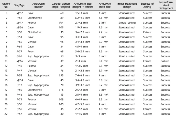
A B
C D
Fig. 1. A case in which the Neuroform stent delivery system failed to advance into the Renegade microcatheter. (A) A working roadmap image shows two well-positioned microcatheters. (B) A working roadmap image shows the dislodging of the microcatheter that was inserted into the aneurysm during the advance of the Neuroform stent delivery system into the Renegade microcatheter.
(C) A switch from Neuroform stent-assisted to Enterprise stent-assisted (black arrow) coiling was performed due to the failure of the Neuroform delivery system to advance into the Renegade microcatheter. (D) A final working angiogram shows partial coiling of the aneurysm.
well positioned in all of the lesions. Failure of the jail- ing technique using the Neuroform EZ stent system developed in three lesions during several steps of the procedure, as follows: 1) the Neuroform EZ stent de- livery system failed to advance into the positioned
Renegade microcatheter (Fig. 1); 2) the Excelsior SL-10, which had been inserted into the cerebral aneurysm, was dislodged from the cerebral aneurysm by the ad- vance of the Neuroform EZ stent system into the Renegade microcatheter; and 3) the Excelsior SL-10
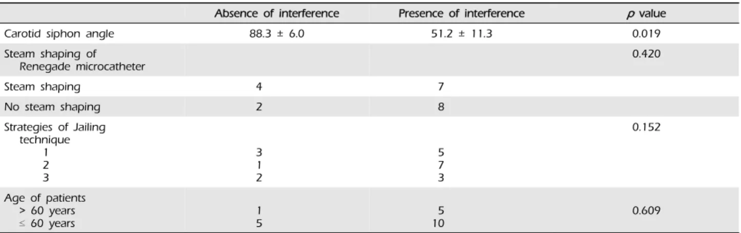
Absence of interference Presence of interference p value
Carotid siphon angle 88.3 ± 6.0 51.2 ± 11.3 0.019
Steam shaping of
Renegade microcatheter 0.420
Steam shaping 4 7
No steam shaping 2 8
Strategies of Jailing technique
12 3
31 2
57 3
0.152
Age of patients
> 60 years
≤ 60 years 1
5 5
10 0.609
Strategy 1: The Renegade microcatheter is positioned first and then the microcatheter is inserted into the cerebral aneurysm. Finally, the Neuroform EZ stent delivery system is advanced into the Renegade microcatheter. Strategy 2: The microcatheter is first inserted into the cerebral aneurysm, and then the Renegade microcatheter is positioned. Finally, the Neuroform EZ stent delivery system is advanced into the Renegade microcatheter. Strategy 3: The Renegade microcatheter is positioned first, and then the Neuroform EZ stent delivery system is advanced into the Renegade microcatheter. Finally, the microcatheter is inserted into the cerebral aneurysm.
Table 2. Factors interfering between microcatheters during jailing technique using the Neuroform EZ stent system.
microcatheter failed to insert into the cerebral aneur- ysm after the Renegade microcatheter had been posi- tioned distal to the aneurysm. The case in which the Neuroform EZ stent delivery system failed to advance into the Renegade microcatheter was switched to a jailing technique using an Enterprise stent system of a closed-cell design (Fig. 1). The other two cases were treated using the conventional sequential technique of stent-assisted coiling with insertion of the microcatheter into the cerebral aneurysm after stent deployment.
Interference between the two microcatheters, possi- bly a major determinant of the success of the jailing technique, developed in six of 21 cerebral aneurysms (28.6%) excluding one lesion into which the micro- catheter was not inserted. Interference between two microcatheters always developed during the step of the procedure in which the Neuroform EZ stent deliv- ery system was advanced into the Renegade micro- catheter regardless of which strategy of the jailing technique was used. Analysis of the factors influencing the interference between two microcatheters revealed that the carotid siphon angle is the only statistically significant variable in univariate analysis (Table 2).
Procedural complications developed in four lesions (16.7%), including one thromboembolism, one stretch- ed coil loop, and contrast leakage in two lesions.
Our procedural morbidity and mortality were 8.3%
and 0%. None of the cases of procedural morbidity (thromboembolism, n = 1; contrast leakage, n = 1) re- sulted in neurological deficits at discharge.
DISCUSSION
Our study demonstrates that the jailing technique using the Neuroform EZ stent system is difficult to apply, especially to cerebral aneurysms with an acute carotid siphon angle. The failure of the jailing techni- que using the Neuroform EZ stent system is explain- able by two influences. One influencing factor in the failure of jailing technique is the Renegade micro- catheter itself. A case in which the Neuroform EZ stent delivery system failed to advance into the Renegade microcatheter suggests that crumpling of the microcatheter in the tortuous vasculature and sub- sequent compromise of the inner lumen adversely in- fluence the jailing technique. Our test of the Renegade microcatheter supported this suggestion that com- promise of the inner lumen by crumpling was ini- tiated by curving the microcatheter at approximately 90 degrees and was not restored by the release of the microcatheter in the distal segment. Therefore, com- promise of the inner lumen of the Renegade micro-
catheter in tortuous vasculature and a lack of sub- sequent restoration of the inner lumen after the re- lease of the tension on the Renegade microcatheter are one of the influencing factors in the failure of the jailing technique using the Neuroform EZ stent system. The Excelsior XT-27 microcatheter (Striker neurovascular, Fremont, CA, USA) is used in clinical settings and may overcome the drawbacks of the Renegade microcatheter such as compromise of the inner lumen. This microcatheter should be studied in the acute carotid siphon angle compared with the Renegade microcatheter. Another factor influencing failure of this technique is interference between two microcatheters (one catheter for coiling and another catheter for stent deployment). In our series, all cases of interference between two microcatheters developed during the advance of the Neuroform EZ stent deliv- ery system into the Renegade microcatheter. Therefore, to reduce interference in the jailing technique using the Neuroform EZ stent system, the Renegade micro- catheter is first navigated distal to the cerebral aneur- ysm, and then the Neuroform EZ stent delivery sys- tem is advanced into the Renegade microcatheter im- mediately before unsheathing the stent. Subsequently, the microcatheter is inserted into the cerebral aneurysm.
In addition, the carotid siphon angle is the only stat- istically significant factor affecting the interference be- tween two microcatheters in the jailing technique us- ing the Neuroform EZ stent system. Interference be- tween two microcatheters may be expected to occur in the carotid siphon angle less than 50 degree. Jailing technique may fail if the carotid siphon angle is about 30 degree. Therefore, in the planning of the coiling of cerebral aneurysms with tortuous vasculature (carotid siphon angle less than 50 degree), the conventional sequential technique, which consists of prior deploy- ment of the stent and subsequent insertion of the mi- crocatheter into the cerebral aneurysm, should be con- sidered for stent-assisted coiling using the Neuroform EZ stent system. Furthermore, in the coiling of cerebral aneurysms in which the insertion of the microcatheter
into the aneurysms is expected to be difficult, the jail- ing technique using a closed-cell stent system, such as the Enterprise stent system, should be considered.
Although our study has a small sample size and is a retrospective design, its significance is focused on the feasibility of the jailing technique using the Neuroform EZ stent system in clinical practice. The jailing technique using the Neuroform EZ stent system should be carefully considered, especially in the coil- ing of cerebral aneurysms with tortuous vasculatures.
CONCLUSIONS
The jailing technique using the Neuroform EZ stent system showed considerable rates of technical failure due to interference between the microcatheters in cerebral aneurysms that were unfavorable to simple coiling. Therefore, the jailing technique using the Neuroform EZ stent system should be used carefully, especially in cerebral aneurysms with tortuous vasculature.
REFERENCES
1. Biondi A, Janardhan V, Katz JM, Salvaggio K, Riina HA, Gobin YP. Neuroform stent-assisted coil emboliza- tion of wide-neck intracranial aneurysms: Strategies in stent deployment and midterm follow-up. Neurosurgery.
2007 Sep;61(3):460-8; discussion 468-9.
2. Heller RS, Malek AM. Parent vessel size and curvature strongly influence risk of incomplete stent apposition in enterprise intracranial aneurysm stent coiling. AJNR Am J Neuroradiol. 2011 Oct;32(9):1714-20.
3. Heller RS, Miele WR, Do-Dai DD, Malek AM. Crescent sign on magnetic resonance angiography revealing in- complete stent apposition: Correlation with diffusion-weighted changes in stent-mediated coil embolization of aneurysms.
J Neurosurg. 2011 Sep;115(3):624-32.
4. Spiotta AM, Wheeler AM, Smithason S, Hui F, Moskowitz S. Comparison of techniques for stent assisted coil em- bolization of aneurysms. J Nuerointerv Surg. 2012 Sep;4(5) :339-44.
5. Wajnberg E, de Souza JM, Marchiori E, Gasparetto EL.
Single-center experience with the Neuroform stent for en- dovascular treatment of wide-necked intracranial aneurysms.
Surg Neurol. 2009 Dec;72(6):612-9.