ORIGINAL ARTICLE
위암 환자에서 림프절 전이와 고식적 수술의 예측에 대한
양전자방출단층촬영에서 18F-Fluorodeoxyglucose 섭취 정도의 유용성
최주영, 심기남, 김성은, 정혜경, 정성애, 유 권
이화여자대학교 의학전문대학원 내과학교실, 의과학연구소
The Clinical Value of 18F-Fluorodeoxyglucose Uptake on Positron Emission
Tomography/Computed Tomography for Predicting Regional Lymph Node Metastasis and Non-curative Surgery in Primary Gastric Carcinoma
Ju Young Choi, Ki-Nam Shim, Seong-Eun Kim, Hye-Kyung Jung, Sung-Ae Jung and Kwon Yoo
Department of Internal Medicine, Ewha Medical Research Institute, Ewha Womans University School of Medicine, Seoul, Korea
Background/Aims: Accurate preoperative detection of regional lymph nodes and evaluation of tumor resectability is critical to determining the most adequate therapy for gastric cancer. The aim of this study is to identify a possible link between 18F-fluorodeoxyglucose (18F-FDG) uptake on PET scan combined with CT scan (PET/CT) and predictions of lymph node metastasis and non-curative surgery.
Methods: This study included 156 gastric cancer patients who underwent preoperative 18F-FDG PET/CT and surgery. In cases with perceptible FDG uptake in the primary tumor or lymph nodes, the maximum standardized uptake value (SUVmax) was calculated.
Results: In multivariate analysis, non-curative surgery (OR, 11.05; 95% CI, 1.10-111.08; p=0.041), tumor size (≥3 cm) (OR, 7.39; 95% CI, 2.41-22.70; p<0.001), and lymph node metastasis (OR, 5.47; 95% CI, 2.05-14.64; p=0.001) were significant independent predictors for 18F-FDG uptake in the primary tumors. Tumor size (tumor size ≥3 cm) (OR, 3.15; 95% CI, 1.16-8.58;
p=0.025) and lymph node metastasis (OR, 3.36; 95% CI, 1.23-9.14; p=0.018) showed significant association with 18F-FDG uptake in lymph node. When the SUVmax of the primary gastric tumor was greater than 3.75, the sensitivity and specificity of PET/CT with regard to the diagnosis of metastatic lymph node were 73.5% and 74.5%. When the SUVmax of the primary gastric tumor was greater than 4.35 and the FDG uptake of lymph nodes was positive, non-curative surgery was predicted with a sensitivity of 58.8% and specificity of 91.6%.
Conclusions: A high FDG uptake of the gastric tumor was related to histologic positive lymph nodes and non-curative surgery.
(Korean J Gastroenterol 2014;64:340-347)
Key Words: Stomach neoplasms; Lymph node; Positron-emission tomography
Received July 16, 2014. Revised October 10, 2014. Accepted October 10, 2014.
CC This is an open access article distributed under the terms of the Creative Commons Attribution Non-Commercial License (http://creativecommons.org/licenses/
by-nc/3.0) which permits unrestricted non-commercial use, distribution, and reproduction in any medium, provided the original work is properly cited.
교신저자: 심기남, 158-710, 서울시 양천구 안양천로 1071, 이화여자대학교 의학전문대학원 내과학교실
Correspondence to: Ki-Nam Shim, Department of Internal Medicine, Ewha Womans University School of Medicine, 1071 Anyangcheon-ro, Yangcheon-gu, Seoul 158-710, Korea. Tel: +82-2-2650-2632, Fax: +82-2-2655-2076, E-mail: [email protected]
Financial support: None. Conflict of interest: None.
INTRODUCTION
The most important step after diagnosis of gastric cancer is accurate staging, which primarily evaluates the tumor resectability. Important factors that determine tumor resect- ability are whether the tumor can be separated from adjacent organs or important blood vessels, the extent of lymph node metastasis, and the presence of peritoneal metastasis or dis- tant organ metastasis. Anatomical imaging has superior spa- tial resolution and is essential for evaluating the extent of lo- cal tumor invasion. The 18F-fluorodeoxyglucose (18F-FDG) PET scan is a useful functional whole-body imaging modality that images various types of malignancies with relatively high sensitivity and specificity in a reasonably rapid time. While CT detects malignant processes based mainly on altered anat- omy, the 18F-FDG PET scan detects a lesion on the basis of abnormal glucose metabolism. The 18F-FDG PET scan has been used in the preoperative staging of gastric cancer with some promising results. In patients with gastric cancers, less than 50% of early gastric cancers (EGC) and 62-98% of ad- vanced gastric cancers (AGC) are detected by 18F-FDG PET scan (AGC).1-6 The 18F-FDG PET scan combined with CT scan (18F-FDG PET/CT) can yield more accurate information by stereographic reconstruction.
The National Comprehensive Cancer Network recently an- nounced that preoperative 18F-FDG PET/CT can be recom- mended as a preoperative staging option for gastric cancer patients; however, the benefits of 18F-FDG PET/CT remain uncertain.7,8
We analyzed information from preoperative 18F-FDG PET/CT for patients with gastric cancer and retrospectively compared this information with the surgical results. The aim of this study is to identify a possible link between 18F-FDG up- take on PET/CT in the primary tumor or local lymph node and predictions of lymph node metastasis and non-curative surgery.
SUBJECTS AND METHODS
1. Subjects
From November 2010 to March 2012, 156 patients diag- nosed with gastric cancer by endoscopic biopsy who had un- dergone both preoperative 18F-FDG PET/CT and surgery were enrolled. The trial protocol was approved by the institu-
tional review board of Ewha Medical Center (ECT 14-07-04).
We collected the preoperative staging data and surgical re- sults for this retrospective study.
2. 18F-FDG PET/CT imaging and interpretation 18F-FDG PET/CT images were obtained using a Siemens Biograph mCT/128 PET/CT scanner (Siemens Medical Solutions, Hoffman Estates, IL, USA). Before administration of 4.81 MBq/kg of 18F-FDG, patients fasted for at least 6 hours to ensure a serum glucose level/150 mg/dL. Low-dose CT images were used for attenuation correction. A semi- quantitative and visual analysis was performed. 18F-FDG up- take was defined as positive for a primary tumor when 18F-FDG uptake in the thickened gastric wall was greater than that of the adjacent gastric wall. Lymph node meta- stases were divided into regional and distant lymph node metastases according to location and were considered pos- itive or negative based on the group as a whole. Additional ab- normal 18F-FDG uptake in the body was documented in order to detect the presence of distant metastases. A focal uptake with the maximum standardized uptake value (SUVmax)
>2.5 was considered pathological.
3. Contrast-enhanced CT imaging and interpretation After fasting for over 6 hours, all of the patients ingested 500 mL of water to distend the stomach before image acquis- ition; 120 mL of contrast media was injected intravenously at a rate of 3 mL/sec using an automatic power injector, and venous phase images involving the diaphragm to perineum were obtained 90 seconds after injection. Radiologists re- viewed all CT scans (Sensation; Siemens Medical Solutions, Erlangen, Germany) for detection of primary tumors and lymph node metastases. The patients were diagnosed with gastric cancer when their gastric wall was thickened or formed a mass that extended beyond the hypoattenuating stripe of the submucosal layer on CT. CT criteria for the diag- nosis of lymph node metastases were as follows: a lymph node greater than 10 mm in the longest diameter, ag- gregations of 3 or more lymph nodes of greater than 7 mm along the long axis, or a lymph node greater than 7 mm in the longest diameter with contrast enhancement. Additional findings that suggested the presence of distant metastasis were recorded.
Table 1. Patient and Tumor Characteristics
Characteristic Value
Age (yr) Male Female
60.1±11.3 59.2±10.1 64.8±13.4 Gender
Male Female
104 (66.7) 52 (33.3) Type of operation
Subtotal gastrectomy Total gastrectomy
127 (81.4) 29 (18.6)
Tumor size (mm) 31.4±25.2
Tumor location (part of the stomach) Upper
Middle Lower Whole
12 (7.7) 65 (41.7)
76 (48.7) 3 (1.9) Depth of invasion
Tis T1 T2 T3 T4
5 (3.2) 89 (57.1) 16 (10.3) 23 (14.7) 23 (14.7) Histology
Tubular adenocarcinoma Well differentiated Moderately differentiated Poorly differentiated Mucinous
Papillary
Signet ring cell carcinoma
113 (72.4) 33/112 (29.5) 43/112 (38.4) 36/112 (32.1) 3 (1.9) 2 (1.3) 38 (24.4) Lymph node metastasis
Negative Positive
102/155 (65.8) 53/155 (34.2) Curability
Curative surgery Non-curative surgery
132 (84.6) 24 (15.4) Extent of lymph node dissection
D0 D1 D2 D3
2 (1.3) 4 (2.6) 129 (82.7)
21 (13.4) Values are presented as mean±SD or n (%).
4. Surgery
An operation was defined as non-curative when open and closed bypass surgery was performed without tumor re- section because of metastatic lesions in other organs or the peritoneum and retroperitoneal lymph nodes, or when non-resectable primary tumors were found during surgery. In addition, palliative resection of primary tumors in which mi- croscopic or macroscopic tumors remained was included in the category of non-curative surgery.
5. Statistical analysis
The statistical software used was the SPSS software ver- sion 13.0 for Windows (SPSS Inc, Chicago, IL, USA). The 18F-FDG uptake rates in primary tumors or local lymph nodes were compared according to the clinicopathological factors using the chi-square test. The independent t-test was per- formed for evaluation of differences in SUVmax between cu- rative and non-curative surgery groups. The sensitivity and specificity of SUVmax for prediction of non-curative surgery were assessed by the receiver operating characteristic (ROC) curve.
RESULTS
1. Patient and tumor characteristics
Clinicopathologic variables are shown in Table 1. A total of 156 patients with gastric cancer underwent gastrectomy. Six patients with distant metastasis underwent D0 or D1 lymph node dissection; 150 patients (96.1%) underwent extended (D2/D3) dissection. The average age of patients was 60 years (range, 40-84 years), and 104 (66.7%) patients were men. The mean tumor size was 31.4±25.2 mm. Tumors were limited to the submucosa in 94 patients (60.3%), and 53 pa- tients (34.2%) had positive lymph nodes at histological examination. Curative surgery was performed in 132 pa- tients (84.6%).
2. Comparison between the status of 18F-FDG uptake and clinicopathologic characteristics
18F-FDG uptake in the primary tumors was observed in 104 patients (66.7%). The incidence of 18F-FDG uptake in the tumor was associated with AGC (AGC vs. EGC, 85.5% vs.
43.6%; p<0.001), tumor size (≥3 cm vs. <3 cm, 86.8% vs.
39.8%; p<0.001), differentiation (moderately to poorly dif- ferentiated vs. well-differentiated adenocarcinoma, 74.7%
vs. 33.3%; p<0.001), lymph node metastases (positive vs.
negative, 86.8% vs. 46.1%; p<0.001), and non-curative sur- gery (non-curative surgery vs. curative surgery, 95.8% vs.
53.8%; p<0.001) (Table 2). Increased 18F-FDG uptake in the lymph node was observed in 43 patients (27.6%). The in- cidence of 18F-FDG uptake in the lymph nodes was higher in the AGC group (AGC vs. EGC, 48.4% vs. 13.8%; p<0.001), larger tumor size (≥3 cm vs. <3 cm, 48.5% vs. 11.4%; p<
0.001), histology (moderately to poorly differentiated vs.
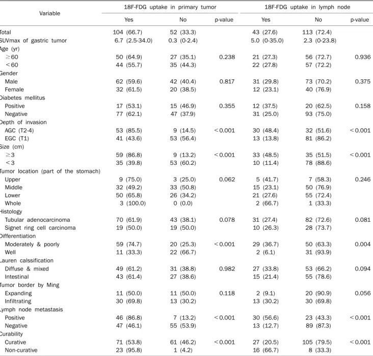
Table 2. Comparison between the Status of 18F-FDG Uptake and Primary Tumor Characteristics in the Gastric Cancers
Variable 18F-FDG uptake in primary tumor 18F-FDG uptake in lymph node
Yes No p-value Yes No p-value
Total 104 (66.7) 52 (33.3) 43 (27.6) 113 (72.4)
SUVmax of gastric tumor 6.7 (2.5-34.0) 0.3 (0-2.4) 5.0 (0-35.0) 2.3 (0-23.8)
Age (yr)
≥60 <60
50 (64.9) 44 (55.7)
27 (35.1) 35 (44.3)
0.238 21 (27.3) 22 (27.8)
56 (72.7) 57 (72.2)
0.936
Gender Male Female
62 (59.6) 32 (61.5)
42 (40.4) 20 (38.5)
0.817 31 (29.8) 12 (23.1)
73 (70.2) 40 (76.9)
0.375
Diabetes mellitus Positive Negative
17 (53.1) 77 (62.1)
15 (46.9) 47 (37.9)
0.355 12 (37.5) 31 (25.0)
20 (62.5) 93 (75.0)
0.158
Depth of invasion AGC (T2-4) EGC (T1)
53 (85.5) 41 (43.6)
9 (14.5) 53 (56.4)
<0.001 30 (48.4) 13 (13.8)
32 (51.6) 81 (86.2)
<0.001
Size (cm)
≥3
<3
59 (86.8) 35 (39.8)
9 (13.2) 53 (60.2)
<0.001 33 (48.5) 10 (11.4)
35 (51.5) 78 (88.6)
<0.001
Tumor location (part of the stomach) Upper
Middle Lower Whole
9 (75.0) 32 (49.2) 50 (65.8) 3 (100.0)
3 (25.0) 33 (50.8) 26 (34.2) 0 (0.0)
0.062 5 (41.7) 15 (23.1) 21 (27.6) 2 (66.7)
7 (58.3) 50 (76.9) 55 (72.4) 1 (33.3)
0.246
Histology
Tubular adenocarcinoma Signet ring cell carcinoma
70 (61.9) 19 (50.0)
43 (38.1) 19 (50.0)
0.078 31 (27.4) 10 (26.3)
82 (72.6) 28 (73.7)
0.081
Differentiation Moderately & poorly Well
59 (74.7) 11 (33.3)
20 (25.3) 22 (66.7)
<0.001 29 (36.7) 2 (6.1)
50 (63.3) 31 (93.9)
0.004
Lauren calssification Diffuse & mixed Intestinal
49 (61.2) 43 (61.4)
31 (38.8) 27 (38.6)
0.982 27 (33.8) 15 (21.4)
53 (66.2) 55 (78.6)
0.094
Tumor border by Ming Expanding
Infiltrating
11 (50.0) 30 (69.8)
11 (50.0) 13 (30.2)
0.118 2 (9.1) 13 (30.2)
20 (90.9) 30 (69.8)
0.056
Lymph node metastasis Positive
Negative
46 (86.8) 47 (46.1)
7 (13.2) 55 (53.9)
<0.001 30 (56.6) 13 (12.7)
23 (43.3) 89 (87.3)
<0.001
Curability Curative Non-curative
71 (53.8) 23 (95.8)
61 (46.2) 1 (4.2)
<0.001 27 (20.5) 16 (66.7)
105 (79.5) 8 (33.3)
<0.001
Values are presented as median (range) or n (%).
18F-FDG, 18F-fluorodeoxyglucose; AGC, advanced gastric cancer; EGC, early gastric cancer.
well-differentiated adenocarcinoma, 36.7% vs. 6.1%; p=0.004), and non-curative surgery (non-curative surgery vs. curative surgery, 66.7% vs. 20.5%; p<0.001) (Table 2).
In multivariate analysis, non-curative surgery was a sig- nificant independent predictor for 18F-FDG uptake in the pri- mary tumors (OR, 11.05; 95% CI, 1.10-111.08; p=0.041).
The median SUVmax of primary tumors in 24 patients who underwent non-curative surgery was 5.6 (range, 0-34.7) and in 132 patients with curative surgery 2.7 (0-21.7). Tumour
larger than 3 cm (OR, 7.39; 95% CI, 2.41-22.70; p<0.001) and lymph node metastasis (OR, 5.47; 95% CI, 2.05-14.64;
p=0.001) were also significant predictors of 18F-FDG uptake in the primary tumors. Tumour larger than 3 cm (OR, 3.15;
95% CI, 1.16-8.58; p=0.025) and lymph node metastasis (OR, 3.36; 95% CI, 1.23-9.14; p=0.018) showed significant association with 18F-FDG uptake in lymph node (Table 3).
Table 3. Significant Predictors of 18F-FDG Uptake by Multiple Regression Analysis
18F-FDG uptake in primary tumor 18F-FDG uptake in lymph node
OR 95% CI p-value OR 95% CI p-value
Depth of invasion T2-4 2.17 0.68-6.92 0.191 1.21 0.41-3.58 0.728
Size (cm) ≥3 7.39 2.41-22.70 <0.001 3.15 1.16-8.58 0.025
Differentiation Moderately & poorly 1.46 0.48-4.41 0.506 1.72 0.66-4.47 0.263
Lymph node metastasis Positive 5.47 2.05-14.64 0.001 3.36 1.23-9.14 0.018
Curability of operation Non-curative 11.05 1.10-111.08 0.041 2.27 0.75-6.88 0.147
18F-FDG, 18F-fluorodeoxyglucose.
Fig. 1. Receiver operator characteristics (ROC) curve of the maximum standardized uptake value (SUVmax) of primary tumor. (A) In ROC for detecting lymph node metastasis, an area under the curve of 0.795 was obtained (95% CI, 0.719-0.871; p<0.001). (B) In ROC for predicting non-curative surgery, area under the curve was 0.801 (95% CI, 0.722-0.879; p<0.001).
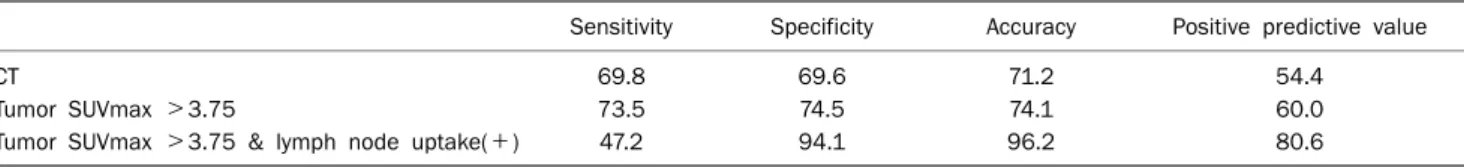
Table 4. Prediction of Lymph Node Metastasis in Patients Who Underwent Operation
Sensitivity Specificity Accuracy Positive predictive value
CT 69.8 69.6 71.2 54.4
Tumor SUVmax >3.75 73.5 74.5 74.1 60.0
Tumor SUVmax >3.75 & lymph node uptake(+) 47.2 94.1 96.2 80.6
SUVmax, the maximum standardized uptake value.
3. Sensitivity and specificity of 18F-FDG uptake for predicting regional lymph node metastasis and non-curative surgery
We assessed the sensitivity and specificity of the SUVmax in detecting lymph node metastasis (area under the curve [AUC] of 0.795, 95% CI, 0.719-0.871, p<0.001) or non-cura- tive surgery (AUC of 0.801, 95% CI, 0.722-0.879, p<0.001) using an ROC curve (Fig. 1). We obtained the most appro- priate SUVmax cutoffs for primary tumor 18F-FDG uptake for prediction of metastatic lymph node (SUVmax ≥3.75) and non-curative surgery (SUVmax ≥4.35).
Tables 4 and 5 show an overview of the calculated sensi- tivity, specificity, accuracy, and positive predictive value of SUVmax in detecting lymph node metastasis or non-curative surgery using SUV cutoffs for primary tumor. When the SUVmax of the primary tumor was greater than 3.75, the sen- sitivity and specificity of PET/CT with regard to the diagnosis of metastatic lymph node groups were 73.5% and 74.5%, re- spectively (Table 4). These values were higher than that ob- tained using CT scanning (the sensitivity, 69.8%; specificity, 69.6%). For patients with a SUVmax of 4.35 or more, non-cu- rative surgery was predicted with a sensitivity of 83.3% and a specificity of 78.0%. When the SUVmax was greater than
Table 5. Prediction of Non-curative Surgery in Patients Who Underwent Operation
Sensitivity Specificity Accuracy Positive predictive value
CT 45.8 93.9 86.5 57.9
Tumor SUVmax >4.35 83.3 78.0 78.8 40.8
Tumor SUVmax >4.35 & lymph node uptake(+) 58.8 91.6 86.5 56.0
SUVmax, the maximum standardized uptake value.
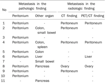
Table 6. Role of CT and 18F-FDG PET/CT in Detecting Distant Metastasis and Peritoneal Carcinomatosis
No
Metastasis in the pathologic finding
Metastasis in the radiologic finding Peritonium Other organ CT finding PET/CT finding
1 Peritonium - Peritoneum Peritoneum
2 Peritonium Colon, small bowel
Peritoneum -
3 Peritonium - - -
4 Peritonium Colon, spleen
Peritoneum Peritoneum
5 Peritonium Colon - -
6 Peritonium Liver - Liver
7 - Small bowel - -
8 Peritonium Pancreas Ovary Ovary
9 Peritonium - Peritoneum -
10 Peritonium - - -
11 - Pancreas - -
-, negative finding.
4.35 and the FDG uptake of lymph nodes was positive, non-curative surgery was predicted with a sensitivity of 58.8%, specificity of 91.6%, accuracy of 86.5%, and a pos- itive predictive value of 56.0%. Specificity, accuracy, and pos- itive predictive values were similar to those of CT scanning (specificity, 93.9%; accuracy, 86.5%; and positive predictive value, 57.9%). However, the sensitivity (58.8%) was higher than that obtained using CT scanning (45.8%) (Table 5).
4. 18F-FDG PET/CT in detecting distant metastasis and peritoneal carcinomatosis
In nine patients, pathological examination of peritoneal le- sions showed malignant cells. Seven patients had meta- static lesion of colon, small bowel, liver, or pancreas.
18F-FDG PET/CT had a low sensitivity (2/9, 22.2%) in detect- ing peritoneal carcinomatosis when compared with that of CT scanning (44.4%). In one case, a focal intense hyper- metabolic lesion in the liver was found by 18F-FDG PET/CT.
However, the lesion was not detected on CT (Table 6).
DISCUSSION
Accurate preoperative staging is critical for determining the most adequate therapy for gastric cancer, and CT scan- ning is the standard imaging modality for preoperative staging. However, as an anatomic imaging method, CT scan- ning is known to have a low sensitivity and specificity at N staging of the disease.1,9,10 In addition, several studies have shown that 18F-FDG PET had a lower sensitivity for detection of lymph node metastasis and had no definite role as a pre- operative imaging method in gastric cancer.11 This is related to the low resolution of 18F-FDG PET (5-7 mm), which does not allow easy differentiation of the perigastric lymph nodes from the primary tumor.6 In recent years, 18F-FDG-PET/CT has been reported to offer several potential advantages over the use of 18F-FDG-PET images or CT images alone.12 18F- FDG PET/CT provides more precise anatomical data along
with metabolic information. What is notable is that in a pre- vious PET study, the “sensitivity for metastasis to lymph no- des” was 17.6% to 46.4% (median 27.5%).1,4-6,13 In this study, we found that the sensitivity for metastasis to lymph nodes in our PET/CT study was rather high, recording 56.6%. Such difference can be explained by the advantage provided by the combination of PET and CT, and the technological develop- ment that has made high-resolution images with an accuracy of below 5 mm possible.
A challenging image obtained by the fusion of 18F-FDG PET and CT would have been regarded as having improved di- agnostic performance for lymph node staging. Many factors, including location, depth, size, macroscopic type, and histo- logical type of the AGC, affect the incidence and distribution of lymph node metastasis.14 Increased 18F-FDG uptake re- lated to rapidly growing and poorly differentiated tumor can reflect the regional lymph node metastasis.15,16 Using the de- rived criterion for lymph node metastasis of SUVmax 3.75 or greater, 18F-FDG PET/CT was predicted with a sensitivity of 73.5%, a specificity of 74.5%, an accuracy of 74.1%, and a
positive predictive value of 60.0%. These values are higher than those obtained with CT scanning (sensitivity, 69.8%;
specificity, 69.6%; accuracy, 71.2%; and positive predictive value, 54.4%).
Therefore, in our study, a metabolic imaging tool like 18F-FDG PET/CT has a superiority over CT in the preoperative N staging. Our results in patients with gastric cancer show that preoperative 18F-FDG PET/CT could provide objective information for use in making decisions regarding treatment strategies such as extended lymph node dissection.
We focused on the prediction of surgical findings by the re- sults of preoperative 18F-FDG PET/CT. The mean SUVmax of the primary tumor in patients who underwent non-curative surgery was significantly higher than that of patients who un- derwent curative surgery. When we defined a mean primary tumor SUVmax of greater than 4.35 and positive uptake of 18F-FDG in lymph nodes as cutoff values for prediction of non-curative surgery, the specificity, accuracy, and positive predictive values were similar to those of CT scanning; how- ever, the sensitivity was higher than that of CT scanning (58.8% vs. 45.8%). Therefore, the SUVmax of the primary tu- mor might be a predictive factor for non-curative surgery and a tool for deciding on the optimal surgical strategy.
In our study, 18F-FDG PET/CT had a low sensitivity (22.2%) in detecting peritoneal carcinomatosis. Pathological exami- nation of peritoneal lesions showed extensive fibrosis around relatively few malignant cells, which could be an explanation for the low 18F-FDG PET/CT sensitivity. The small size of the peritoneal lesions (<5 mm) could be another reason for the low detection rate. Little is known about the role of 18F-FDG PET/CT in detection of distant metastasis. One series re- ported a sensitivity and specificity of 85% and 74% for de- tection of liver metastasis; 67% and 88% for lung metastasis;
24% and 76% for ascites; 4% and 100% for pleural carcino- matosis, and 30% and 82% for bone.17
PET with 18F-FDG is useful for detection of malignancy.
However, benign processes can also result in false-positive 18F-FDG uptake. Physiologic 18F-FDG uptake can be chal- lenging, particularly in the abdomen and pelvis because of multiple structures with variable physiologic 18F-FDG up- take (e.g., bowel) and because 18F-FDG is excreted through the urinary collecting system.18,19 We had a patient who was without ovarian metastases but showed 18F-FDG uptake on PET/CT. Ames et al.20 presented a case in which 18F-FDG up-
take in a normal ovary was misinterpreted initially as a meta- static lesion. Change in the ovary associated with ovulation is the likely explanation for the increased 18F-FDG accumu- lation in the ovary. Lerman et al.21 observed increased 18F-FDG uptake in the ovaries of 21 of 112 premenopausal patients without known gynecologic malignancy using PET/CT. Fifteen of these patients were imaged near the time of ovulation as determined by the presence of functional ovarian cysts.
Our study has a limitation. The number of enrolled patients might have been too small to confirm the clinical validity of 18F-FDG PET/CT for gastric cancer. Therefore, studies with larger populations should be planned in order to confirm the correlation between preoperative 18F-FDG PET/CT and sur- gical findings.
In conclusion, it is difficult to assert that PET/CT scanning is far superior to CT scanning in prediction of non-curative sur- gery or lymph node metastasis. However, we did find that a higher SUVmax of primary gastric cancer is correlated with both non-curative surgery and lymph node metastasis.
Therefore, information from the preoperative 18F-FDG PET/CT might be helpful to physicians in deciding the extent of lymphadenectomy and optimal treatment modalities.
REFERENCES
1. Kim SK, Kang KW, Lee JS, et al. Assessment of lymph node meta- stases using 18F-FDG PET in patients with advanced gastric cancer. Eur J Nucl Med Mol Imaging 2006;33:148-155.
2. Yeung HW, Macapinlac H, Karpeh M, Finn RD, Larson SM.
Accuracy of FDG-PET in gastric cancer. Preliminary experience.
Clin Positron Imaging 1998;1:213-221.
3. Stahl A, Ott K, Weber WA, et al. FDG PET imaging of locally ad- vanced gastric carcinomas: correlation with endoscopic and histopathological findings. Eur J Nucl Med Mol Imaging 2003;
30:288-295.
4. Mochiki E, Kuwano H, Katoh H, Asao T, Oriuchi N, Endo K.
Evaluation of 18F-2-deoxy-2-fluoro-D-glucose positron emis- sion tomography for gastric cancer. World J Surg 2004;28:
247-253.
5. Chen J, Cheong JH, Yun MJ, et al. Improvement in preoperative staging of gastric adenocarcinoma with positron emission tomography. Cancer 2005;103:2383-2390.
6. Yun M, Lim JS, Noh SH, et al. Lymph node staging of gastric can- cer using (18)F-FDG PET: a comparison study with CT. J Nucl Med 2005;46:1582-1588.
7. Ajani J, Bekaii-Saab T, D'Amico TA, et al. Gastric cancer clinical practice guidelines. J Natl Compr Canc Netw 2006;4:350-366.
8. Hur H, Kim SH, Kim W, Song KY, Park CH, Jeon HM. The efficacy
of preoperative PET/CT for prediction of curability in surgery for locally advanced gastric carcinoma. World J Surg Oncol 2010;8:86.
9. Bentrem D, Gerdes H, Tang L, Brennan M, Coit D. Clinical correla- tion of endoscopic ultrasonography with pathologic stage and outcome in patients undergoing curative resection for gastric cancer. Ann Surg Oncol 2007;14:1853-1859.
10. Burke EC, Karpeh MS, Conlon KC, Brennan MF. Laparoscopy in the management of gastric adenocarcinoma. Ann Surg 1997;
225:262-267.
11. Dassen AE, Lips DJ, Hoekstra CJ, Pruijt JF, Bosscha K. FDG-PET has no definite role in preoperative imaging in gastric cancer. Eur J Surg Oncol 2009;35:449-455.
12. Bombardieri E, Aktolun C, Baum RP, et al. FDG-PET: procedure guidelines for tumour imaging. Eur J Nucl Med Mol Imaging 2003;30:BP115-BP124.
13. Mukai K, Ishida Y, Okajima K, Isozaki H, Morimoto T, Nishiyama S. Usefulness of preoperative FDG-PET for detection of gastric cancer. Gastric Cancer 2006;9:192-196.
14. Akagi T, Shiraishi N, Kitano S. Lymph node metastasis of gastric cancer. Cancers (Basel) 2011;3:2141-2159.
15. Duhaylongsod FG, Lowe VJ, Patz EF Jr, Vaughn AL, Coleman RE, Wolfe WG. Lung tumor growth correlates with glucose metabo-
lism measured by fluoride-18 fluorodeoxyglucose positron emission tomography. Ann Thorac Surg 1995;60:1348-1352.
16. Adler LP, Blair HF, Williams RP, et al. Grading liposarcomas with PET using [18F]FDG. J Comput Assist Tomogr 1990;14:960-962.
17. Yoshioka T, Yamaguchi K, Kubota K, et al. Evaluation of 18F-FDG PET in patients with advanced, metastatic, or recurrent gastric cancer. J Nucl Med 2003;44:690-699.
18. Shreve PD, Anzai Y, Wahl RL. Pitfalls in oncologic diagnosis with FDG PET imaging: physiologic and benign variants. Radiograph- ics 1999;19:61-77.
19. Cook GJ, Maisey MN, Fogelman I. Normal variants, artefacts and interpretative pitfalls in PET imaging with 18-fluoro-2-deoxy- glucose and carbon-11 methionine. Eur J Nucl Med 1999;26:
1363-1378.
20. Ames J, Blodgett T, Meltzer C. 18F-FDG uptake in an ovary con- taining a hemorrhagic corpus luteal cyst: false-positive PET/CT in a patient with cervical carcinoma. AJR Am J Roentgenol 2005;185:1057-1059.
21. Lerman H, Metser U, Grisaru D, Fishman A, Lievshitz G, Even-Sapir E. Normal and abnormal 18F-FDG endometrial and ovarian uptake in pre- and postmenopausal patients: assess- ment by PET/CT. J Nucl Med 2004;45:266-271.