종골 관절내 골절의 수술적 치료
- 임상 결과와 관련된 예후인자에 관한 분석 -
성균관대학교 의과대학 삼성서울병원 정형외과학 교실 정성수・서재곤・박윤수・이종서・황태규
= Abstract =
Intra-articular Fracture of the Calcaneus
: Analysis of Result of Operative Treatment and Prognostic Factor
Sung-Soo Chung, M.D., Jai-Gon Seo, M.D., Youn-Soo Park, M.D., Chong-Suh Lee, M.D., Tae-Kyu Hwang, M.D.
Department of Orthopaedic Surgery, Samsung Medical Center, Sung Kyun Kwan University, Seoul, Korea
The calcaneus is the most commonly fractured tarsal bone, but the appropriate care of calcaneal fracture continue to be an unsolved dilemma. As technology in imaging has improved, operative treatment is more suggested.
The purpose of this study is to evaluate the results of operative treatment in intra-articular calcaneal fracture and to analyse the results in accordance with various prognostic factors.
We analysed retrospectively 13 patients, 17 intra-articular calcaneal fractures undergone operative treatment. Mean follow-up period was 27 months (range:13 ~ 44 months). There were 11 males and 2 females with 41 year old mean age (range:18 ~ 63 years old).
Clinical assessment used the modified Creighton-Nebraska health foundation assessment sheet for fracture of the calcaneus.
We obtained excellent result in 7 cases (41.2%), good in 2 cases (11.8%), fair in 7 cases (41.2%) and poor in 1 case (5.8%). Clinically age and body weight, radiologically B..
ohler angle, Vol.11, No.4, October, 1998
※통신저자: 정성수
서울특별시강남구일원동5 0번지( 1 3 5 - 2 3 0 ) 삼성서울병원정형외과
Tel : (02) 3410 - 0385 Fax : (02) 3410 - 0388
* 본논문의요지는 1 9 9 8년 4월대한골절학회제 2 4차춘계학술대회에서구연되었음.
* 본논문은 9 8년삼성서울병원임상연구비보조로이루어졌음.
서 론
종골은족근골중가장흔하게 골절되는 것으로관 절내 골절 환자에 대한 치료에는 많은 논란이 있다.
최근수술적으로치료하여더 좋은결과을보고하는 저자들이있으나예후에영향을미치는인자에대해 서는여전히상반된의견들이제시되고있고, 논란이 많은것으로알려져있다. 이에저자들은수술적으로 치료한종골관절내골절환자의치료결과를후향적 으로조사하여그결과에미치는예후인자에대하여 알아보고자하였다.
연구대상 및 방법
1 9 9 4년 1 1월부터 1 9 9 7년 4월까지수술적으로치료 하였던 종골 관절내 골절 환자중 1년 이상 추시가능 한총 1 3명 1 7례를대상으로하였다.
1. 성별및연령분포
남녀 비는 남자가 1 1명 여자가 2명이었고 연령분 포는 최저 1 8세부터 최고 6 3세까지평균연령이 4 1세 였다.
2. 골절의원인
전체 1 3명( 1 7례)중 7 6 . 9 %인 1 0명( 1 4례)에서 추락사 고가 원인이었으며, 23.1%인 3명( 3례)에서는 교통사 고가원인이었다.
3. 골절의분류
골절의 분류에 있어서 저자들은 족관절과 종골의 전후방및측면의단순방사선사진과종골의축면방 사선사진을이용하여기존의 분류법중 널리이용되 는 E s s e x - L o p r e s t i5 )분류의 설상형(tongue type)과 관절 함몰형(joint depression type)에 거골하 관절면의 분쇄 가 있는 경우를W a t s o n - J o n e s2 9 )분류를 참고하여 분쇄 형(comminution type)이라하여 추가하여 새로이분류 하였다.
전체 1 7례중 설상형은6례, 관절함몰형은7례였고, 분쇄형은4례였다.
4. 동반손상
척추를 포함한 다른 부위의 골절이 5례였고 고관 절탈구가 1례, 기타다른장기의손상은 3례(간열상, 신장좌상, 혈흉)였다.
5. 수술방법및재활
수술은 설상형 골절의 경우 6례 모두 E s s e x - L o p r e s t i5 )의 경피적 중축 금속정 고정술을 시행하였 고관절함몰형골절의경우 7례중 6례는관혈적정복 및 스테이플을 이용한 내고정술을, 1 례는 E s s e x - L o p r e s t i의경피적중축금속정고정술을 시행하였다.
그리고분쇄형골절의경우 4례중 3례는관혈적정복 및 스테이플을 이용한 내고정술을, 1 례는 E s s e x - L o p r e s t i의경피적중축금속정고정술을시행하였다 . 골이식은관절함몰형골절의3례, 분쇄형골절의 3례 에서시행하였다.
술후 체중부하는 단순 방사선 사진에서 골유합을 fibulo-calcaneal distance and subtalar joint discrepancy are related to the prognosis of intra- articular calcaneal fracture following operative treatment. Postoperative complications are limping (2 case), heel pain (3 cases), hump bump of calcaneus (1 case) and subtalar arthritis (1case).
In conclusion, on the basis of our results, there is a relationship between anatomical abnormalities of the heel and a poor clinical outcome. Therefore, in operative treatment of intra- articular fracture of calcaneus, we recommend anatomical reduction, if possible, not only of the subtalar joint but also of the B..ohler angle and fibulo-calcaneal distance.
Key Words : Calcaneus, Intra-articular fracture, Operative treatment, Prognostic factor
확인한 6주에서 1 2주 사이에 시작하였다. 술후 추시 기간은 최저 1년 1개월에서최고 3년 8개월로평균 2 년 3개월이었다.
6. 예후판정
예후 판정은 단순 방사선 소견 및 임상 소견을 토 대로 하였으며 단순 방사선 소견에는 B..o h l e r2 )각, c r u c i a l1 0 )각, 종골의 높이와 너비, 비골-종골간 거리 그리고 거골하관절부조화를포함시켰으며술후추 시관찰에서거골하관절염의여부도포함하였다.
거골하 관절의 부조화의 경우 요수근 관절( r a d i o - carpal joint)의 조화도( c o n g r u i t y )에 적용되는 K n i r k와 J u p i t e r1 4 )의분류법을적용하였다(Table 1).
추적 관찰후 임상적 결과는C r e i g h t o n - N e b r a s k a
health foundation의종골골절평가표를수정하여적용
Table 1. Scoring system for posterior subtalar j o i n t *
Articular incongruity
Grade (mm step-off) Arthritic changes
0 0 to 1 None
1 1 to 2 Slight joint-space narrowing 2 2 to 3 Marked joint space narrowing
Osteophyte formation
3 > 3 Bone-on-bone
Osteophyte formation Cyst formation
* The criteria of Knirk-Jupiter for congruity of radiocarpal joint14).
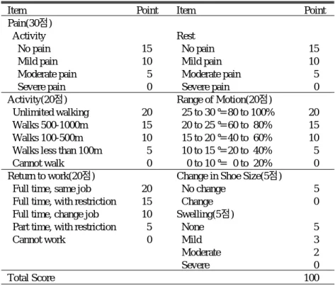
Table 2. Assessment sheet for fracture of the calcaneus*
Item Point Item Point
Pain(30점)
Activity Rest
No pain 15 No pain 15
Mild pain 10 Mild pain 10
Moderate pain 5 Moderate pain 5
Severe pain 0 Severe pain 0
Activity(20점) Range of Motion(20점)
Unlimited walking 20 25 to 30°= 80 to 100% 20
Walks 500-1000m 15 20 to 25°= 60 to 80% 15
Walks 100-500m 10 15 to 20°= 40 to 60% 10
Walks less than 100m 5 10 to 15°= 20 to 40% 5
Cannot walk 0 0 to 10°= 0 to 20% 0
Return to work(20점) Change in Shoe Size(5점)
Full time, same job 20 No change 5
Full time, with restriction 15 Change 0
Full time, change job 10 Swelling(5점)
Part time, with restriction 5 None 5
Cannot work 0 Mild 3
Moderate 2
Severe 0
Total Score 100
* Modification from Creighton-Nebraska health foundation assessment sheet for fracture of the calcaneus1).
A score of 90 to 100 points in judged to be an excellent result ; 80 to 89 points, a good result ; 65 to 79 points, a fair result ; and 64 points or fewer, a poor result.
하였다(Table 2).
결 과
장기추시를통한임상적결과판정에서전체 1 7례 중우수( E x c e l l e n t )가 7례로 4 1 . 2 %였고양호( G o o d )는 2 례로 11.8%, 보통( F a i r )은 7례로 4 1 . 2 %였으며 불량 ( P o o r )은 1례로 5 . 8 %였다.
그중골절분류상설상형은 5례중 3례에서, 관절함 몰형은 7례중 4례에서그리고 분쇄형은 5례중 2례에 서양호이상의결과를보였다.
수상당시의평균연령은양호이상의군에서3 1 . 4세 로 보통이하의 군의 평균연령인 4 3 . 6세 보다 작았으 며(p<0.05), 평균체중도6 0 . 6 k g으로 6 5 . 4 k g의보통이하 의군보다작았다(p<0.05) (Table 3).
최종추시의 Bohler 각은 양호이상의 군에서 2 6 . 7도..
로 보통이하의 군의 B..ohler 각 1 1 . 4도 보다 컸으며 (p<0.05), crucial 각은양호이상의군에서 1 1 7 . 5도로보 통이하의 군의 1 1 8 . 1도와 유의한 차이는 없었다
Table 3. Correlation between clinical result and age and body weight.
>80 <79
Age(years) 31.4 43.6*
Body weight(kg) 60.6 65.4*
>80 : Excellent or Good *p<0.05
<79 : Fair or Poor
Table 4. Correlation between clinical result and radiological measurements.
Radiological finding >80 <79 B..
ohler angle 26.7° 11.4°*
crucial angle 117.5° 118.1°
Calcaneal height(mm) 37.0 36.8
Calcaneal width(mm) 38.6 38.5
Fibulo-calcaneal distance(mm) 17.4 13.0*
Subtalar joint discrepancy(Grade) 0.8 2.0*
>80 : Excellent or Good *p<0.05
<79 : Fair or Poor
Fig 1. 43year old male patient sustained intra-articular calcaneal fracture of left foot by fall down from 2m height.
Fig A. Preoperative X-ray shows tongue type intra- articular calcaneal fracture with -20oB..ohler angle and 140ocrucial angle. Subtalar joint discrepancy is not seen..
Fig B. Immediate postoperative X-ray after closed reduction and axial fixation with 3.2mm steinmann Fig C.Postoperative 13months X-ray , Bpin. ..
ohler angle is 16o, crucial angle is 125oand Subtalar joint arthritis is not seen.
A C
B
(Table 4) (Fig 1-A,B,C).
종골 높이(calcaneal height)와너비(calcaneal width)는 양호이상의군에서각각 3 7 . 0 m m와 3 8 . 6 m m로보통이 하의 군의각각 36.8mm, 38.5mm와 유의한 차이는없 었다(Table 4).
비골-종골간거리(fibulo-calcaneal distance)는양호이 상의군에서 1 7 . 4 m m로보통이하의군의 1 3 . 0 m m보다 컸으며(p<0.05), 거골하 관절 부조화(subtalar joint d i s c r e p a n c y )는 양호이상의 군에서 Grade 0.8로 G r a d e
2 . 0의보통이하의군보다작았다(p<0.05) (Table 4).
술후합병증으로는종골부동통이 3례, 파행이2례 였으며, 1례에서 거골하 관절염이 관찰되었다(Fig 2- A,B,C,D). 그리고종골외측면의골돌출이설상형골 절을 Essex-Lopresti 방법으로치료한환자 1례에서발 생하여지속적인통증을보였으며, 돌출된골의제거 후통증의감소를보였다.
A
C D
B
Fig 2. 31 year old male patient sustained intra-articular calcaneal fracture of both feet by fall down from 4m height.
Fig A. Preoperative X-ray of left foot shows joint depression type intra-articular calcaneal fracture with 21o B..
ohler angle and 140ocrucial angle. Subtalar joint discrepancy is Grdae 2.
Fig B. Postoperative 17months X-ray after open reduction and internal fixation with staple. B..
ohler angle is 34o, crucial angle is 125oand Subtalar joint arthritis is not seen.
Fig C.Preoperative X-ray of right foot shows comminution type intra-articular calcaneal fracture with 9o B..
ohler angle and 130ocrucial angle. Subtalar joint discrepancy is Grade 3.
Fig D.Postoperative 17months X-ray after open reduction and internal fixation with staple and bone graft.
B..
ohler angle is 30o, crucial angle is 115oand Subtalar joint arthritis is seen.
고 찰
족부 골절 중 가장 많은 종골 골절은 해부학적 복 잡성과골절양상의다양성으로 인해치료방법을결 정하기어려운골절중하나이다.
종골골절은거골하관절의 침범여부에따라크게 관절외골절과 관절내골절로분류하며관절내골절 도 양상에 따라 세부적으로 분류된다. 단순 방사선 촬영에 의한 종골 관절내 골절의 분류로는 E s s e x - Lopresti, Rowe 및 Paley 등의방법이있으나골절의치 료 방향 및 최종 예후와는 일치하지 않는다
5 , 1 1 , 2 1 , 2 3 , 2 5 , 2 6 ). 저자들의 경우에는 E s s e x - L o p r e s t i5 )의 이 차골절선에따른분류 , 즉설상형(tongue type)과관절 함몰형(joint depression type)에 거골하 후관절면의 분 쇄가있는경우를 W a t s o n - J o n e s29) 분류에따라분쇄형 (comminution type)이라 하여 추가하여 분류하였는데 이는거골하후관절의분쇄가심할수록수술적으로 정확한해부학적인정복이어려워그예후가좋지않 을것으로생각하였기때문이다.
관절내종골골절은치료에있어다양한방법이소 개되고있으나이를요약하면정복하지않고조기운 동 시키는 방법1 6 , 1 9 , 2 4 ), 도수 정복후 고정하는 방법
5 , 1 3 ), 관혈적 정복후 골이식 또는 내고정하는 방법
3 , 1 8 , 2 2 ), 일차적인 관절 고정술6 , 1 2 , 2 0 )을 실시하는 경우
등이 있다. 그러나 치료 방법에 따른결과는항상 일 치하는 것이 아니라고 여러 저자들은 말하고 있다.
일반적으로해부학적정복및 견고한고정과관절의 조기운동이좋은 결과의가능성을높인다는원칙과 관절내의 전위된 골편은 비수술적 방법으로는 결코 정복될수없으며따라서정상적인기능으로회복될 가능성이 희박하다는 점에서 적극적인 수술적 치료 를선호하는경향이있다.
S a l a m a24) 등에 따르면 골절의 정복이나 고정 없이 시행한관절의조기능동적운동의장점은 합병증의 발생이적고치료기간이단축되며수술에따른부작 용과석고고정에따른불편이없다고하였다 .
K i n g1 3 )에따르면 E s s e x - L o p r e s t i의경피적중축금속 정 고정술은 Bo h l e r각을 보존시켜 후방 관절면의 조..
화를회복시킴으로써좋은치료효과를거둘수있다 고 하였으며, Maxfield1 5 )와 H a r d i n g8 )등 여러 저자들
2 1 , 2 6 )은종골관절내골절을다른관절의 관절내골절
과 같이 치료하여 정확한 해부학적 정복 및 조기 관 절운동에주안점을두었고 H a r r i s8 )는종골관절내골 절은거골하관절이강직되고관절염에따른동통을 일으키는 요인이므로 조기 거골하 관절고정술을 주 장하였으며, Thompson2 8 )은 종입방 관절과 거골주상 골관절도같이고정하는삼관절고정술이좋다고하 였다.
저자들의 경우수술은관혈적정복및내고정하였 던 경우와 Essex-Lopresti 방법으로 정복 후 S t e i n m a n n p i n을 이용하여 경피적고정하였던 경우가있었으며 골이식은관절함몰형골절의3례, 분쇄형골절의 3례 에서시행하였다.
H a m m e s f a h r와 F l e m i n g7 )은 설상형이 관절함몰형에 비해 양호한 결과를 보인다고 하였고, Paley와 H a l l1 7 ) 은분쇄정도가심할수록그결과가불량하다고하였 다. 저자들의 경우에는 종골 관절내 골절의 수술적 치료후 추시 관찰한 결과 설상형과 관절함몰형간의 예후에는유의한차이를발견할수없었고 , 분쇄형의 경우설상형과관절함몰형에비해불량한예후를보 였다.
M a x f i e l d와 M c D e r m o t t1 5 )에 의하면 관혈적 정복으 로치료한종골관절내골절의최종결과는단순방사 선사진에나타난골절의형태와는관계없다고하였 고 종골의 측면 사진상 측정이 가능한 B..o h l e r각과 c r u c i a l각은 손상의 정도를 평가할 수는 있지만 관절 내 침범 정도와 위치를 정확하게 평가할 수 없다고 하였다. 저자들의경우최종추시결과 Bo h l e r각의크..
기가 작을 수록 불만족스러운 결과를 보였으며, c r u c i a l각의 크기는 결과에 미치는 영향이 크지 않을 것으로보였다.
P a l e y와 H a l l1 7 )은 비골-종골간 거리가 작을수록 그 리고종골의너비가클수록최종결과가불량하다고 하였다. 저자들의경우에는비골-종골간거리가클수 록양호한것으로나타났고종골의너비에따른결과 의차이는크지않은것으로생각되었다.
C r o s b y와 F i t z g i b b o n4 )에의하면종골관절내골절에 서가장중요한요인은종골의후방거골하관절면의 손상정도라 하였고 P a l e y와 H a l l1 7 )도 거골화 관절면 의부조화와관절염의정도가심할수록예후가불량 한것으로보고하였다. 저자들의경우에도수상당시 거골화관절면의부조하가많을수록불만족스런결
과를보였으며최종추시방사선촬영상후방관절면 에관절염이있는환자에서결과가더불량한것으로 나타났다.
결 론
종골 골절시 수술적 치료로 종골의 후방관절면, Bo h l e r각, 비골-종골간 거리 등의 모양을 해부학적으..
로복원할수있으면후방관절의관절염의발생이방 지될 수 있고 치료의 결과도 우수할 것으로 생각된 다. 따라서종골골절의치료에서심한분쇄형골절이 아닌경우에는종골모양특히후방관절면의해부학 적인복원을위해적극적인수술적치료를하는것이 좋은결과를가져올수있다고생각한다.
REFERENCES
1) 송광순, 강형철, 민병우, 손국진: 전산화 단층촬 영을이용한종골관절내골절의분류및 치료결 과 판정, 대한정형외과학회지 , 31 : 606-614, 1 9 9 6 .
2) B..
ohler L : Diagnosis, pathology and treatment of fracture of the os calcis. J Bone Joint Surg, 13 : 75- 89, 1931.
3) Burdeaux BD : Reduction of calcaneal fracture by the McReynolds medial approach technique and its experimental basis. Clin Orthop, 7-103, 1983.
4) Crosby LA and Fitzgibbon T : Computerized tomography scanning of acute intra-articular fracture of the calcaneus. J Bone Joint Surg, 72-A : 852-859, 1 9 9 0 .
5) Essex-Lopresti P : Mechanism, reduction technique.
and results in fracture of the os calcis. British J Surg, 39 : 395-419, 1985.
6) Gallie WE : Subtalar arthrodesis in fractures of the os calcis. J Bone Joint Surg, 25 : 731-736, 1943.
7) Hammesfahr, R. and Fleming, L.L. : Calcaneal fractures : a good prognosis. Foot and Ankle, 2 : 161- 171, 1981.
8) Harding, D. and Waddel, J.P. : Open reduction in depressed fractures of the os calcis. Clin. Orthop, 199 : 124-131, 1985.
9) Harris, R.I. : Fractures of os calcis : Treatment by early subtalar arthrodesis. Clin Orthop, 30 : 100-110, 1 9 6 3 .
10) Harty M : Anatomic consideration in injuries of the calcaneus. Orthop Clin N Am, 4 : 179-183, 1973.
11) Heckman JD : Fracture and dislocations of the foot.
In Fractures in adult. Rockwod and green. 3rd ed.
Philadelphia, J B Lippincott Co : 2103-2131, 1991.
12) Kalamchi A and Evans JG : Posterior subtalar fusion : A preliminary report on a modified Gallie’s procedure. J Bone Joint Surg, 59-B : 287-289, 1977.
13) King RE : Axial pin fixation of fractures of the os calcis (Method of Essex-Lopresti). Orthop Clin N A m , 4 : 185-188, 1973.
14) Knirk JL, Jupiter JB : Intra-articular fractures of the distal end of the radius in young adults. J Bone Joint Surg, 68-A : 647-659, 1986.
15) Maxfield, J.E. and McDermott, F.J. : Experiences with palmar open reduction of fractures of the calcaneus. J Bone Joint Surg, 37-A : 99-106, 1955.
16) O’Connel F, Mital MA and Rowe CR : Evaluation of mothern management of fractures of the os calcis.
Clin Orthop, 83 : 214-223,1972.
1 7 ) Paley D and Hall : Intra-articular fractures of the calcaneus. J Bone Joint Surg, 75-A : 342-354, 1993.
18) Palmer I : The mechanism and treatment of fractures of the calcaneus.Open reduction with the use of cancellous grafts. J Bone Joint Surg, 30-A : 2-8, 1948.
19) Park JC II : Tthe nonreductive treatment for fractures of the os calcis Ortho Clin N Am, 4 : 193- 195, 1973.
20) Pennel GF and Yadav MP : Operative treatment of comminuted fractures of the os calcis. Orthop Clin N A m, 4 : 197-221, 1973.
21) Ross ERS and Peddy P : Current controversies in intra-articular calcaneal fractures. Inter J of Orthop T r a u m a, 4 : 52-56, 1994.
22) Ross SDK and Sowerby MRR : The operative treatment of fractures of the os calcis. Clin Orthop, 199 : 132-143, 1985.
23) Rowe CR, Sakellarides H, Freeman P and Sorbie C : Fractures of the os calcis - A long-term follow-up study of one hundred forty six patient. J A M A, 184 : 920-923, 1963.
24) Salama R, Benamara A and Weissman SL : Functional treatment of intra-articular fractures of the calcaneus. Clin Orthop, 83 : 214-223, 1972.
25) Sanders R, Hansen ST and McReynolds IS : Trauma to the calcaneus and its tendon. In Disorders of the foot and ankle. Jahss, M.H.(ed). Philadelphia, W B Saunders Co : 2333-2338, 1991.
26) Smith RW and Staple TW : Computerized tomography(CT) scanning technique for the hindfoot. Clin Orthop, 177 : 34-38, 1983.
27) Stephenson, A.R. : Treatment of displaced intra- articular fractures of the calcaneus using medial and lateral approaches, internal fixation, and early motion. J Bone Joint Surg, 69-A : 115-130, 1987.
28) Thompson, K.R. : Treatment of comminuted fractures of the calcaneus by triple arthrodesis.
Orthop Clin N Am, 4 : 189-191, 1973.
29) Wilson JN, ed. : Watson-Jones Fractures and Joint I n j u r i e s . 5th ed. Edinburgh, London and New York, Churchill Livingstone, 2 : 1157-1175, 1976.