282
Correspondence to: Byoung Yoon Ryu, Department of Surgery, College of Medicine, Hallym University, 153, Gyo-dong, Chuncheon 200- 704, Korea. Tel: 033-252-9970, Fax: 033-243-6413, E-mail: byryu@
hallym.or.kr
Received January 21, 2009, Accepted April 3, 2009
Multilevel Duodenal Injury after Blunt Trauma
Department of Surgery, College of Medicine, Hallym University, Chuncheon, Korea
Jeong Hee Han, M.D., Sung Il Hong, M.D., Hae Sung Kim, M.D.,
Byoung Yoon Ryu, M.D., Hong Ki Kim, M.D.
Duodenal trauma is an uncommon injury associated with significant mortality and morbidity. Upper gastrointestinal radiological studies and computed tomography may lead to the diagnosis of blunt duodenal trauma.
Exploratory laparotomy remains as the ultimate diagnostic test if a high suspicion of duodenal injury continues even in the face of absent or equivocal radiographic signs. The majority of duodenal injuries may be managed by simple repair of the injured site. More complicated injuries require more sophisticated techniques. Here, we report a case of multilevel blunt duodenal injury successfully managed with duodenal diverticulization, Roux-en-Y gastrojejunostomy and catheter duodenostomy. (J Korean Surg Soc 2009;77:282-286)
Key Words: Duodenal injury, Duodenal diverticulization, Roux-en-Y gastrojejunostomy
INTRODUCTION
Injuries of the duodenum are uncommon, although not rare, because of its retroperitoneal location. Duodenal injuries represent approximately 3% to 5% of all abdominal injuries.(1) Penetrating trauma accounts for 78% of all duodenal injuries, whereas blunt trauma accounts for 22%.(1) Mortality and morbidity from duodenal injuries are related to the causative agent, location, presence of associated injuries, and type of surgical repair. However, the interval from injury to operation plays the most significant role in determining the incidence of morbidity and mortality. Lucas and Ledgerwood(2) reported a mortality rate of 40% in patients who were not operated within the first 24 hours after injury, in contrast to a mortality rate of 11% among those patients operated less than 24 hours after the injury. The incidence of associated injuries ranges from 30% to 87% in several series, with the liver being the most common associated organ injured
(17%). In penetrating trauma, associated injuries to the inferior vena cava (35%) and pancreas (10∼53%) are predominant. Over the last two decades, there has been a decline in the overall mortality in these injuries with an overall current mortality between 10.5% and 14%.
Blunt duodenal injury at more than one site, discussed in this report, can be a rare surgical entity. In our review over the past 20 years, a few reports have specifically described blunt duodenal injury at multiple levels. The management options for duodenal injuries are several methods and are determined by the injury level, the extent of injury and the patient’s overall condition. We report a case of multilevel blunt duodenal injury managed success- fully with duodenal diverticulization, Roux-en-Y gastrojeju- nostomy and catheter duodenostomy.
CASE REPORT
A 21-year-old man was crushed by a push cart during working. His chief complaint was abdominal pain and he was hemodynamically stable, with a pulse of 72 beats/min and a blood pressure of 120/80 mmHg. Diffuse abdominal tenderness and rebound tenderness was revealed in physical examination. Blood tests showed increased white cell count
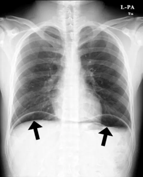
Fig. 1. Thoracic radiograph shows pneumoperitoneum in the bilateral subphrenic areas (arrows).
Fig. 2. Abdomen computed tomography scan shows the findings of pneumoperitoneum (arrows).
Fig. 3. Duodenal disruption (75% of the circumference, grade 3) is present on the first part of duodenum (arrow).
Fig. 4. Duodenal perforation with multiple injury. Duodenal disruption (75% of the circumference, grade 3) is present on the first part of duodenum. The serosa of the third and fourth part of duodenum is detached from pancreas and the segments devasculized.
(24.3×10³/L) and level of hemoglobin was 15 g/dl. The level of serum amylase(s) and lipase(s) were elevated (156 and 959 IU/L). Thoracic radiographs and abdominal computed tomography showed the findings of pneumo- peritoneum (Fig. 1, 2). We take diagnostic exploratory laparotomy. On operation field, 1st part of duodenum was perforated; grade III, about 75% laceration of the whole circumference (Fig. 3). The serosa of duodenal 3rd & 4th parts was detached from pancreas and the segments were devasculized (Fig. 4).
Duodenal resection of 1st part was performed. Then
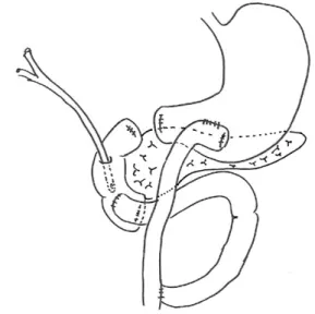
segmental resection of devasculized duodenal 3rd & 4th parts was performed. Both ends of diverticulized duodenal segment were closed. Side to side Roux-en-Y gastrojeju- nostomy and side to side duodenojejunostomy using afferent jejunal loop were performed. Catheter duodeno- stomy was performed for duodenal decompression (Fig. 5).
The patient recovered without evidence of leak and the
Fig. 5. Operative management. Duodenal diverticulization is performed with the duodenal resection of the 1st, 3rd, 4th parts. Side to side Roux-en-Y gastrojejunostomy was per- formed and side-to-side duodenojejunostomy using afferent jejunal loop was performed. Catheter duodenostomy using foley catheter was performed.
complications. The patient was discharged after post operative 14 days.
DISCUSSION
Blunt abdominal injuries such as direct blow to the epigastrium result bowel trauma, and they account for 25%
of all duodenal traumas, while the remaining 75% of duodenal trauma are due to penetrating trauma.(1,3) Isolated blunt duodenal injuries are very rare since they are commonly associated with lesions of other abdominal or thoracic organs, including major vessels.(4) Considering the deep and relatively protected anatomical site of the duodenum, it is likely that when the situation of the trauma is able to injury the duodenum, other intraab- dominal organs are usually involved. Thus, if any traumatic lesion of the duodenum is detected, injuries to other structures have to be ruled out.(1,3-5)
The diagnosis of duodenal injury is difficult unless a high index of suspicion is maintained. While the history and clinical findings of steering wheel or seat belt marks on the anterior abdominal wall may make the clinician
more suspicious of a duodenal injury, the physical examina- tion can be extremely misleading because of its retroperi- toneal location. Retroperitoneal duodenal perforations are usually subtle on presentation, although tachycardia, right upper quadrant tenderness, vomiting, and a progressive rise in temperature and heart rate are common findings.
Intraperitoneal perforations will manifest typical signs of peritonitis. In retroperitoneal perforations, signs of perito- nitis usually develop once duodenal contents extravasate into the peritoneal cavity. This process may require several hours. Delay in diagnosis and management clearly increases the morbidity and mortality.(2) Diagnostic peritoneal lavage is positive in 35% of patients of duodenal injury, and usually owing to associated injuries; however, a negative lavage findings does not exclude duodenal injury. Snyder et al.(6) reported that the serum amylase was elevated in 53% of their patients with proven duodenal injuries.
Serum amylase determination is not helpful in the diagnosis of duodenal injuries, because those patients who had amylase elevation almost always had other signs that warranted operation.(7) Like diagnostic peritoneal lavage, focused assessment with sonography for trauma investiga- tion is only helpful if free peritoneal fluid is present.(8) Sonography can be also performed initially to rule out injuries of the intraabdominal organs and vessels, but it is inadequate to detect lesions in the pancreaticoduodenal area.(4) Thus, computed tomography (CT) scan with both oral and intravenous contrast medium is of paramount importance; in this way it may be possible to demonstrate the extravasation of oral or intravenous contrast medium in the presence of a laceration. The development of multidetector CT has improved the ability to examine and detect duodenal injuries. However, in some cases finding on CT scan can be negative at admission, or subtle because of small amount of unexplained fluid existed and unusual bowel morphology, duodenal injury can be underestimated and dismissed.(9) For these reasons, subtle findings on abdominal CT should be an indication for laparotomy or explorative laparoscopy.
The surgical management of duodenal injuries is verified depending on the number of basic criteria: the anatomical
relationship to the ampulla of Vater; the characteristics of the injury (simple laceration or wall destruction); the involved circumference; and associated injury to the biliary tract, pancreas, or other major structure. In operative terms, the duodenum can be divided into two parts; an upper part, with the complex anatomical structures within it (the common bile duct and sphincter) and the pylorus, and a lower part. The lower part can be treated like small bowel; diagnosis and operative management are relatively simple, including debridement, closure, resection, and reanastomosis of the bowel. Although most duodenal injuries can be managed with primary repair, a certain subset of high-risk patients are more at risk of duodenal dehiscence. Approximately 80% of duodenal injuries can be safely primarily repaired, while the remaining usually requires more complex procedures, such as pyloric exclusion, duodenoduodenostomy, and duodenojejunos- tomy.(1,4,10) Pancreaticoduodenectomy is rarely and it might be performed in case of massive disruption of the duodenopancreatic complex.(11) Duodenal injury should be always adequately explored at laparotomy, through a wide Kocher maneuver and exposing all the duodenal portions.(4)
Unfavorable prognostic factors of duodenal injury are the involvement of common bile duct and pancreas, blunt duodenal trauma, and an involvement of more than 75%
of the duodenal circumference. Additional unfavorable prognostic factors were represented by delay of treatment after the first 24 hours from onset of the trauma, and lesions located in the first and second portions of the duodenum.(4,10)
Protection of a primary duodenal repair site is important to decrease the risk of duodenal suture dehiscence.
Approximately 10 liters of gastric, biliary, pancreatic and duodenal secretions pass daily through the duodenum. The rich proteolytic enzyme’s content and the great volume by itself may lead to a breakdown of suture lines, with subsequent fistula, which can lead to peritonitis and sepsis.
To secure the closed duodenal wound, many methods for diversion of gastric flow have been suggested such as duodenal diverticulization, antrectomy, vagotomy and
end-to-side gastrojejunostomy.(4) Pyloric exclusion without antrectomy plus vagotomy and biliary diversion has been also proposed.
In the face of duodenal injuries at two separated sites with the disruption of the first portion of the duodenum, primary repair alone appeared to be an inadequate therapy.
Because duodenal injuries were found in multiple levels in 1st, 3rd and 4th of the duodenum, we prudently consi- dered pancreaticoduodenectomy. However, the pancreas was intact. The patient’s condition was stable, thus it allowed us to proceed with sophisticated surgery. We managed the disruption of the first part of the duodenum by duodenal diversion. Duodenal diversion was accom- plished with duodenal diverticulization by segmental resec- tion of duodenal first part because of the devasculized third and fourth part of the duodenum. Then we elected to exclude the third and fourth part of the duodenum, too.
Resection of the devasculized lower part of the duodenum was done and side to side Roux-en-Y duodenojejunostomy reconstruction was performed by side to side anastomosis of Roux limb using diverticulized duodenal segment and following side to side gastrojejunostomy using afferent jejunal loop. Catheter duodenostomy was performed. In this case we recognized that duodenal diverticulization is the good surgical option in the multilevel duodenal injury without associated pancreas injury if it accompanied by optimal drainage operation, thus avoiding pancreaticoduo- denectomy.
REFERENCES
1) Carrillo EH, Richardson JD, Miller FB. Evolution in the management of duodenal injuries. J Trauma 1996;40:1037-6.
2) Lucas CE, Ledgerwood AM. Factors influencing outcome after blunt duodenal injury. J Trauma 1975;15:839-46.
3) Moore EE, Cogbill TH, Malangoni MA, Jurkovich GJ, Champion HR, Gennarelli TA, et al. Organ injury scaling, II:
pancreas, duodenum, small bowel, colon, and rectum. J Trauma 1990;30:1427-9.
4) Jurkovich GJ, Bulger EM. Duodenum and pancreas. In: Moore EE, Feliciano DV, Mattox KL, editors. Trauma. 5th ed. New York: McGraw-Hill; 2004. p.709-34.
5) Huerta S, Bui T, Porral D, Lush S, Cinat M. Predictors of
morbidity and mortality in patients with traumatic duodenal injuries. Am Surg 2005;71:763-7.
6) Snyder WH 3rd, Weigelt JA, Watkins WL, Bietz DS. The surgical management of duodenal trauma. Precepts based on a review of 247 cases. Arch Surg 1980;115:422-9.
7) Lucas CE, Dulchavsky SA, Ledgerwood AM. Pancreaticoduo- denal injury. In: Hurst JM, editor. Common Problems in Trauma. Chicago: Year Book Medical Publishers; 1987. p.204- 12.
8) Yutan E, Waitches GM, Karmy-Jones R. Blunt duodenal rupture: complementary roles of sonography and CT. AJR Am
J Roentgenol 2000;175:1600.
9) Miller LA, Shanmuganathan K. Multidetector CT evaluation of abdominal trauma. Radiol Clin North Am 2005;43:1079-95.
10) Lopez PP, Benjamin R, Cockburn M, Amortegui JD, Schulman CI, Soffer D, et al. Recent trends in the management of combined pancreatoduodenal injuries. Am Surg 2005;71:847- 52.
11) Asensio JA, Petrone P, Roldan G, Kuncir E, Demetriades D.
Pancreaticoduodenectomy: a rare procedure for the manage- ment of complex pancreaticoduodenal injuries. J Am Coll Surg 2003;197:937-42.