Is the anatomical resection necessary for single hepatocellular carcinoma smaller than 3 cm?:
single-center experience of liver resection for a small HCC
Sungwook Shin, Tae-Seok Kim, Jeong Woo Lee, Keun Soo Ahn, Yong Hoon Kim, and Koo Jeong Kang
Department of Surgery, Dongsan Medical Center, Keimyung Univsersity School of Medicine, Daegu, Korea
Backgrounds/Aims: The superiority of anatomical resection (AR) for a small HCC remains controversial. In this study, we investigated the clinical outcomes after AR and non-anatomical liver resection (NAR) for single HCC smaller than 3 cm and the risk factors for HCC recurrence. Methods: A total of 116 consecutive patients who underwent liver re- section for single HCC (<3 cm) between Jan 2006 and Dec 2015 were included in this study. The medical records of these patients were reviewed and analyzed retrospectively. Results: There was no significant difference in tumor recurrence and survival between AR and NAR group. Multivariate analysis showed that hepatitis B (p=0.035, HR=8.72), presence of satellite nodule (p=0.029, HR=3.97) and microvascular invasion (MVI) (p=0.039, HR=2.79) were in- dependent risk factors for early recurrence within 1 year. The overall recurrence was independently related to the presence of satellite nodule (p=0.001, HR=4.98) and background liver cirrhosis (p=0.032, HR=1.96). In patients with MVI, HCC recurrence was significantly more frequent in width of safety margin <1 cm group than ≥1 cm group (p=0.049). Conclusions: The outcomes of NAR are comparable with those of AR in single HCC smaller than 3 cm.
The presence of satellite nodule, MVI and hepatitis B are the independent risk factors for early recurrence, however overall recurrence is correlated with background liver cirrhosis and the presence of satellite nodule rather than pathobio- logic factors in single HCC smaller than 3 cm. Hepatic resection with sufficient margin (≥1 cm) is recommended for decreasing risk of recurrence in patients with suspected MVI. (Ann Hepatobiliary Pancreat Surg 2018;22:326-334) Key Words: Hepatocellular carcinoma; Hepatic resection; Anatomical resection; Non-anatomical resection
Received: September 5, 2018; Revised: September 28, 2018; Accepted: October 4, 2018 Corresponding author: Tae-Seok Kim
Division of Hepatobiliary & Pancreatic surgery, Department of Surgery, Keimyung University School of Medicine, 56 Dalseong-ro, Jung-gu, Daegu 41931, Korea
Tel: +82-53-250-7488, Fax: +82-53-250-7322, E-mail: [email protected]
Copyright Ⓒ 2018 by The Korean Association of Hepato-Biliary-Pancreatic Surgery
This is an Open Access article distributed under the terms of the Creative Commons Attribution Non-Commercial License (http://creativecommons.org/
licenses/by-nc/4.0) which permits unrestricted non-commercial use, distribution, and reproduction in any medium, provided the original work is properly cited.
Annals of Hepato-Biliary-Pancreatic Surgery ∙ pISSN: 2508-5778ㆍeISSN: 2508-5859
INTRODUCTION
With the advance in imaging technologies and im- proved HCC surveillance, the incidence of early stage tu- mor has been improved gradually and effective treatment of early stage HCC has become increasingly important.1 Although HCC surveillance and advance in treatment technologies have led to improved patient survival, the rate of recurrence is still high. Hepatic resection (HR) and radiofrequency ablation (RFA) are now widely used to treat patients with early stage HCC. Although it remains debatable which treatment has superiority over other, HR has been regarded as the first-line treatment for patients with early stage HCC and preserved hepatic function due to its acceptable mortality, morbidity and long-term
outcomes.2-4
Anatomical resection (AR) is defined as resection of the tumor including tumor-bearing portal vein territories.5 Theoretically, HCC has a high propensity to invade the intrahepatic vascular structures and spreads mainly through the portal venous system rather than by adjacent diffusion;6-8 thus, AR is regarded as effective treatment to avoid intrahepatic metastasis and recurrence. However, AR needs to sacrifice a large amount of liver parenchyma and is therefore significantly unfavorable for treating a liver that has an underlying disease. On the contrary, non-anatomical resection (NAR) is the conventional lim- ited resection focused on achieving a non-tumoral liver parenchyma rim, without consideration of the Glisson’s pedicles.9,10 While some studies have reported the superi-
ority of AR over NAR,10-13 other studies have shown no prognostic difference between AR and NAR,8,14,15 and some meta-analyses have also reported conflicting con- clusions.16-18 Therefore, the ideal treatment for HCC re- mains debatable.
A Japanese nationwide study demonstrated that AR is only beneficial for HCC of 2 to 5 cm, and not for very small HCC less than 2 cm.19,20 Usually, HCC smaller than 2 cm is accepted as very early HCC and larger than 5 cm as large HCC. However, size range of 2 to 5 cm can be ambiguous and HCC of 2 to 3 cm is also accepted as early stage HCC in BCLC staging system. Therefore, we compared the outcomes following AR and NAR in pa- tients with solitary HCC smaller than 3 cm and inves- tigated the prognostic risk factors for HCC recurrence in these patients.
MATERIALS AND METHODS
A total of 154 patients underwent liver resection for single HCC less than 3 cm at Keimyung University Dongsan Medical Center in Daegu between January 2006 and December 2015. Among these patients, 38 patients who were treated for recurred HCC after primary treat- ment previously and had no clinical follow-up data after resection were excluded and 116 patients who underwent HR for primary treatment were enrolled in this study. The medical records of these patients were reviewed retro- spectively and the following data were collected for each patient. AR was performed in 53 patients (45.7%) and NAR was performed in 63 patients (54.3%).
Tumor size was defined as the largest diameter of the tumor in the specimen. Single HCC was defined based on the preoperative imaging studies regardless of post- operative pathologic results including satellite nodules and/or vascular invasion. AR was defined as the system- atic resection of hepatic segments according to the seg- mental and sectional anatomy along the hepatic vascula- ture based on Couinaud’s classification. For the AR, Glissonean approach method and Indocyanine Green (ICG) dye injection method21 were performed in each patient. NAR was defined as liver resection without re- gard to the Couinaud’s classification. The extent of hep- atic resection (AR/NAR) was determined by surgeon’s preference considering tumor location and patients’
condition.
The routine follow-up program consisted of physical examination, computed tomography (CT) and laboratory tests including alpha-fetoprotein (AFP) and PIVKA-II (Protein induced vitamin K antagonist-II) level every three month for the first year, and then every six months for next five years, thereafter annually for patients who have neither recurrence nor metastasis. Recurrence was defined as the appearance of a new lesion compatible with HCC in radiologic examination during follow-up period. With regard to the type of recurrence, we considered as margin- al recurrence located in same segment of liver or near the resection margin within 1 cm, and as multicentric occur- rence located in another segment or more than 1 cm from resection margin.
For the investigation of risk factors predicting tumor re- currence, various cut-off points of tumor markers, which were used in previous studies, were validated using our data. However, there was no significant cut-off point to predicting tumor recurrence in this study. So, the cut-off points for AFP 500 and PIVKA-II 100, which were near- est to the statistical significance, were chosen for stat- istical analysis.
Statistical analysis was performed with IBM SPSS ver- sion 18.0 program (SPSS Inc., Chicago, IL, USA). The Mann-Whitney U-test was used to evaluate differences in the continuous variables. Categorical variables were com- pared by Fisher`s exact test. Cumulative survival curves were analyzed by using the Kaplan-Meier method, and significance was determined by log-rank test. To inves- tigate the prognostic factors predicting tumor recurrence, univariate and stepwise multivariate regression analysis was performed using a Cox proportional hazard model with p<0.05 considered statistically significant.
RESULTS
Comparison of demographics and
clinicopathologic features between resection groups
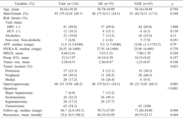
The demographics and clinical features of the 116 pa- tients are summarized in Table 1. Among these patients 92 patients were male (79.3%) and 24 patients were fe- male (20.7%). The mean age of patients was 56.4 years.
All of these patients were classified as Child-Pugh A in
Table 1. Clinical characteristics of patients underwent anatomical resection and non-anatomical resection
Variables (%) Total (n=116) AR (n=53) NAR (n=63) p
Age, mean 56.42±10.26 56.74±10.09 56.16±10.48 0.764
Male/Female (%) 92 (79.3)/24 (20.7) 40 (75.5)/13 (24.5) 52 (82.5)/11 (17.5) 0.368 Risk factors (%)
Viral status
HBV (+) 81 (69.8) 37 (69.8) 44 (69.8) 1.000
HCV (+) 12 (10.3) 8 (15.1) 4 (6.3) 0.139
Alcoholics 23 (19.8) 7 (13.2) 16 (25.4) 0.11
Non-viral, Non-alcoholic 7 (6.0) 2 (3.8) 5 (7.9) 0.451
AFP, median (range) 11.9 (1.3-6540) 9.2 (1.7-6540) 12.06 (1.3-1725.5) 0.79
PIVKA-II, median (range) 26.55 (4-1448) 27.55 (4-1448) 25.96 (4-405) 0.739
MELD, mean 8.04±2.61 7.67±1.23 7.98±1.70 0.268
Preop ICG, mean 15.2±7.97 14.15±5.39 16.15±9.62 0.187
Tumor size, mean 2.28±0.51 2.36±0.47 2.21±0.47 0.106
Tumor location (%) 0.023
Protrusion 27 (23.3) 8 (15.1) 19 (30.2)
Peripheral 69 (59.5) 31 (58.5) 38 (60.3)
Medial 20 (17.2) 14 (26.4) 6 (9.5)
Open/Laparoscopy 60 (51.7)/56 (48.3) 40 (75.5)/13 (24.5) 20 (31.7)/43 (68.3) 0.001
Opeartion <0.001
Major hepatectomy 7 (6.0) 7 (13.2)
Sectionectomy 26 (22.5) 26 (49.1)
Segmentectomy 20 (17.2) 20 (37.7)
Tumorectomy 63 (54.3) 63 (100)
Follow-up, median (range) 66.7 (6.4-143.3) 70.71±37.85 71.20±34.80 0.944
Recurrence, mean (month) 25.6 (0.5-140.2) 44.32±35.89 49.37±33.17 0.444
AR, Anatomical resection; NAR, Noon-anatomical resection; HBV, Hepatitis B virus; HCV, Hepatitis C virus; AFP, Alpha-feto protein; PIVKA-II, Protein induced vitamin K antagonist-II; MELD, Model for end-stage liver disease
preoperative liver function assessment. The median pre- operative AFP and PIVKA-II were 11.9 ng/ml and 26.5 mIU/mL, and the mean preoperative ICG R-15 level was 15.2%. The mean tumor size was 2.3 cm. Among these patients, 40 patients (64%) had tumors measuring less than 2 cm and 76 patients (36%) had tumors measuring between 2 and 3 cm. Tumors were located protrusively in 27 patients (23.3%), peripherally in 69 patients (59.5%), and medially in 20 patients (17.2%). AR was performed more frequently for medial located tumor than NAR, however there was no significant difference be- tween AR and NAR group in demographics except tumor location.
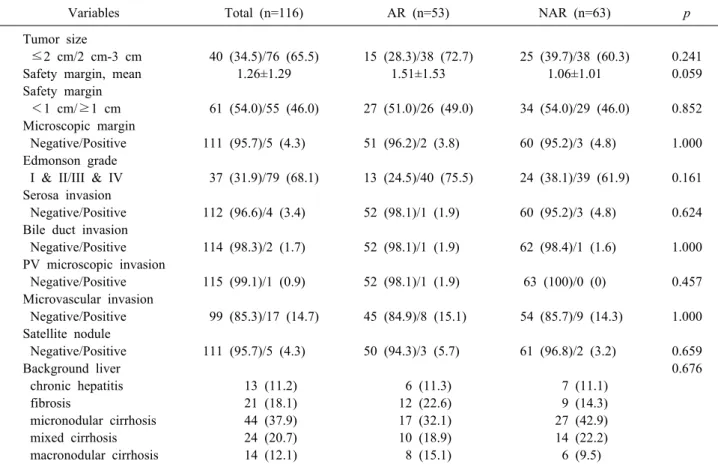
Pathological features of the patients in this study were summarized in Table 2. Mean free margin was 1.26 cm and Resection margin was grossly free from tumor in all patients, however microscopic involvement was identified in 5 patients (4.3%). Portal vein micro invasion was iden- tified in only 1 patient (0.9%), microscopic vascular in- vasion (MVI) in 17 patients (14.7%), and satellite nodule
was detected in 5 patients (4.3%). Underlying liver cir- rhosis was identified in 82 patients (70.7%). There was no significant difference between AR and NAR group in pathologic features.
Survival & recurrence after resection
The median follow-up period was 66.65 months (range, 6.4-143.3). During a follow-up period, tumor recurrence occurred in 65 patients (56.0%) and median time to re- currence was 25.6 months after surgery, respectively. At the time of last follow-up, 19 patients (16.4%) had died of liver-related disease including tumor progression. There was no in-hospital mortality. For the entire cohort of 116 patients, 1-, 3- and 5-year overall survival rates were 99, 92 and 84% (Fig. 1A) and recurrence-free survival rates were 84, 63 and 47%, respectively (Fig. 1B).
In comparison of the clinicopathologic characteristics between AR and NAR group, there was no significant dif- ferences in demographics and pathologic findings except tumor location (Table 1 and 2). The 1-, 3- and 5-year
Table 2. Pathologic results of patients underwent anatomical resection non-anatomical resection
Variables Total (n=116) AR (n=53) NAR (n=63) p
Tumor size
≤2 cm/2 cm-3 cm 40 (34.5)/76 (65.5) 15 (28.3)/38 (72.7) 25 (39.7)/38 (60.3) 0.241
Safety margin, mean 1.26±1.29 1.51±1.53 1.06±1.01 0.059
Safety margin
<1 cm/≥1 cm 61 (54.0)/55 (46.0) 27 (51.0)/26 (49.0) 34 (54.0)/29 (46.0) 0.852 Microscopic margin
Negative/Positive 111 (95.7)/5 (4.3) 51 (96.2)/2 (3.8) 60 (95.2)/3 (4.8) 1.000 Edmonson grade
I & II/III & IV 37 (31.9)/79 (68.1) 13 (24.5)/40 (75.5) 24 (38.1)/39 (61.9) 0.161 Serosa invasion
Negative/Positive 112 (96.6)/4 (3.4) 52 (98.1)/1 (1.9) 60 (95.2)/3 (4.8) 0.624 Bile duct invasion
Negative/Positive 114 (98.3)/2 (1.7) 52 (98.1)/1 (1.9) 62 (98.4)/1 (1.6) 1.000 PV microscopic invasion
Negative/Positive 115 (99.1)/1 (0.9) 52 (98.1)/1 (1.9) 63 (100)/0 (0) 0.457 Microvascular invasion
Negative/Positive 99 (85.3)/17 (14.7) 45 (84.9)/8 (15.1) 54 (85.7)/9 (14.3) 1.000 Satellite nodule
Negative/Positive 111 (95.7)/5 (4.3) 50 (94.3)/3 (5.7) 61 (96.8)/2 (3.2) 0.659
Background liver 0.676
chronic hepatitis 13 (11.2) 6 (11.3) 7 (11.1)
fibrosis 21 (18.1) 12 (22.6) 9 (14.3)
micronodular cirrhosis 44 (37.9) 17 (32.1) 27 (42.9)
mixed cirrhosis 24 (20.7) 10 (18.9) 14 (22.2)
macronodular cirrhosis 14 (12.1) 8 (15.1) 6 (9.5)
AR, Anatomical resection; NAR, Non-anatomical resection; PV, Portal vein
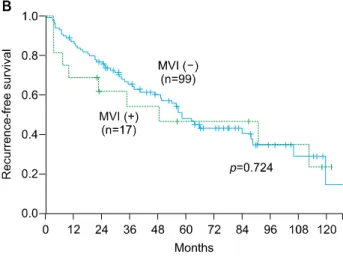
Fig. 1. Overall survival (A) and recurrence-free survival (B) rates after hepatic resection for single HCC smaller than 3 cm (n=113).
overall survival rates were 100, 88 and 81% in AR group, and 98, 95 and 86% in NAR group, respectively (Fig.
2A). There was no significant difference in overall surviv- al between AR and NAR group (p=0.78). The 1-, 3- and 5-year recurrence-free survival rates were 75, 56 and 43%
in AR group, and 90, 70 and 51% in NAR group, re-
spectively (Fig. 2B). There was also no significant differ- ence in recurrence-free survival between AR and NAR group (p=0.455).
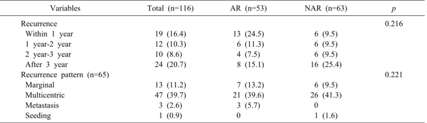
Although there was no significant difference between groups in recurrence time and pattern, tumor recurrences within 1 year after resection were more frequent in AR
Table 3. Comparison of recurrence time and pattern
Variables Total (n=116) AR (n=53) NAR (n=63) p
Recurrence 0.216
Within 1 year 19 (16.4) 13 (24.5) 6 (9.5)
1 year-2 year 12 (10.3) 6 (11.3) 6 (9.5)
2 year-3 year 10 (8.6) 4 (7.5) 6 (9.5)
After 3 year 24 (20.7) 8 (15.1) 16 (25.4)
Recurrence pattern (n=65) 0.221
Marginal 13 (11.2) 7 (13.2) 6 (9.5)
Multicentric 47 (39.7) 21 (39.6) 26 (41.3)
Metastasis 3 (2.6) 3 (5.7) 0
Seeding 1 (0.9) 0 1 (1.6)
AR, Anatomical resection; NAR, Non-anatomical resection
Fig. 2. Overall survival (A) and recurrence-free survival (B) rates according to the extent of surgery (AR/NAR) in patients with single HCC smaller than 3 cm.
group and after 3 years were more frequent in NAR group (Table 3).
Prognostic factor analysis for early and overall recurrence
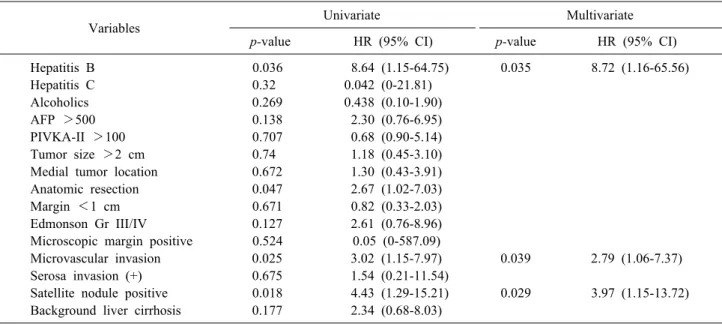
In patients with HCC smaller than 3 cm, hepatitis B, anatomical resection, microvascular invasion, and pres- ence of satellite nodule were associated with early re- currence (within 1 year) after resection in the univariate analysis, however high level of tumor markers, tumor size, and microscopic margin positive were not associated with early recurrence In multivariate analysis, hepatitis B (hazard ratio (HR) 8.72; p=0.035), satellite nodule (HR 3.97; p=0.029) and MVI (HR 2.79; p=0.039) were in- dependent risk factors for early recurrence (within 1 year) after resection (Table 4). In the aspect of overall re- currence after resection for HCC smaller than 3 cm, satel-
lite nodule (HR 6.21; p=0.005) and background liver cir- rhosis (HR 1.96; p=0.032) were revealed as independent risk factors in multivariate analysis (Table 5).
Relation between safety margin, microvascular invasion and recurrence
In terms of recurrence, width of safety margin (margin
<1 cm) was not correlated with HCC recurrence after surgery in single HCC smaller than 3 cm (Fig. 3A).
Although MVI was the independent prognostic factors for early recurrence (within 1 year), overall recurrence was not significantly affected by MVI (Fig. 3B). However, time of recurrence was significantly different in patients with MVI according to the width of safety margin (<1 cm vs ≥1 cm, p=0.049), on the other hand, there was no difference in patients without MVI according to the width of safety margin (Fig. 3C).
Table 4. Risk factors for early recurrence (within 1 year) in patients with single HCC smaller than 3 cm
Variables Univariate Multivariate
p-value HR (95% CI) p-value HR (95% CI)
Hepatitis B 0.036 8.64 (1.15-64.75) 0.035 8.72 (1.16-65.56)
Hepatitis C 0.32 0.042 (0-21.81)
Alcoholics 0.269 0.438 (0.10-1.90)
AFP >500 0.138 2.30 (0.76-6.95)
PIVKA-II >100 0.707 0.68 (0.90-5.14)
Tumor size >2 cm 0.74 1.18 (0.45-3.10)
Medial tumor location 0.672 1.30 (0.43-3.91)
Anatomic resection 0.047 2.67 (1.02-7.03)
Margin <1 cm 0.671 0.82 (0.33-2.03)
Edmonson Gr III/IV 0.127 2.61 (0.76-8.96)
Microscopic margin positive 0.524 0.05 (0-587.09)
Microvascular invasion 0.025 3.02 (1.15-7.97) 0.039 2.79 (1.06-7.37)
Serosa invasion (+) 0.675 1.54 (0.21-11.54)
Satellite nodule positive 0.018 4.43 (1.29-15.21) 0.029 3.97 (1.15-13.72) Background liver cirrhosis 0.177 2.34 (0.68-8.03)
HR, Hazard ratio; AFP, Alpha-fetoprotein; PIVKA-II, Protein induced vitamin K antagonist-II
Table 5. Risk factors for overall recurrence in patients with single HCC smaller than 3 cm
Variables Univariate Multivariate
p-value HR (95% CI) p-value HR (95% CI)
HBV (+) 0.115 1.56 (0.90-2.69)
HCV (+) 0.296 0.61 (0.25-1.53)
Alcoholics 0.68 0.88 (0.48-1.61)
AFP >500 0.999 1.00 (0.46-2.20)
PIVKA-II >100 0.467 0.59 (0.14-2.44)
Tumor size >2 cm 0.935 1.02 (0.61-1.70)
Medial tumor location 0.667 1.16 (0.59-2.29) Non-anatomic resection 0.456 0.83 (0.51-1.36)
Margin <1 cm 0.53 1.17 (0.72-1.92)
Edmonson Gr III/IV 0.306 1.33 (0.77-2.30)
Microscopic margin positive 0.741 0.82 (0.26-2.63) Microvascular invasion 0.532 1.23 (0.64-2.38)
Serosa invasion (+) 0.323 1.80 (0.56-5.76)
Satellite nodule positive 0.002 4.24 (1.66-10.80) 0.001 4.98 (1.93-12.87) Background liver cirrhosis 0.057 1.81 (0.98-3.34) 0.032 1.96 (1.06-3.64) HR, Hazard ratio; AFP, Alpha-fetoprotein; PIVKA-II, Protein induced vitamin K antagonist-II
DISCUSSION
Liver resection has been accepted as the gold standard treatment for solitary HCC in patients with well-preserved liver function.22 However, the superiority of AR has been still controversial. Recently, with the technical improve- ment, laparoscopic liver resection has been widely applied for the treatment of HCC and NAR can be performed more easily than AR with laparoscopy especially in cases
of small HCC which is protruded or located peripherally.
For this reason, the proportion of laparoscopic resection was significantly higher in NAR group than AR group in this study.
Previously several retrospective studies reported the su- periority of AR for HCC in the aspect of recurrence and survival.9,11-13,23 However, most of these studies have sig- nificant selection bias of patients, the difference of re- served liver function which is a significant postoperative
Fig. 3. Recurrence-free survival rates according to the width of safety margin (A) and presence of micro- vascular invasion (B). (C) The different recurrence-free survival according to the width of safety margin in MVI (–) and MVI (+) group.
prognostic factor in recurrence and survival. On the con- trary, other retrospective studies which tried to control confounding factors and eliminate selection bias, reported the comparable outcomes between AR and NAR in soli- tary HCC less than 4 cm8 and 3 cm.15 In this study, we only included the patients with well-preserved liver func- tion (Child-Pugh class A) and there were no differences in preoperative characteristics and postoperative patho- logic results between AR group and NAR group except tumor location and the proportion of laparoscopic surgery.
Our results showed that the outcomes of NAR in terms of recurrence pattern, recurrence-free survival, and overall survival were not different from those in AR (Table 3, Fig. 2). In univariate analysis for identifying prognostic factors predicting early recurrence (within 1 year), HCC recurrences within 1 year were more frequently occurred in patients underwent AR than NAR. We presumed that this result is caused by the disparity between groups in proportion of tumor size. Although there was no statistical significance between AR and NAR group in mean tumor
size (2.36 vs 2.21, p=0.106) and the proportion of patients who had HCC of 2 to 3 cm (72.7% vs 60.3%, p=0.241), there were more patients who had HCC of 2 to 3 cm in AR group. And this might make the significant difference in early recurrence rate between groups in univariate analysis.
Instead of the extent of hepatic resection (AR/NAR), multivariate analysis identified that hepatitis B, the pres- ence of satellite nodule and MVI were the independent risk factors for early (within 1 year) recurrence of tumor.
There are several studies about correlation between hep- atitis B viral infection and early recurrence of HCC.24-27 But the mechanism by which HBeAg positivity could pos- sibly enhance tumor recurrence remains unclear. The pos- sible explanation is that positive HBeAg may associate with active inflammation in liver parenchyma to promote intrahepatic metastasis by changing tumor microenviron- ment, finally resulting in early recurrence within a short time after hepatectomy. The other possibility is positive HBeAg may associated with increased synchronic multi-
centric tumor. MVI has been accepted as powerful prog- nostic factors predicting recurrence and survival in large and multiple HCC. However, several studies reported that MVI had no significant impact on overall survival in pa- tients with early HCC, whereas MVI had significant im- pact on recurrence.19,28,29 These data correlate with our re- sults up to a point. Our results showed that MVI was not correlated with overall recurrence and survival, while ear- ly recurrence (within 1 year) was significantly affected by MVI. In our data, background liver cirrhosis had more significant impact on overall survival along with the pres- ence of satellite nodule rather than MVI and viral activity.
These results suggest that the prognosis after resection can be more affected by underlying liver status rather than pathobiological factors of tumor in early stage HCC, an observation supported by previous study which reported the importance of liver status as an independent prog- nostic factor predicting recurrence after resection.8,15,30
The significance of safety margin in recurrence and sur- vival after liver resection for HCC remains controversial.
Several studies reported no relation between the safety margin and prognosis,8,31-33 while other studies suggested that safety margin less than 1 cm had an negative effect on long-term prognosis.34-36 Our data suggested that the width of safety margin has no impact on survival and HCC recurrence after resection in entire cohort of patients with single HCC smaller than 3 cm. However, the impact of safety margin on tumor recurrence was different be- tween MVI (+) and MVI (–) group when the patients were categorized according to the presence of MVI. In patients without MVI, there was no significant difference in HCC recurrence between safety margin <1 cm and safety mar- gin ≥1 cm group. On the other hand, tumor recurrence (especially early recurrence) was more frequently ob- served in safety margin <1 cm group in patients with MVI (p=0.049). Based on these results, we can suggest that hepatic resection with adequate margin over 1 cm might be required for decreasing risk of early recurrence in patients with single HCC smaller than 3 cm because MVI can’t be identified preoperatively. However, pres- ence of MVI was detected in only 17 patients (14.7%) with small HCC in this study and this makes it difficult to confirm this result. Therefore, further studies with large sample size are required to confirm this result.
The limitations of this study include its retrospective
nature, small volume cohort from single center and se- lected population. Furthermore, most of patients in this study showed favorable biologic behavior and some fac- tors which accepted as powerful prognostic factors were observed in very few patients. this could raise the possi- bility of statistical error. However, similar liver function in all patients (well-preserved liver function) and no dif- ference in clinical and pathologic findings between groups can minimize the risk of selection bias. In addition, when considering the less aggressive biologic nature of early HCC, our results might be accepted as a meaningful data.
In conclusion, the outcomes of NAR were comparable with those of AR in patients with single HCC smaller than 3 cm and well-preserved liver function. Although MVI was not correlated with overall survival and recurrence, early recurrence was significantly affected by MVI, and the outcome of resection with safety margin <1 cm was shown worse than safety margin ≥1 cm. Therefore, wide resection with adequate safety margin is recommended in patients with single HCC smaller than 3 cm. In terms of overall recurrence, back ground liver status such as cir- rhosis and viral activity has more significant impact on prognosis rather than pathobiologic behavior in single HCC smaller than 3 cm.
REFERENCES
1. Takayasu K, Arii S, Sakamoto M, Matsuyama Y, Kudo M, Kaneko S, et al. Impact of resection and ablation for single hy- povascular hepatocellular carcinoma ≤2 cm analysed with pro- pensity score weighting. Liver Int 2018;38:484-493.
2. European Association for the Study of the Liver. EASL clinical practice guidelines: liver transplantation. J Hepatol 2016;64:433- 485.
3. Duan C, Liu M, Zhang Z, Ma K, Bie P. Radiofrequency ablation versus hepatic resection for the treatment of early-stage hep- atocellular carcinoma meeting Milan criteria: a systematic review and meta-analysis. World J Surg Oncol 2013;11:190.
4. Bruix J, Sherman M; American Association for the Study of Liver Diseases. Management of hepatocellular carcinoma: an update. Hepatology 2011;53:1020-1022.
5. Popescu I, Câmpeanu I. [Surgical anatomy of the liver and liver resection. Brisbane 2000 Terminology]. Chirurgia (Bucur) 2009;
104:7-10. Romanian.
6. Nakashima T, Kojiro M. Pathologic characteristics of hep- atocellular carcinoma. Semin Liver Dis 1986;6:259-266.
7. Makuuchi M, Imamura H, Sugawara Y, Takayama T. Progress in surgical treatment of hepatocellular carcinoma. Oncology 2002;62 Suppl 1:74-81.
8. Kang CM, Choi GH, Kim DH, Choi SB, Kim KS, Choi JS, et al. Revisiting the role of nonanatomic resection of small (< or
=4 cm) and single hepatocellular carcinoma in patients with
well-preserved liver function. J Surg Res 2010;160:81-89.
9. Regimbeau JM, Kianmanesh R, Farges O, Dondero F, Sauvanet A, Belghiti J. Extent of liver resection influences the outcome in patients with cirrhosis and small hepatocellular carcinoma.
Surgery 2002;131:311-317.
10. Tan Y, Zhang W, Jiang L, Yang J, Yan L. Efficacy and safety of anatomic resection versus nonanatomic resection in patients with hepatocellular carcinoma: a systemic review and meta- analysis. PLoS One 2017;12:e0186930.
11. Yamazaki O, Matsuyama M, Horii K, Kanazawa A, Shimizu S, Uenishi T, et al. Comparison of the outcomes between anatomi- cal resection and limited resection for single hepatocellular carci- nomas no larger than 5 cm in diameter: a single-center study.
J Hepatobiliary Pancreat Sci 2010;17:349-358.
12. Wakai T, Shirai Y, Sakata J, Kaneko K, Cruz PV, Akazawa K, et al. Anatomic resection independently improves long-term sur- vival in patients with T1-T2 hepatocellular carcinoma. Ann Surg Oncol 2007;14:1356-1365.
13. Eguchi S, Kanematsu T, Arii S, Okazaki M, Okita K, Omata M, et al. Comparison of the outcomes between an anatomical subsegmentectomy and a non-anatomical minor hepatectomy for single hepatocellular carcinomas based on a Japanese nationwide survey. Surgery 2008;143:469-475.
14. Tanaka K, Shimada H, Matsumoto C, Matsuo K, Nagano Y, Endo I, et al. Anatomic versus limited nonanatomic resection for solitary hepatocellular carcinoma. Surgery 2008;143:607-615.
15. Tomimaru Y, Eguchi H, Marubashi S, Wada H, Kobayashi S, Tanemura M, et al. Equivalent outcomes after anatomical and non-anatomical resection of small hepatocellular carcinoma in patients with preserved liver function. Dig Dis Sci 2012;57:1942- 1948.
16. Cucchetti A, Cescon M, Ercolani G, Bigonzi E, Torzilli G, Pinna AD. A comprehensive meta-regression analysis on outcome of anatomic resection versus nonanatomic resection for hep- atocellular carcinoma. Ann Surg Oncol 2012;19:3697-3705.
17. Ye JZ, Miao ZG, Wu FX, Zhao YN, Ye HH, Li LQ. Recurrence after anatomic resection versus nonanatomic resection for hep- atocellular carcinoma: a meta-analysis. Asian Pac J Cancer Prev 2012;13:1771-1777.
18. Tang YH, Wen TF, Chen X. Anatomic versus non-anatomic liver resection for hepatocellular carcinoma: a systematic review.
Hepatogastroenterology 2013;60:2019-2025.
19. Shindoh J, Andreou A, Aloia TA, Zimmitti G, Lauwers GY, Laurent A, et al. Microvascular invasion does not predict long- term survival in hepatocellular carcinoma up to 2 cm: reappraisal of the staging system for solitary tumors. Ann Surg Oncol 2013;
20:1223-1229.
20. Jung DH, Hwang S, Lee YJ, Kim KH, Song GW, Ahn CS, et al. Small hepatocellular carcinoma with low tumor marker ex- pression benefits more from anatomical resection than tumors with aggressive biology. Ann Surg 2017. doi: 10.1097/SLA.
0000000000002486. [in press]
21. Ahn KS, Kang KJ, Park TJ, Kim YH, Lim TJ, Kwon JH. Benefit of systematic segmentectomy of the hepatocellular carcinoma:
revisiting the dye injection method for various portal vein branches. Ann Surg 2013;258:1014-1021.
22. Hwang S, Lee YJ, Kim KH, Ahn CS, Moon DB, Ha TY, et al. The impact of tumor size on long-term survival outcomes af-
ter resection of solitary hepatocellular carcinoma: single-in- stitution experience with 2558 patients. J Gastrointest Surg 2015;
19:1281-1290.
23. Hasegawa K, Kokudo N, Imamura H, Matsuyama Y, Aoki T, Minagawa M, et al. Prognostic impact of anatomic resection for hepatocellular carcinoma. Ann Surg 2005;242:252-259.
24. Sun HC, Zhang W, Qin LX, Zhang BH, Ye QH, Wang L, et al. Positive serum hepatitis B e antigen is associated with higher risk of early recurrence and poorer survival in patients after cura- tive resection of hepatitis B-related hepatocellular carcinoma. J Hepatol 2007;47:684-690.
25. Sun HC, Tang ZY, Ma ZC, Qin LX, Wang L, Ye QH, et al.
The prognostic factor for outcome following second resection for intrahepatic recurrence of hepatocellular carcinoma with a hep- atitis B virus infection background. J Cancer Res Clin Oncol 2005;131:284-288.
26. Ikeda K, Arase Y, Kobayashi M, Saitoh S, Someya T, Hosaka T, et al. Significance of multicentric cancer recurrence after po- tentially curative ablation of hepatocellular carcinoma: a long- term cohort study of 892 patients with viral cirrhosis. J Gastroenterol 2003;38:865-876.
27. Portolani N, Coniglio A, Ghidoni S, Giovanelli M, Benetti A, Tiberio GA, et al. Early and late recurrence after liver resection for hepatocellular carcinoma: prognostic and therapeutic impli- cations. Ann Surg 2006;243:229-235.
28. Yamashita Y, Tsuijita E, Takeishi K, Fujiwara M, Kira S, Mori M, et al. Predictors for microinvasion of small hepatocellular carcinoma ≤2 cm. Ann Surg Oncol 2012;19:2027-2034.
29. Roayaie S, Obeidat K, Sposito C, Mariani L, Bhoori S, Pellegrinelli A, et al. Resection of hepatocellular cancer ≤2 cm:
results from two Western centers. Hepatology 2013;57:1426- 1435.
30. Vauthey JN, Lauwers GY, Esnaola NF, Do KA, Belghiti J, Mirza N, et al. Simplified staging for hepatocellular carcinoma.
J Clin Oncol 2002;20:1527-1536.
31. Lise M, Bacchetti S, Da Pian P, Nitti D, Pilati PL, Pigato P.
Prognostic factors affecting long term outcome after liver re- section for hepatocellular carcinoma: results in a series of 100 Italian patients. Cancer 1998;82:1028-1036.
32. Nonami T, Harada A, Kurokawa T, Nakao A, Takagi H. Hepatic resection for hepatocellular carcinoma. Am J Surg 1997;173:288- 291.
33. Chau GY, Lui WY, Tsay SH, King KL, Loong CC, Chiu JH, et al. Prognostic significance of surgical margin in hepatocellular carcinoma resection: an analysis of 165 Childs' A patients. J Surg Oncol 1997;66:122-126.
34. Kosuge T, Makuuchi M, Takayama T, Yamamoto J, Shimada K, Yamasaki S. Long-term results after resection of hep- atocellular carcinoma: experience of 480 cases. Hepatogastroen- terology 1993;40:328-332.
35. Fuster J, García-Valdecasas JC, Grande L, Tabet J, Bruix J, Anglada T, et al. Hepatocellular carcinoma and cirrhosis. Results of surgical treatment in a European series. Ann Surg 1996;223:
297-302.
36. Izumi R, Shimizu K, Ii T, Yagi M, Matsui O, Nonomura A, et al. Prognostic factors of hepatocellular carcinoma in patients un- dergoing hepatic resection. Gastroenterology 1994;106:720-727.