Letter to the Editor
Vol. 26 No. 4, 2014 549
Received July 26, 2013, Revised September 16, 2013, Accepted for publication September 21, 2013
Corresponding author: Kwang Hyun Cho, Department of Dermatology, Seoul National University Hospital, 101 Daehak-ro, Jongno-gu, Seoul 110-744, Korea. Tel: 82-2-2072-2412, Fax: 82-2-742-7344, E-mail: khcho@snu.ac.kr
This is an Open Access article distributed under the terms of the Creative Commons Attribution Non-Commercial License (http://
creativecommons.org/licenses/by-nc/3.0) which permits unrestricted non-commercial use, distribution, and reproduction in any medium, provided the original work is properly cited.
clinical and histopathological features, we diagnosed all these cases as DNS. Furthermore, we performed excision only in cases of considerable DN that either presented un- usual clinicopathological features or where the patient strongly wanted the DN to be removed. For the others, close observation was recommended.
Concerning the various definitions, there is considerable debate about the number of DN. Most previous reports agreed that large numbers of DN should be included in the diagnostic criteria. However, Elder et al.3 described DNS as a wide spectrum of clinical phenotype from a single DN in a patient without a personal and family history of melanoma to familial atypical multiple mole- melanoma syndrome. As we have followed Elder’s de- finitions, we classified even 5 DN as DNS.
In analyzing previous cases1,2, all 10 cases could not be analyzed according to asymmetry, border, and color because of a lack of description. However, their size was 3∼45 mm, except in 1 case. Although the DN in 1 pa- tient had a minimum size of 3 mm, as the number of DN was as many as 406, we considered this patient as having
DNS.
Considering that there are no definite clinicopathological diagnostic criteria of DNS in the West, DNS in Korean patients may be underdiagnosed, as previous Korean reports suggest. In addition, a difference in the number of moles between Asian and Western patients with DNS may encourage changing the diagnostic threshold of DN, and more importantly, DNS.
REFERENCES
1. Roh JY, Lee SY, Kye YC, Cinn YW, Kim SN. A case of dysplastic melanocytic nevus. Korean J Dermatol 1988;26:
447-452.
2. Shin SB, Lee DW, Lee JY, Cho BK. Clinical and pathologic findings of dysplastic nevus: review of 15 cases. Korean J Dermatol 2000;38:1055-1062.
3. Elder DE, Green MH, Guerry D 4th, Kraemer KH, Clark WH Jr. The dysplastic nevus syndrome: our definition. Am J Der- matopathol 1982;4:455-460.
http://dx.doi.org/10.5021/ad.2014.26.4.549
A Case of Mucinous Nevus Clinically Mimicking Nevus Lipomatosus Superficialis
Eun Jee Kim, Seong-Jin Jo, Kwang Hyun Cho
Department of Dermatology, Seoul National University College of Medicine, Seoul, Korea
Dear Editor:
Mucinous nevus is a very rare entity and can be classified as both a cutaneous mucinosis and a connective tissue nevus1. The term “mucinous nevus” was proposed beca- use of its nevoid appearance and the characteristic pattern of mucin deposits in the papillary dermis1. We report the
case of a mucinous nevus diagnosed in a young Korean man.
A previously healthy 24-year-old man visited our clinic because of confluent flesh-colored to brownish non-firm nodules on his left lower back, with a zosteriform dis- tribution (Fig. 1). The skin lesions had been present since
Letter to the Editor
550 Ann Dermatol
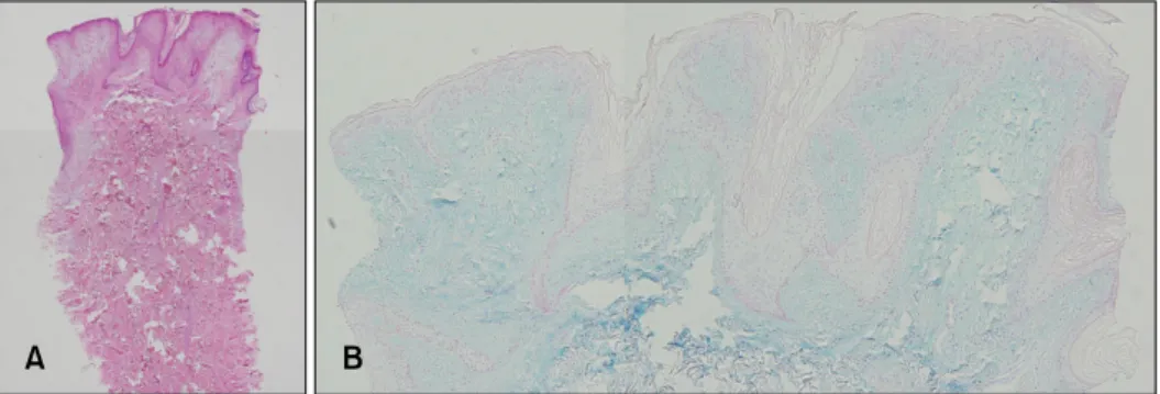
Fig. 2. (A) The band-like deposition of mucin was mainly observed in the superficial dermis, with papillo- matosis, hyperkeratosis, and elon- gation of the rete ridge in the epi- dermis (H&E, ×40). (B) Mucin de- posited in the papillary dermis stained positive to alcian blue at pH 2.5 (×100).
Fig. 1. Confluent flesh-colored to brownish non-firm papules and nodules on the left lower back, with a zosteriform distribution.
adolescence without any symptom; however, they slowly grew in size. He had no history of trauma, and the lesion was not treated before. He also denied any familial his- tory. Biopsy was taken, with the first impression of nevus lipomatosus superficialis (NLS) according to clinical fea- tures. Histologically, the findings consisted of a band-like deposit of mucin mainly in the superficial dermis, with papillomatosis, hyperkeratosis, and elongation of the rete ridge in the epidermis (Fig. 2A). The mucin deposited in the dermis stained positive with alcian blue at pH 2.5 (Fig.
2B). Finally, the lesion was confirmed as a mucinous nevus (epidermal-connective tissue nevus of proteoglycan [epidermal-CTNP]). For the treatment, a first-stage opera- tion was done without any complication, and a second- stage operation is planned 4 months later. Most of the lesions were removed with the first-stage operation and no recurrence has occurred.
Mucinous nevus is a neoplastic hamartoma and a rare form of primary cutaneous mucinosis2. Mucinous nevus clinically presents as grouped brownish papules and con- fluent plaques, usually with a unilateral, linear, zosteri-
form, or grouped distribution. It usually appears at birth or in early childhood and mainly occurs on the back2. Al- though most cases are sporadic, the possibility of familial association has been suggested2. The nevoid feature of mucinous nevus needs to be clinically differentiated from epidermal nevus or NLS. In mucinous nevus, the band- like deposit of mucin is limited mainly in the superficial dermis, like in our case3. However, recently, a case of mucinous nevus with mature fat cells in the upper dermis similar to NLS has been reported, making the diagnosis more confusing3. Mucinous nevus is divided into two his- topathologic types: CTNP type and combined epidermal- CTNP type4. The difference between the two types lies in whether the epidermis is normal or shows hyperkeratosis and acanthosis, with elongation of the rete ridge indi- cating epidermal nevus4. After reviewing the histopatho- logic changes of the reported cases of mucinous nevus, Chi et al.4 found that approximately half of the mucinous nevus cases were CTNP. Here, we report a rare case of mucinous nevus, the epidermal-CTNP type, presenting si- milar to NLS.
REFERENCES
1. Chen CW, Tsai TF, Chen YF, Hung CM. Familial mucinous nevus. Pediatr Dermatol 2008;25:288-289.
2. Perez-Crespo M, Lopez-Navarro N, Betlloch I, Herrera E, Ni- veiro M, Gallego E. Acquired and familial mucinous nevus.
Int J Dermatol 2011;50:1283-1285.
3. Song BH, Park S, Park EJ, Kwon IH, Kim KH, Kim KJ. Mu- cinous nevus with fat: an unusual case report and literature review. Am J Dermatopathol 2012;34:e146-e148.
4. Chi CC, Wang SH, Lin PY. Combined epidermal-connective tissue nevus of proteoglycan (a type of mucinous nevus): a case report and literature review. J Cutan Pathol 2009;36:
808-811.