ISSN 2234-3806 • eISSN 2234-3814
https://doi.org/10.3343/alm.2019.39.1.105 www.annlabmed.org 105
Ann Lab Med 2019;39:105-108
https://doi.org/10.3343/alm.2019.39.1.105
Letter to the Editor
Diagnostic Genetics
Compound Heterozygous Pathogenic Variants of the 15-Hydroxyprostaglandin Dehydrogenase Gene in a
Patient With Hypertrophic Osteoarthropathy: First Case in Korea
Mi Ra Ryu, M.D.1, Ji Hyun Yang, M.D.2, Sang Youl Rhee, M.D.3, Ahra Cho, M.D.4, Seong Yoon Kim , M.D.2, and Chang-Seok Ki , M.D.5
1Department of Laboratory Medicine and Genetics, Samsung Medical Center, Sungkyunkwan University School of Medicine, Seoul, Korea; 2Division of Cardiology, Department of Internal Medicine, Korea Association of Health Promotion, Seoul, Korea; 3Department of Endocrinology and Metabolism, Kyung Hee University School of Medicine, Seoul, Korea; 4Division of Molecular Genetics, Department of Laboratory Medicine, Seoul Clinical Laboratories, Yongin, Korea; 5Green Cross Genome, Yongin, Korea
Dear Editor,
Hereditary or primary hypertrophic osteoarthropathy (PHO) is an autosomal recessive disorder characterized by excessive pro- liferation of skin and bone cells at the distal parts of the extremi- ties, resulting in clubbing of fingers and toes, periostitis of long tubular bones, and arthritis [1]. Pathogenic variants in the 15-hy- droxyprostaglandin dehydrogenase (HPGD) gene and solute car- rier organic anion transporter family member 2A1 gene (SLCO2A1) have been identified in hypertrophic osteoarthropathy, primary, autosomal recessive 1 (PHOAR1; Online Mendelian Inheritance in Man [OMIM] #259100) and hypertrophic osteoarthropathy, primary, autosomal recessive 2 (PHOAR2; OMIM #614441), re- spectively [2, 3]. Some differences exist between the clinical phe- notypes of PHOAR1 and PHOAR2: 1) pachydermia and cutis gy- rate are more frequent and severe in PHOAR2 than in PHOAR1;
2) a certain proportion of PHOAR2 exhibits gastrointestinal hem- orrhage, which is not observed in PHOAR1; 3) higher urinary prostaglandin E2 (PGE2) levels are observed in PHOAR2 (ap- proximately 10-fold increase) than in PHOAR1 (approximately 4-fold increase); and 4) onset is usually around puberty in PHO AR2, whereas onset is often around birth in PHOAR1 [4]. In Korea, several cases of PHO have been reported, all harboring SLCO2A1 mutations [5, 6]. We report the first case in Korea of a patient with PHO carrying compound heterozygous pathogenic variants in the HPGD gene.
A 21-year-old military soldier was referred to the outpatient clinic of the Armed Forces Capital Hospital, Seongnam, Korea, with a suspected diagnosis of acromegaly in February 2011. He had abnormal thickening of both hands and feet, as well as digi- tal clubbing, which is an unusual finding in patients with acro-
Received: February 6, 2018 Revision received: April 26, 2018 Accepted: August 27, 2018
Corresponding author: Seong Yoon Kim, M.D.
https://orcid.org/ 0000-0003-0490-0447
Division of Cardiology, Department of Internal Medicine, Korea Association of Health Promotion, 335 Hwagok-ro, Gangseo-gu, Seoul 07649, Korea Tel: +82-2-2600-2109, Fax +82-2-2696-4500, E-mail: [email protected] Co-corresponding author: Chang-Seok Ki, M.D.
https://orcid.org/0000-0001-7679-8731
Green Cross Genome, 107 Ihyeon-ro, Giheung-gu, Yongin 16924, Korea Tel: +82-31-260-9600, Fax: +82-31-260-9087
E-mail: [email protected]
© Korean Society for Laboratory Medicine
This is an Open Access article distributed under the terms of the Creative Commons Attribution Non-Commercial License (http://creativecommons.org/licenses/by-nc/4.0) which permits unrestricted non-commercial use, distribution, and reproduction in any medium, provided the original work is properly cited.
1 / 1 CROSSMARK_logo_3_Test
2017-03-16 https://crossmark-cdn.crossref.org/widget/v2.0/logos/CROSSMARK_Color_square.svg
Ryu MR, et al.
HPGD-related hypertrophic osteoarthropathy
106 www.annlabmed.org https://doi.org/10.3343/alm.2019.39.1.105 megaly. The digital clubbing required him to visit a tertiary hos-
pital when he was six years old, but the cause of clubbing was not found. He denied any medical history, including that of gas- trointestinal hemorrhage, any affected family members, or a sud- den increase in height. Upon physical examination, digital club- bing of the fingers (Fig. 1A) and abnormal thickening of the feet (Fig. 1B) were noted; however, forehead pachydermia was not evident. Laboratory test results were within the reference range (blood chemistry, complete blood cell count, erythrocyte sedi- mentation rate, and C-reactive protein) as were hormonal test results (total-triiodothyronine, free-thyroxine, thyroid-stimulating hormone, and insulin-like growth factor 1). The chest X-ray showed no abnormalities, whereas the hand X-ray showed soft tissue swelling of the fingers without apparent acroosteolysis (Fig. 1C).
No other long bones showed abnormalities. Bone scintigraphy and sella magnetic resonance imaging did not reveal any re- markable findings. Secondary causes of hypertrophic osteoar- thropathy were ruled out using echocardiography, pulmonary function test, ultrasonography of the abdomen, and esophago- duodenoscopy. Urinary PGE2 was 2,626 ng, approximately 4-fold
higher than the upper normal range (reference range: 400–620 ng/24-hour urine).
The patient was suspected to have PHOAR1 rather than PHO AR2, and we decided to perform sequence analysis of the HPGD gene.
After obtaining informed consent, genomic DNA was extracted from peripheral blood leukocytes of the patient and both par- ents. All seven coding exons and their flanking intronic regions were amplified and sequenced using an ABI 3730xl genetic an- alyzer (Applied Biosystems, Foster City, CA, USA) and the Big- Dye Terminator v3.1 Cycle Sequencing Kit (Applied Biosystems) with primers designed by the authors (available upon request).
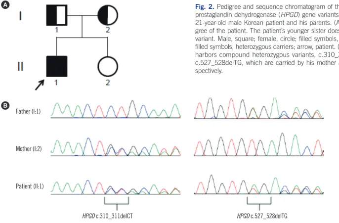
HPGD sequences were analyzed using Sequencher software (Gene Codes Corp., Ann Arbor, MI, USA) and compared with the reference sequence (NM_000860.5). The patient was com- pound heterozygous for a 2-bp deletion (NM_000860.5:c.310_
311delCT) in exon 3 and a 2-bp deletion (c.527_528delTG) in exon 6, and both parents were heterozygous carriers of each variant (Fig. 2).
The c.310_311delCT variant has been predicted to cause a truncated protein (p.Leu104Alafs*3) and was reported as a patho-
8 140
8 140
Fig. 1. Clinical and radiological findings of a 21-year- old male Korean patient: (A) digital clubbing, (B) ab- normal thickening of the feet, and (C) hand X-ray showing soft tissue swelling of the fingers.
8 140
8 140
A B
C
R
Ryu MR, et al.
HPGD-related hypertrophic osteoarthropathy
https://doi.org/10.3343/alm.2019.39.1.105 www.annlabmed.org 107
genic variant in two pairs of Turkish siblings [7, 8], two Chinese siblings [9], and seven sporadic Chinese patients [9]. The c.527_
528delTG variant is predicted to lead to a truncated protein (p.Val176Glufs*11) by Mutalyzer 2.0.28. It is absent from the Genome Aggregation Database (http://gnomad.broadinstitute.
org) and the Korean Reference Genome database (http://152.
99.75.168/KRGDB/), but it can be classified as a pathogenic variant according to the guidelines of the American College of Medical Genetics and Genomics and the Association for Molec- ular Pathology [10].
Previously reported patients carrying c.310_311delCT homo- zygous variants presented common clinical PHOAR1 findings:
digital clubbing, periostosis, acroosteolysis, joint swelling, and palmoplantar hyperkeratosis. Tüysüz et al. [7] described that the Turkish boys’ father and grandfather also had mild digital clubbing and palmar hyperkeratosis. Carriers of HPGD gene mutations have been known to present some signs of PHO [1].
However, Erken et al. [8] showed that the Turkish siblings’ rela- tives who were carriers did not exhibit signs of PHO. Neither par- ent of our patient showed any signs of PHO.
Although our patient had abnormal thickening of both hands and feet before school age, he neither experienced hand pain nor exhibited apparent acroosteolysis. Variable clinical pheno-
10 145
Fig. 2. Pedigree and sequence chromatogram of the 15-hydroxyprostaglandin dehydrogenase 146
(HPGD) gene variants identified in a 21-yr-old male Korean patient and his parents. (A) 147
Family pedigree of the patient. The patient’s younger sister does not have any variant. Male, 148
square; female, circle; filled symbols, affected; half-filled symbols, heterozygous carriers;
149
arrow, patient. (B) The patient harbors compound heterozygous variants, c.310_311delCT 150
and c.527_528delTG, which are carried by his mother and father, respectively.
151
Fig. 2. Pedigree and sequence chromatogram of the 15-hydroxy- prostaglandin dehydrogenase (HPGD) gene variants identified in a 21-year-old male Korean patient and his parents. (A) Family pedi- gree of the patient. The patient’s younger sister does not have any variant. Male, square; female, circle; filled symbols, affected; half- filled symbols, heterozygous carriers; arrow, patient. (B) The patient harbors compound heterozygous variants, c.310_311delCT and c.527_528delTG, which are carried by his mother and father, re- spectively.
A
B
10 145
Fig. 2. Pedigree and sequence chromatogram of the 15-hydroxyprostaglandin dehydrogenase 146
(HPGD) gene variants identified in a 21-yr-old male Korean patient and his parents. (A) 147
Family pedigree of the patient. The patient’s younger sister does not have any variant. Male, 148
square; female, circle; filled symbols, affected; half-filled symbols, heterozygous carriers;
149
arrow, patient. (B) The patient harbors compound heterozygous variants, c.310_311delCT 150
and c.527_528delTG, which are carried by his mother and father, respectively.
151
Father (I:1)
Mother (I:2)
Patient (II:1)
HPGD c.310_311delCT HPGD c.527_528delTG
types between PHOAR1 and PHOAR2, as well as within the same disease subtype, have not been completely elucidated;
thus, further studies are necessary to elucidate the genotype- phenotype correlation.
Authors’ Disclosures of Potential Conflicts of Interest
No potential conflicts of interest relevant to this article were re- ported.
REFERENCES
1. Castori M, Sinibaldi L, Mingarelli R, Lachman RS, Rimoin DL, Dallapi- ccola B. Pachydermoperiostosis: an update. Clin Genet 2005;68:477- 86.
2. Zhang Z, Xia W, He J, Zhang Z, Ke Y, Yue H, et al. Exome sequencing identifies SLCO2A1 mutations as a cause of primary hypertrophic os- teoarthropathy. Am J Hum Genet 2012;90:125-32.
3. Uppal S, Diggle CP, Carr IM, Fishwick CW, Ahmed M, Ibrahim GH, et al. Mutations in 15-hydroxyprostaglandin dehydrogenase cause primary hypertrophic osteoarthropathy. Nat Genet 2008;40:789-93.
4. Li SS, He JW, Fu WZ, Liu YJ, Hu YQ, Zhang ZL. Clinical, biochemical, and genetic features of 41 Han Chinese families with primary hypertro- phic osteoarthropathy, and their therapeutic response to etoricoxib: re- sults from a six-month prospective clinical intervention. J Bone Miner
Ryu MR, et al.
HPGD-related hypertrophic osteoarthropathy
108 www.annlabmed.org https://doi.org/10.3343/alm.2019.39.1.105 Res 2017;32:1659-66.
5. Kim HJ, Koo KY, Shin DY, Kim DY, Lee JS, Lee MG. Complete form of pachydermoperiostosis with SLCO2A1 gene mutation in a Korean fami- ly. J Dermatol 2015;42:655-7.
6. Lee S, Park SY, Kwon HJ, Lee CH, Kim OH, Rhee Y. Identification of the mutations in the prostaglandin transporter gene, SLCO2A1 and clinical characterization in Korean patients with pachydermoperiostosis. J Kore- an Med Sci 2016;31:735-42.
7. Tüysüz B, Yilmaz S, Kasapcopur Ö, Erener-Ercan T, Ceyhun E, Bilguvar K, et al. Primary hypertrophic osteoarthropathy caused by homozygous deletion in HPGD gene in a family: changing clinical and radiological findings with long-term follow-up. Rheumatol Int 2014;34:1539-44.
8. Erken E, Köroğlu Ç, Yildiz F, Özer HT, Gülek B, Tolun A. A novel reces- sive 15-hydroxyprostaglandin dehydrogenase mutation in a family with primary hypertrophic osteoarthropathy. Mod Rheumatol 2015;25:315- 21.
9. Yuan L, Chen L, Liao RX, Lin YY, Jiang Y, Wang O, et al. A common mu- tation and a novel mutation in the HPGD gene in nine patients with pri- mary hypertrophic osteoarthropathy. Calcif Tissue Int 2015;97:336-42.
10. Richards S, Aziz N, Bale S, Bick D, Das S, Gastier-Foster J, et al. Stan- dards and guidelines for the interpretation of sequence variants: a joint consensus recommendation of the American College of Medical Genet- ics and Genomics and the Association for Molecular Pathology. Genet Med 2015;17:405-24.