MRCP는 T2강조영상기법에 의해 담췌관내의 담즙을 고신 호강도로 영상화한 기법으로 투사영상과 단면영상의 장점을 결 합하여 담췌관에 대한 높은 진단적영상을 제공하며 내시경역 행췌담관조영술 (Endoscopic Retrograde Cholangio- pancreatography; 이하 ERCP로 줄임)과는 달리 조영제의 사 용없이 비침습적인 방법으로 영상을 획득할 수 있다는 장점을 가지고 있다(1-4).
또한 초음파나 ERCP에 비해 검사자에 대한 의존도가 낮고 담췌관의 해부학적 다양성이나 폐쇄에 관계없이 담췌관 전체 를 보여줄 수 있어 진단의 정확도나 유용성에 대한 연구가 활 발히 진행되고 있다(5).
그러나 대부분의 보고가 담관내결석을 대상으로 한 것이었 으며 다른 검사 특히 ERCP와의 비교 연구나 다른 기법간의 비교 연구는 다수 보고되어 있으나(3, 6) 담낭내결석을 대상
으로 고속스핀에코기법인 단발포고속스핀에코기법과 Sensitivity encoding(이하 SENSE)기술을 이용한 3차원고속 스핀에코기법 간의 비교 연구는 발표된 바가 없었다. 이에 저 자들은 담낭내결석의 진단에 있어 MRCP의 검출감도를 알아 보고, 연구에 사용된 단발포고속스핀에코기법과 SENSE기술을 이용한 3차원고속스핀에코기법을 비교하고자 하였다.
대상과 방법
2003년 8월부터 1년간 MRCP를 시행한 141명의 환자를 대 상으로 하였다. 남자가 77명, 여자가 58명이었고 환자들의 평 균연령은 54.9세(18-91세)이었다.
자기공명영상은 1.5 T 초전도형자기공명영상기기(Philips Medical Systems, Intera, The Netherlands)를 이용하였다. 먼 저 단발포고속스핀에코기법을 이용하여 호흡정지상태에서 강 한 T2강조의 원천절편영상(source image)횡단면과 관상면영
담낭내결석의 진단에 있어 자기공명담췌관조영술의 검출감도:
단발포고속스핀에코(SS-TSE)기법과 SENSE 기술을 이용한 3차원고속스핀에코(3D-TSE)기법의 비교
1김주애・윤은주・최철순・윤대영・박상준・서영란・이유진・문증희
목적: 담낭내결석에 대한 자기공명담췌관조영술(Magnetic Resonance Cholangiopancre- atography; 이하 MRCP로줄임)의 검출감도에 대해 알아보고 연구에 사용된 단발포고속스핀에 코기법과 Sense encoding(이하 SENSE)기술을 이용한 3차원고속스핀에코기법을 비교하고자 하였다.
대상과 방법: 2003년 8월부터 1년간 MRCP를 시행한 141명의 환자를 대상으로 하였다. MRCP
영상은 단발포고속스핀에코기법에 의한 원천절편횡단면영상과 관상면영상, 그리고 3차원고속 스핀에코기법에 의한 원천절편관상면영상과 최대강도투사프로그램을 이용한 삼차원영상을 얻 었다. 최종진단은 수술과 초음파, 전산화단층촬영술(Computed Tomography; 이하 CT로 줄 임) 검사소견을 기준으로 하여 담낭내결석의 유무에 대한 두 관찰자의 판독 결과를 비교 분석 하였다. 또 4가지 다른 기법의 각각의 영상에서 담낭내결석 유무를 두 관찰자가 합의하에 판 독하여 비교 분석하였다.
결과: 총 141명의 대상 중 135명이 연구에 포함되었으며 담낭내결석이 확인된 경우는 69명 (51%)이었다. 담낭내결석의 발견 결과는 정확도 85%였으며 두 관찰자간의 일치도는 우수하 였다(κ=0.94). 또 4가지 다른 기법의 각각의 영상에서 담낭내결석은 단발포고속스핀에코기법 과 3차원고속스핀에코기법의 원천절편영상에서 가장 잘 보였고 다음으로 단발포고속스핀에코 기법의 관상면영상, 3차원고속스핀에코기법의 삼차원영상 순이었다(p <0.01).
결론: 담낭내결석에 대한 MRCP의 검출감도는 비교적 우수하며 특히 정확도가 우수한 원천절 편영상을 숙지하는 것이 진단에 도움이 될 것으로 생각된다.
1한림대학교 의과대학 방사선과학교실
이 논문은 2005년 8월 3일 접수하여 2005년 9월 16일에 채택되었음.
상을 얻었다. 원천절편횡단면영상은 간내담관말초부부터 총담 관원위부까지 3-5 mm 간격으로 20개의 영상을 얻었고, 간문 부를 중심으로 하여 12도 간격으로 총 12개의 관상면영상을 얻었다. 이 때 TR/TE는 9000/1200 msec였고, 숙임각(flip angle)은 90°, 영상면적(field of view, FOV)은 250mm, 자료 획득행렬 (matrix)은 256×512였으며, 절편두께(slice thickness)는 40 mm, 신호평균화수 (number of signal average, NSA)는 2였다. 총 영상획득시간은 평균 1분 21초였 다.
두번째로, 호흡유발상태에서 SENSE기술을 이용한 3차원고 속스핀에코기법을 시행하기위해 환자의 복부에 호흡감지장치 를 부착한 후 호흡운동에 의한 복부움직임이 최소가 되는 일 정한 호흡주기에만 자료획득을 제한하고 호흡수에 따라 TR이 조절되도록 하여, 고속스핀에코펄스로 T2 강조(TR/TE=
1800/700 msec)에서 총 64개의 관상면영상을 얻었다. 호흡 운동에 의한 인공음영발생을 보상하기 위해 2회의 신호평균화 를 하였다. 숙임각은 90°, 영상면적은 260 mm, 자료획득행렬 은 256×512, 절편두께는 1 mm, TSE 인자는 110이고, start- up echo는 47, Water fat shift(WFS)는 0.517이었다. 지방억 제 방 법 으 로 Spectra Presaturation with Inversion Recovery(SPIR)를 실행했고, SENSE 인자를 2로 하였을 때 영상획득시간은 1분 57초였다. 이렇게 얻은 강한 T2강조영상 을 최대강도투사 프로그램을 이용해 삼차원으로 재구성하여 12도 간격으로 총 12개의 영상을 만들었다.
영상의 분석은 MRCP상에서 담낭벽과 예각을 이루는 둥글 거나 난형의 검은 저신호강도가 담낭에 존재할 경우를 결석이 라고 정의하였고 두 명의 방사선과 의사가 최종진단을 모르는 상태에서 서로 독립적으로 4가지 영상을 종합적으로 보아 담 낭내결석의 유무를 판독하였다. 최종진단은 수술소견 (n=58) 과 다른 방사선학적 검사소견(초음파; n=93, CT; n=91)을 기 준으로 하였다.
두 관찰자의 판독결과에 대한 정확도를 구하였고, 판독결과 에 대한 두 관찰자간의 일치도는 교차분석을 이용하여 κ상수 를 구하였으며, κ상수가 0.75이상일때 일치도가 매우 우수한 것으로 평가하였다. 또 4가지 다른 기법의 각각의 영상에서 담 낭내결석 유무를 두 명의 방사선과 의사가 합의하에 판독하였 고 판독결과에 대한 각각의 정확도를 구한 후 chi-square (
x
2) test를 이용하여 비교, 분석하였다.(Fig. 1).
4가지 다른 기법의 각각의 결과로 단발포고속스핀에코기법 의 경우 원천절편횡단면영상은 정확도 85%를 보였고 관상면 영상은 정확도 70%를 보였다. 또한 3차원 고속스핀에코기법 의 경우 원천절편관상면영상은 정확도 86%를 보였고, 최대강 도투사프로그램을 이용한 삼차원영상에서는 정확도 56%를 보 였다.
MRCP의 4가지 다른 기법을 각각 비교하였을 때 담낭내결 석은 단발포고속스핀에코기법의 원천절편횡단면영상과 3차원 고속스핀에코기법의 원천절편관상면영상에서 가장 잘 보였고 다음으로 단발포고속스핀에코기법의 관상면영상, 3차원 고속 스핀에코기법의 삼차원영상 순이었다 (p<0.01) (Table 1) (Fig. 2).
고 찰
초음파와 CT는 간담도계의 평가에 있어 전통적으로 사용된 비침습적 검사방법으로 간실질의 이상에 대한 묘사와 담도의 확장을 평가하는데 있어서 비교적 정확한 방법으로 알려져 있 다(7). 초음파와 CT의 담낭내결석 진단에 대한 정확도는 결 석의 성분과 크기에 가장 많은 영향을 받는 것으로 알려져 있 으며 각각 84-100%와 66-93%로 보고되어져 왔다(8-10).
특히 초음파는 담낭내결석의 진단에 있어 높은 정확도를 보이 며 저하된 간 기능이나 조영제에 의한 부작용에 따른 위험이 없고 급성 질환에서 빠르고 쉽게, 비침습적으로 시행될 수 있 다는 장점을 가지고 있다(8).
MRCP는 1986년 Dooms 등에 의해 처음 알려진 이후로 그 기술이 향상되어져 왔으며, 많은 연구들에서 담관내결석에 대 한 진단의 민감도가 81-100%, 특이도 85-100%, 정확도 89- 100%로 ERCP와 유사한 결과를 보여 주고 있다(11). 또한 비 침습적인 검사로서 경구 혹은 정맥을 통한 조영제의 주입없이 질 좋은 담췌관계의 투사영상과 단면영상을 제공할 뿐만 아니 라 담관외 부위의 병변에 대한 평가가 가능하다. 이외에도 검 Table 1. Comparison of Four Different MR Techniques in the Evaluation of Gallstones
SSTSE 3DTSE
source axial coronal source coronal MIP
image image image image
Accuracy (%) 85 70 86 56
A B
C D
E
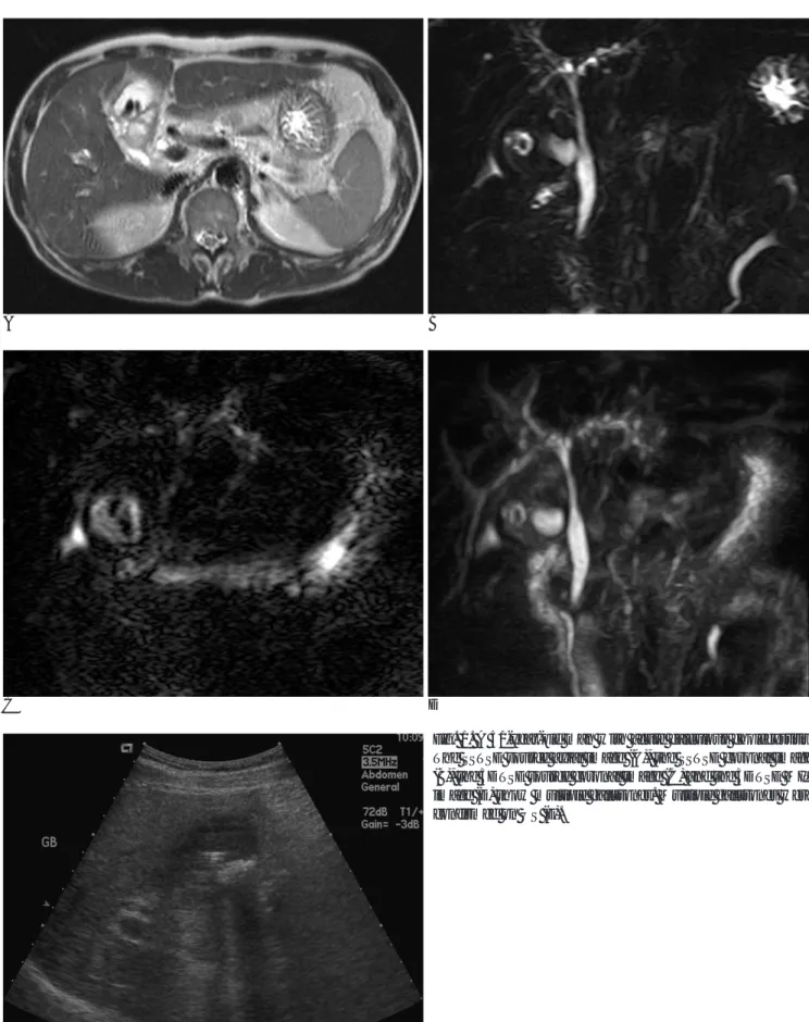
Fig. 1. A 51-year-old man with acute calculous cholecystitis.
The SSTSE source axial image (A), the SSTSE coronal image (B), the 3DTSE source coronal image (C) and the 3DTSE MIP image (D) show multiple gallstones. Multiple gallstones were confirmed on US (E).
A B
C D
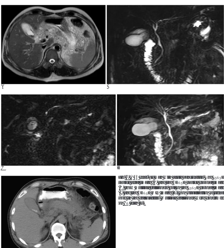
Fig. 2. A 33 year-old man with acute pancreatitis. The SSTSE source axial image (A) and the 3DTSE source coronal image (C) show multiple gallstones, but on the SSTSE coronal image (B) and the 3DTSE MIP image (D), non visualization of signal void in the gallbladder. Multiple gallstones were confirmed on CT scan (E).
로 지적되어 왔다(4). 이에 따라 다양한 펄스배열순을 이용한 MRCP가 시행되었고, 각각의 기법들과 이들의 비교에 대한 연 구가 보고되었으며, 최근에는 HASTE기법과 단발포급속스핀 에코기법이 움직임에 의한 인공물의 영향을 줄이고 공간해상 력을 높이며 신호 대 잡음비를 증가시킨다는 장점이 보고되었 다(15, 16). 또한 HASTE기법은 single-shot RARE기법과 비 교하였을 때 담낭내결석의 진단에 유용한 것으로 보고되었다 (6).
저자들의 연구에서 단발포고속스핀에코기법과 SENSE기술 을 도입한 3차원고속스핀에코기법을 이용하였을 때, 각각의 기 법에 따른 담낭내결석의 진단에 대한 정확도는 큰 차이를 보 이지 않았다. 그러나 두 기법 모두에서 원천절편영상의 정확도 가 이를 재구성한 단발포고속스핀에코기법의 관상면영상과 3 차원고속스핀에코기법의 삼차원영상에 비해 통계적으로 유의 하게 높았다. 이는 관상면영상이나 최대강도투사에 의해 재구 성된 삼차원영상이 담관암에서와 같이 전체적인 삼차원영상을 판독할 때는 도움이 되나 영상판두께에 따라 간혹 병변부위가 투사영상 내에 포함되지 않을 수도 있기 때문으로 생각되며 이 연구의 결과에서 보여주듯이 이러한 점을 보완하기 위해서는 원천절편영상에 대한 면밀한 판독이 필요하다(17).
그러나 이 연구는 이전의 보고들이 대부분 담관내결석을 대 상으로 한 것이어서 결과를 비교할 만한 기존의 보고가 없다 는 것과 수술을 시행한 경우를 제외하고 초음파나 CT 소견에 서 양성인 경우를 기준으로 하였기 때문에 담낭벽의 주름, 콜 레스테롤용종, 담낭과 인접한 석회화, 장관내가스 등과 같은 위 양성 또는 위음성인 경우도 포함될 수 있다는 제한점을 가지 고 있다.
결론적으로 담낭내결석에 대한 MRCP의 검출감도는 비교적 우수하며 특히 단발포고속스핀에코기법과 SENSE기술을 이용 한 3차원고속스핀에코기법 모두에서 정확도가 가장 우수한 원 천절편영상을 반드시 숙지하는 것이 진단에 도움이 될 것으로 생각된다.
참 고 문 헌
1. Coakley F, Schwartz L. Magnetic resonance cholangiopancreatog- raphy. J Magn Reson Imaging 1999;9:157-162
2. Gallix BP, Rengent D, Bruel JM. Use of magnetic resonance cholangiography in the diagnosis of choledocholithiasis. Abdom Imaging 2001;26:21-27
3. Boraschi P, Neri E, Braccini G, Gigoni R, Caraemlla D, Perri G.
Choledocholithiasis: diagnostic accuracy of MR cholangiopancre- atography. Three-year experience. Magn Reson Imaging 1999;17:
1245-1253
4. Aube C, Delorme B, Yzet T, Burtin P, Jerome L, Pessaux P, et al.
MR cholangiopancreatography versus endoscopic sonography in suspected common bile duct lithiasis: a prospective, comparative study. AJR Am J Roentgenol 2005;184:55-62
5. Bret PM, Reinhold C. Magnetic resonance cholangiopancreatogra- phy. Endoscopy 1997;29:472-486
6. Lee M-G, Jeong Y-K, Kim M-H, Lee S-G, Kang E-M, Chien D, et al.
MR cholangiopancreatography of pancreaticobiliary disease: com- paring single-shot RARE and multislice HASTE sequences. AJR Am J Roentgenol 1998;171:1539-1545
7. D. Pickuth. Radiologic diagnosis of common bile duct stones.
Abdom Imaging 2000;25:618-621
8. Hessler PC, Hill DS, Detorie FM, Rocco AF. High accuracy sono- graphic recognition of gallstones. AJR Am J Roentgenol 1980;136:
517-520
9. Havrilla TR, Reich NE, Haaga JR, Seidelmann FE, Cooperman AM, Alfidi RJ. Computed tomography of the gallbladder. AJR Am J Roentgenol 1978;130:1059-1067
10. Barakos JA, Ralls PW, Lapin SA, Jonson MB, Radin DR, Coletti PM, et al. Cholelithiasis: Evaluation with CT. Radiology 1987;162:415-418
11. Varghese JC, Liddell RP, Farrell MA, Murray FE, Osborne DH, Lee MJ. Diagnostic accuracy of magnetic resonance cholangiopan- creatography and ultrasound compared with direct cholangiogra- phy in the detection of cholodecholithiasis. Clinical Radiology 2000;55:25-35
12. Reinhold C, Taourel P, Bret PM, Cortas GA, Mehta SN, Barkun AN, et al. Choledocholithiasis: evaluation of MR cholangiography for diagnosis. Radiology 1998;209:435-442
13. Calvo MM, Bujanda L, Heras I, Calderon A, Cabriada JL, Orive V, et al. Magnetic Resonance cholangiography versus ultrasound in the evaluation of the Gallbladder. J Clin Gastroenterol 2002;34:233- 236
14. 오형진, 이재문, 정승은, 김응국, 한성태. 복강경하 담낭절제술 시행 전 자기공명 담도조영술의 유용성. 대한방사선의학회지 2000;42:
497-503
15. Becker CD, Grossholz M, Becker M, Mentha G, De Peyer R, Terrier F. Choledocholithiasis and bile duct stenosis: diagnostic ac- curacy of MR cholangiopancreatography. Radiology 1997;205:523- 530
16. 윤은주, 최철순, 윤대영, 윤영철, 박상준, 서영란 등. 자기공명담췌 관조영술: 단발포고속스핀에코(SS-TSE)기법과 SENSE 기술을 이용 한 3차원고속스핀에코(3D-TSE)기법의 영상비교. 대한방사선의학 회지 2003;49:483-488
17. 박성원, 백승연, 강병철, 이정식. 담석진단에 있어서 자기공명담관 조영술의 정확도 및 관찰자간의 일치도에 대한 평가. 대한방사선의 학회지 2001;44:577-582
J Korean Radiol Soc 2006;54:97-102
Address reprint requests to : Eun Joo Yun, M.D., Department of Radiology, Hallym University, Kangdong Sacred Heart Hospital, 445 Gil-Dong, Kangdong-gu, Seoul, 134-701, Korea.
Tel. 82-2-2224-2305 Fax. 82-2-488-7370 E-mail: [email protected]
The Detection of Gallstones on MR Cholangiopancreatography:
Comparison between the Single-Shot Turbo Spin-Echo Pulse Sequence and the Three-Dimensional Turbo Spin-Echo Pulse Sequence with the SENSE Technique
1Ju Ae Kim, M.D., Eun Joo Yun, M.D., Chul Soon Choi, M.D., Dae Young Yoon, M.D., Sang Joon Park, M.D., Young Lan Seo, M.D., Yu-Jin Lee, M.D., Jeung Hee Moon, M.D.
1Department of Radiology, Hallym University College of Medicine
Purpose: We wanted to evaluate the detectability of gallstones on magnetic resonance cholangiopancreatogra- phy (MRCP) and to compare the accuracy between the single-shot turbo spin-echo (SSTSE) sequence and the three-dimensional turbo spin-echo (3DTSE) sequence with the sensitivity encoding (SENSE) technique.
Materials and Methods: A total of 141 patients who had undergone MRCP for a year period since August, 2003 were involved in the study. The source axial-SSTSE, coronal-SSTSE, source coronal-3D TSE and maximum in- tensity projection (MIP)-3DTSE images were obtained. Based on the operative findings and the findings of the ultrasound and CT examinations, the results of the reading by two investigators for the presence of gallstones were compared and analyzed.
Results: Among 141 patients, 135 patients were included in the study. 69 cases (51%) were found to have gall- stones. In terms of detection of gallstones, the accuracy was 85%. The reading by one investigator greatly ac- corded with that of the other investigator (κ=0.94). As a result of comparing the four kinds of images obtained with the different techniques, it was found that gallstones were seen best on the source axial-SSTSE and source coronal-3DTSE images; the coronal-SSTSE image was the next best image and the MIP-3DTSE image followed (p<0.01).
Conclusion: The detectability of gallstones on MRCP was relatively excellent and the source axial-SSTSE and source coronal-3DTSE imagings should be included for the detection of gallstones.
Index words :Magnetic resonance (MR) Gallbladder
Gallstone