INTRODUCTION
An epithelioid hemangioendothelioma is an extremely rare tumor classified as an intermediate grade vascular tumor.
This tumor can arise in soft tissue and deep organs such as the lung and liver, and in rare cases, within bone (1). In this article, we describe a case of epithelioid hemangioendothelio- ma of the femur that was mistaken for a benign lesion, such as a simple bone cyst and fibrous dysplasia, because the tu- mor showed an unusual cystic appearance on MR imaging which to the best of our knowledge has not yet been reported in the current literature.
CASE REPORT
A 77-year-old female presented with pain and swelling in her right thigh after falling down and complained of resting pain of the right thigh for 2 years before the time she was in-
jured. A plain radiography showed a comminuted fracture with displacement in the middle of the femur shaft and an underlying osteolytic lesion of the femur shaft. She did not have any underlying disease or history of cancer. She had total knee replacement arthroplasty of the right knee 11 years ago and the radiograph obtained at that time was not available.
Radiography of the right femur demonstrated an osteolytic lesion involving almost the entire right femur, except for the head and trochanters. The lesion was confined to be in the medullary cavity. Cortical bone was markedly thinned, but there was neither cortical disruption nor a periosteal reaction.
The lesion showed a lobulated contour and was septated into multiple lobules by thin bony septae. The border was well de- marcated and slightly sclerotic (Fig. 1).
MR images of the right thigh showed a mass, measuring about 2.8 × 2.4 × 29 cm, replacing most of the normal medul- la of the femur. The mass appeared to arise from the medul- lary cavity and extend to the adjacent cortex. There was no
J Korean Soc Radiol 2011;65(6):607-611
Received June 3, 2011; Accepted September 19, 2011 Corresponding author: Jung-Ah Choi, MD Department of Radiology, Seoul National University Bundang Hospital, 166 Gumi-ro, Bundang-gu, Seongnam 463-707, Korea.
Tel. 82-31-787-7609 Fax. 82-31-787-4011 E-mail: [email protected]
Copyrights © 2011 The Korean Society of Radiology
An epithelioid hemangioendothelioma is an intermediate grade tumor between hemangioma and angiosarcoma that frequently shows marked enhancement be- cause it is a vascular tumor. Herein, we describe a rare case of a malignant epitheli- oid hemangioendothelioma of the femur that was mistaken as a benign lesion such as a simple bone cyst or fibrous dysplasia because the tumor had a benign cystic appearance on MRI and its imaging findings showed a histopathologic correlation.
Index terms Bone Neoplasms
Bone Neoplasms, Diagnosis
Bone Neoplasms, Magnetic Resonance Hemangioendothelioma, Epithelioid
Epithelioid Hemangioendothelioma of the Femur with Benign Cystic Appearance
1낭성 종괴의 자기공명영상 소견을 보인 대퇴골의 상피양혈관내피세포종1
Yeo Goon Kim, MD
2, Jung-Ah Choi, MD
1,2, Jin-Haeng Chung, MD
3, Joo Han Oh, MD
4, Heung Sik Kang, MD
1,21Department of Radiology, Seoul National University Bundang Hospital, Seongnam, Korea
2Department of Radiology and Institute of Radiation Medicine, Seoul National University College of Medicine, Seoul, Korea Departments of 3Pathology, 4Orthopedic Surgery, Seoul National University Bundang Hospital, Seongnam, Korea
mass was isointense to muscle on the T1-weighted image and hyperintense on the T2-weighted image. The rim and septum were hyperintense on the T1-weighted image and hypoin- tense on the T2-weighted image. After injecting gadolinium, the rim and septum were enhanced, whereas the mass itself did not show any enhancement (Fig. 2). The radiologic differ- apparent soft tissue involvement, but focal cortical disruption
was noted on a few axial images. The margin of the lesion was distinct and lobulated. The mass was septated by incomplete thin septa. Femoral shaft fracture and associated findings such as intraosseous hemorrhage and soft tissue edema were found around the fracture site. Regarding signal intensity, the
Fig. 1. On the right femur AP (A) and lateral (B) views, an elongated, radiolucent, slightly expansile lesion is seen at right femur shaft, extending from the proximal metadiaphysis to the distal metaphysic area. A pathologic fracture is noted at distal 2/3 of the femur (arrowheads).
Note.-AP = anteroposterior A
A
B
B C
Fig. 2. MR images of right femur.
A. On a coronal T2-weighted image (TR/TE = 3,734/100), the lesion extends from the proximal metadiaphysis area spanning from just below the intertrochanteric area to the distal femur up to the prosthesis. The lesion shows high signal intensity with focal intermediate to low signal inten- sity foci and numerous septae (arrowheads). A fracture is noted at distal two-thirds of femur.
B. On a sagittal T1-weighted image (TR/TE = 421/7), the lesion is shown to have intermediate signal intensity similar to muscle.
C. On a coronal post-contrast fat suppression T1-weighted image (TR/TE = 600/6), the lesion shows thin peripheral enhancement and enhance- ment of the septae (arrowheads).
Note.-TR = repetition time, TE = echo time
lung are the most frequently involved sites, but EH can be found in soft tissues and bones. The peak incidence occurs in the second and the third decades, but EH can occur at almost any age (7-76) (2). Unlike EH in other locations, osseous EH has a predilection for occurrence in males (male to female ra- tio is 2 : 1).
Osseous EH involves skull, axial skeleton, and the lower ex- tremities in most cases. The most frequently involved long bones are the tibia (23% of cases), femur (18%), and humerus (13%) (3). In over 50% of cases, lesions are multifocal, espe- cially with involvement of the lower extremity. Multifocal le- sions have a tendency to involve bones of the same region.
Synchronous involvement of contiguous bones is common, such as the tibia and fibula, but in some cases, separated tu- mor foci are present in distant bones.
Clinically, local pain and swelling that last weeks to years are the most common chief complaints. A pathologic fracture can be associated in 10% of patients (3). Constitutional symp- toms, such as hemolytic anemia and consumption coagulopa- thy are reported but rare (4).
Radiologically, EH presents as osteolysis without mineral- ization. Calcifications and periosteal reaction are rare. Lesions may be well defined or poorly demarcated. Expansile remod- eling, cortical disruption and soft tissue extension can be seen. Most of them develop in the metaphysis and diaphysis, but the epiphysis can be involved. Multifocal neoplastic foci in cortical or medullary bones can appear as a bubble-like ential diagnoses included a simple bone cyst, fibrous dyspla-
sia with cystic degeneration, or cystic angiomatosis.
The surgical plan was made to perform curettage, because the tumor was thought to be a benign lesion. On pathologic evaluation, there were aberrant vessel formations and extrav- asated red blood cells within the tumor. No involvement of skeletal muscle and adipose tissue was noted and increased cellularity and moderate nuclear pleomorphism were seen with mitosis of up to 12/10 high power field (Fig. 3). Surpris- ingly, necrosis was present in less than 10% of the tumor. The initial differential diagnosis included angiosarcoma. Immu- nohistochemistry results revealed a malignant tumor with ex- pression of endothelial markers CD31 and CD34. The final histopathologic diagnosis was malignant epithelioid heman- gioendothelioma of bone. The patient underwent adjuvant ra- diotherapy after pathologic confirmation, and showed no evi- dence of disease at 2 years of follow-up.
DISCUSSION
Epithelioid hemangiothelioma (EH) is an extremely rare vascular tumor. EH is classified as an intermediate-grade vas- cular tumor, whose clinical course and prognosis are some- where between hemangioma and angiosarcoma. In 1982, Weiss and Enzinger first used the term epithelioid hemangio- endothelioma to describe a vascular tumor originating from the endothelial cell of epithelioid appearance (1). Liver and
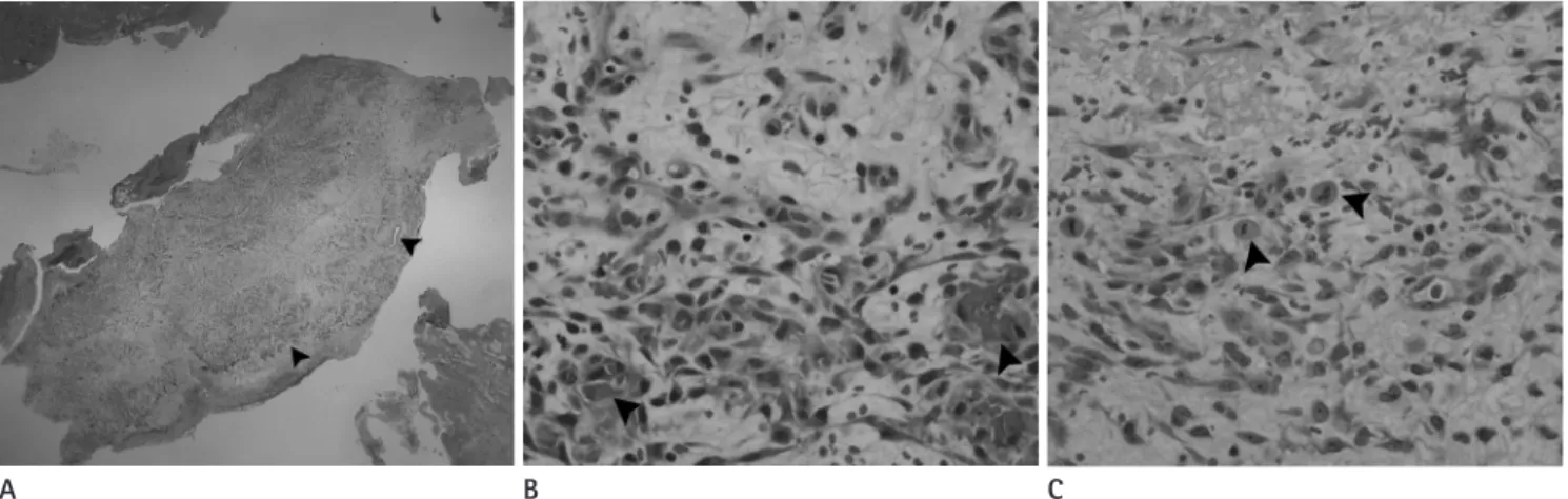
A B C
Fig. 3. Histopathologic specimen images.
A. On histopathologic examination, endothelial lining at the margin of the tumor and numerous vessels with RBCs (arrowheads) are seen in fragments of tissue obtained by curettage (H&E, × 10).
B. Aberrant vessel formation and extravasated RBCs (arrowheads) are characteristic (H&E, × 400).
C. Atypical mitoses (arrowheads) are common (H&E, × 400).
Note.-RBC = red blood cell
centric disease.
In our case, EH showed a cystic appearance with rim and septal enhancement, which is unusual in a vascular tumor. Ra- diologists should be aware of this cystic benign-appearing ma- lignant bone tumor, as this benign cystic appearance can lead to misdiagnosis and improper treatment of the patient.
REFERENCES
1. Weiss SW, Enzinger FM. Epithelioid hemangioendothelioma:
a vascular tumor often mistaken for a carcinoma. Cancer 1982;50:970-981
2. Vigorita VJ, Ghelman B, Bernard Ghelman. Vascular/Mes- enchymal tumors. In Orthopaedic pathology. Philadelphia:
Lippincott Williams & Wilkins, 1999;399-400
3. Ignacio EA, Palmer KM, Mathur SC, Schwartz AM, Olan WJ.
Epithelioid hemangioendothelioma of the lower extremity.
Radiographics 1999;19:531-537
4. Larochelle O, Périgny M, Lagacé R, Dion N, Giguére C. Best cases from the AFIP: epithelioid hemangioendothelioma of bone. Radiographics 2006;26:265-270
5. Murphey MD, Fairbairn KJ, Parman LM, Baxter KG, Parsa MB, Smith WS. From the archives of the AFIP. Musculosk- eletal angiomatous lesions: radiologic-pathologic correla- tion. Radiographics 1995;15:893-917
6. Dorfman HD, Czerniak B. Vascular lesions. In Bone tumors.
Mosby 1998;769-795
7. Kleer CG, Unni KK, McLeod RA. Epithelioid hemangioen- dothelioma of bone. Am J Surg Pathol 1996;20:1301-1311 8. Kabukçuogˇ lu F, Kabukçuogˇ lu Y, Livaogˇ lu A, Ozagˇ ari A,
Armagˇ an R, Kuzgun U. [Epithelioid hemangioendothelioma of bone]. Acta Orthop Traumatol Turc 2006;40:324-328 enhanced CT scan. The signal intensity on MR imaging is not
specific. EH has been reported to show low to intermediate signal intensity on T1-weighted image and high signal inten- sity on T2-weighted images. After injecting gadolinium con- trast agent, the mass has been reported to show homogeneous enhancement (5). The presence of flow voids, suggestive of vascular channels, may represent a neoplasm of vascular ori- gin, but this finding does not indicate hemangioendothelio- ma, but rather should suggest other diagnosis such as heman- giopericytoma (5).
The gross features of EH is a bright red hemorrhagic tumor with irregular scalloped borders. Microscopically, the EH usu- ally consists of an anastomosing cord of epithelioid cells that occasionally form poorly defined vascular channels. Cells are plump with abundant, granular, eosinophilic cytoplasm. The vacuolization of cytoplasm is characteristic, presenting at- tempts to form a primitive vascular lumen. The nuclei are round with prominent nucleoli. The individual epitheliod cell cytoplasmic lumen may contain red blood cells. Mitotic activ- ity is usually low with 1 to 2 mitoses per 10 high power fields.
Epithelioid cells express endothelial markers such as factor VIII-related antigen, Ulex europaeus lectin, as well as CD31 and CD34 (6).
The clinical course and prognosis of EH is somewhere be- tween that of hemangioma and angiosarcoma. Most cases show locally destructive indolent behavior, but the prognosis of EH of bone is hardly predictable because prognosis cannot be made on the basis of histologic grade alone (4). Visceral involvement seems to be the most important indicating poor prognosis (7). There is no established standard treatment of EH of bone. The number, size, location, and presence of me- tastasis determine the treatment. For localized disease, wide
낭성 종괴의 자기공명영상 소견을 보인 대퇴골의 상피양혈관내피세포종1
김여군
2· 최정아
1,2· 정진행
3· 오주한
4· 강흥식
1,2상피양혈관내피세포종은 혈관종과 혈관육종 사이의 중증도 악성종양으로, 혈관성 종양이므로 균질하고 분명한 조영증 강을 보이는 것으로 알려져 있다. 저자들은 대퇴골에 발생한 혈관내피세포종이 MR에서 낭성 종괴로 보여 양성종양과의 감별이 어려웠던 증례를 경험하여 이 증례의 영상 소견과 병리 소견을 비교하여 보고하고자 한다.
1분당서울대학교병원 영상의학과, 2서울대학교 의과대학 영상의학과학교실, 분당서울대학교병원 3병리과, 4정형외과