천대퇴동맥(superficial femoral artery, 이하 SFA)의 동 맥경화성 협착 또는 폐쇄의 치료 방법으로 풍선 카테터를 이용 한 경피적경혈관성형술(percutaneous transluminal angioplasty, 이하 PTA)은 높은 성공률을 보이고 있다. 하지 만, 병변의 협착 범위가 길거나 완전 폐색인 경우, 석회성 협 착, 한쪽으로 치우친 병변, 그리고 PTA 후 발생한 동맥벽의 박리 등은 재발률과 합병증의 발생률이 높은 것으로 알려져 있 다(1-3). 최근에는 혈관의 개통률 향상을 위해 PTA와 함께 스텐트 삽입을 이용한 치료가 시행되고 있으며 3년 개통률이
60% 이상으로 높은 것으로 보고되고 있다(2, 4, 5). 그러나 천대퇴동맥 자체의 해부학적, 혈류역학적 특징과 스텐트의 형 태학적 변화 때문에, 스텐트 삽입을 이용한 치료의 유용성은 논란이 되고 있다(2, 3, 6).
최근 보고들에 따르면 천대퇴동맥에 삽입된 스텐트 자체의 골절이 비교적 흔하게 발생하며, 재협착의 중요한 원인이 될 수 있다고 한다(7-9). 하지만, 대부분이 다양한 병변의 길이와 여러 종류의 스텐트를 대상으로 한 연구로써 비교 연구에 제한 이 있었다. 본 연구는 단일 스텐트를 이용한 연구로써 15 cm 이상 폐쇄 병변을 대상으로 하여 골절의 위험인자 및 재협착과 의 연관성에 대하여 알아보고자 하였다.
천대퇴동맥 스텐트 골절 발생의 위험요인 및 임상적 연구:
Prote ′ ge ′GPS 스텐트
1이다운∙김재규∙정혜두∙허태욱∙임남열∙오현준∙장남규2∙최수진나3
목적: 천대퇴동맥 스텐트(Prote′ge′GPS) 골절의 발생빈도를 평가하고, 스텐트 골절과 원인 인 자 및 임상 양상의 연관성에 대해 알아보고자 하였다.
대상과 방법: 천대퇴동맥에 발생한 동맥경화성 폐쇄 또는 협착을 Prote′ge′GPS 스텐트 삽입으
로 치료하였던 환자 38명 중 스텐트 모양의 추적관찰이 가능하였던 17 명을 대상으로 하였다 (남자 15명, 여자 2명, 평균연령 72.8세). 총 17개의 천대퇴동맥에 43개의 스텐트가 삽입되었 고, 병변의 평균 길이는 약 26.4 cm (15-50 cm) 이었다. 시술에 사용되었던 혈관 조영기기를 이용하여 전후, 측면 단순 영상을 획득 후 골절을 평가하였으며, 동시에 색 도플러 초음파, 카테 터혈관조영술 또는 CT혈관조영술을 이용하여 스텐트 내부 혈류를 평가하였다. 스텐트의 길이, 위치 및 수 등에 따른 골절 발생률의 차이를 분석하였으며 골절과 개통률의 차이도 알아보고자 하였다.
결과: 스텐트의 골절은 43개의 스텐트 중 14개(30.2%)에서 발생하였다. 10개의 스텐트 (71.4%)가 천대퇴동맥의 상부 1/3, 4개의 스텐트(28.6%)가 하부 1/3에서 골절이 발생하였 다. 단일 strut 골절이 10예(71.4%), 2개 이상의 strut 골절이 4예(28.6%) 있었다. 병변 및 삽입된 스텐트의 길이가 길수록 스텐트 골절의 발생빈도가 높았다(각각p=0.021, 0.012). 관 절과 가까이 스텐트가 삽입될수록 골절 발생빈도가 높았으며 오금동맥까지 연장하여 스텐트가 삽입될수록 하부 1/3에 골절이 잘 발생하였다(hip joint; p=0.01, knee joint: 0.029, insertion in popliteal a.: 0.015). 스텐트 개수와 골절간의 유의한 상관관계는 보이지 않았 으며(p=0.126), 골절 여부에 따른 혈관 개통률은 의의 있는 차이를 보이지 않았다(p=0.44).
결론: 천대퇴동맥에 삽입했던 Prote′ge′GPS 스텐트는 추적기간에 비교적 많은 골절이 발생하 였고, 병변과 스텐트의 길이 및 관절과의 거리와 유의한 상관관계를 보였다. 골절 여부와 개통 률 사이에 의미 있는 관계는 없었다.
1전남대학교 의과대학 영상의학교실
2화순전남대학교병원 영상의학교실
3전남대학교 의과대학 외과학교실
이 논문은 2010년 4월 26일 접수하여 2010년 8월 2일에 채택되었음.
대상과 방법
연구대상
2006년 8월부터 2008년 8월까지 CT 및 혈관조영상 천대퇴 동맥에 국한되어 폐쇄 병변을 보인 환자 중 Prote′ge′GPS 자 가 확장성 니티놀 스텐트(eV3, Minneapolis, MN, USA)를 삽입한 38명 중 추적관찰 기간이 3개월 이상, 병변의 길이가 15 cm 이상, 그리고 투시 촬영 및 단순 촬영을 이용해 스텐트 모양의 추적 관찰이 가능한 17명을 대상으로 하였다. 남녀 비 는 15:2이었으며 평균연령은 72.8세(58-89세)였다.
Rutherford-Becker category의 분류에 의한 환자들의 임상 증상은 간헐성 파행 3명, 휴식기 동통 8명, 하지 괴저 6명이었 다.
시술
스텐트 일차 삽입 기준은 완전 폐쇄, 90% 이상의 협착 또는 동맥벽의 박리가 있는 경우이며, 이차 삽입 기준은 먼저 PTA 를 시행한 후, 남아 있는 협착이 30% 이상이거나 동맥벽의 박 리가 의심되는 경우였다. 17명의 환자에서 총 17개의 천대퇴동 맥에 43개의 스텐트를 삽입하였다. 시술의 기술적인 성공 여부 는 스텐트 내경이 30% 이상의 협착부위 없이 완전히 확장되 고, 조영 검사 상 병변 하방으로 혈류가 잘 유지되는 것으로 하 였으며, 시술 직후 모든 환자에서 혈관의 확장과 병변 하방으 로 잘 유지되는 혈류를 확인할 수 있었다. 기술적으로 스텐트 삽입이 실패한 경우는 없었다. 임상적으로는 Rutherford- Becker category가 최소 한 단계 이상 상승하는 것으로 평가 하였다.
추적 조사
추적 관찰기간은 재발한 경우에는 재협착이 진단된 날로 하 였고 재발하지 않은 경우에는 마지막 검사일로 하였으며 6개 월에서 29개월로 평균 12.9개월이었다. 추적 관찰은 혈관 조 영기기인 Multistar T.O.P (Siemens, Erlangen, Germany)를 이용하여 스텐트를 전후, 측면에서 촬영하여 골 절 양상을 분석하였으며 추가로 색도플러초음파, 카테터혈관 조영술, 또는 CT혈관조영술을 이용하여 스텐트 내부의 혈류를 평가하였다. 골절의 분류는 Cardiovascular Institute of the South (CIS)에서 발표한 stent fracture classification system의 분류법을 따랐다(Fig. 1)(7).
모든 환자는 시술 후 아스피린(325 mg/일)과 Ticlid (500 mg/bid)를 3개월간 복용하였다. 재협착은 임상적으로 재발이 의심된 경우에 추적 혈관 조영술상 치료받은 동맥의 직경이 30% 이상 감소하였거나, 색도플러 검사 상 PSVR (peak systolic velocity ratio)이 2배 이상 되거나 도플러 파형이 폐 쇄 소견을 보인 경우로 정의하였다(10).
분석
환자의 연령, 성별, 병변 및 스텐트의 길이, 스텐트의 수와 스텐트 골절의 위험을 분석하였다. 관절과 가까이 스텐트가 위 치할수록 골절위험이 클 것이라는 가정 하에 혈관조영술시 대 퇴골두(femoral head)와 대퇴골과간함요(intercondylar fossa)를 경계지표로 이용하였으며 이들과 스텐트와의 간격을
Fig. 1. Nitinol stent fracture classification system from cardio- vascular institute of the south (7).
Type I A single strut fracture only
Type II Multiple single nitinol stent fractures that can occur at different sites
Type III Multiple nitinol stent fractures resulting in complete transverse linear fracture, but with- out stent displacement
Type IV A complete transverse linear type III fracture with stent displacement.
Type I Type II Type III Type IV
A B
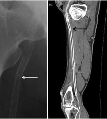
Fig. 2. 64-year-old man with right leg claudication.
A. Type I single fracture (arrow) occur at overlapping zone in the proximal superficial femoral artery 5 months after impan- tation.
B. Note the reocclusion of the superficial femoral artery.
측정하였다. 상부 1/3 골절의 위험 요인으로 총대퇴동맥에 삽 입 여부 및 대퇴골두와 스텐트 간의 거리를 분석하였고 하부 1/3 골절의 위험 요인으로 오금동맥에 삽입 여부 및 대퇴골과 간함요와 스텐트 간의 거리를 분석하였다.
통계
총 43개의 스텐트를 대상으로 스텐트 골절과 위험 요인과의 관계를 Logistic regression analysis를 이용하여 알아보았고 골절과 재발률을 Kaplan-Meier method 및 log-rank test를 이용하여 분석하였다.
결 과
병변의 평균 길이는 26.4 cm (15-50 cm)이였다. 2007
TransAtlantic Inter-Society Consensus (TASC)에 의한 병변의 분류는 Type C 8명, Type D 9명이었으며, Type D 병변은 모두 오금동맥까지 병변이 확장되어 있었다. 일차 스텐 트 삽입 동맥은 8개, 이차 스텐트 삽입 동맥은 나머지 9개였 다. 1개의 동맥에 평균 2.53 개의 스텐트가 삽입되었으며(1 개: 1명, 2개: 7명, 3개: 8명, 4개: 1명), 천대퇴동맥에 삽입 된 스텐트의 길이는 평균 27.5 cm (8-38 cm)이었다. 17개의 동맥 중 11개의 동맥에서 천대퇴동맥 전체에 걸쳐 스텐트가 삽입되었고, 1개의 동맥에서 중부 1/3, 5개의 동맥에서 상부 와 중부 1/3에 스텐트가 삽입되었다. 삽입된 43개 스텐트 중 14개(30.2%, 11/17명 환자)에서 골절이 발생하였다. 14개 골 절 중 10개(71.4%)는 1형 단순 골절(single strut fracture), 4개(28.6%)는 II형 다중 단순 골절(multiple single fracture)이었다(Figs. 2, 3). 특히 10개(71.4%)의 골절이
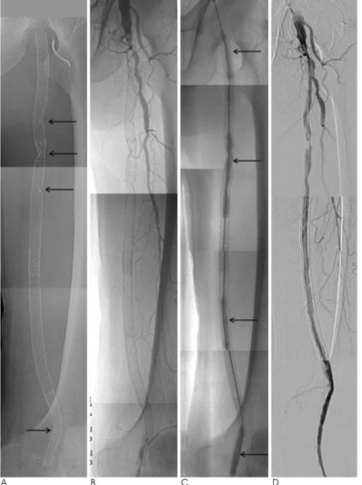
A B C D
Fig. 3. A, B. 72-year-old man with type I and II stent fractures (arrows) with total occlusion of SFA at 16 months af- ter placement of four stents in the SFA and the popliteal artery.
C, D. After balloon angioplasty (ar- rows) following thrombolysis with in- fusion of UK 240,000 U, SFA is re- canalized with residual stenosis.
천대퇴동맥의 상부 1/3에서 발생하였으며, 4개(28.6%)가 하 부1/3에서 발생하였다.
스텐트 골절 발생의 위험요인을 분석한 결과는 표 1과 같다.
병변 및 삽입된 스텐트의 길이가 길수록 스텐트 골절의 발생빈 도가 높았으나(각각p = 0.021, 0.012) 삽입된 스텐트의 수 와의 유의한 연관관계는 없었다(p = 0.126). 오금동맥에 연 장하여 삽입될수록, 대퇴골과간함요와 가까이 삽입될수록 하 부 골절이 잘 발생하였다(p = 0.015, 0.029). 대퇴골두와 가 까이 삽입될수록 상부 골절이 잘 발생(p = 0.012) 하였으나 총대퇴동맥 삽입 여부와 골절 발생 빈도와는 통계학적으로 유
의한 상관관계가 없었다(p = 0.402).
2개의 스텐트에서 스텐트 겹침 부위에 골절이 발생하였으며 (Fig. 2) 나머지 12개의 스텐트에서 골절부위와 관계없이 재 협착이 발생하였다(Fig. 3). 전체 17명의 환자 중 7명의 환자 에서 시술 후 증상의 호전(Rutherford- Becker category가 1 단계 이상 상승)을, 3명의 환자에서는 증상이 유지되었으며, 5명의 환자에서 절단 수술을, 2 명의 환자에서 우회로 조성 수 술을 받았다.
Kaplan-Meier 방법을 통해 분석한 6개월 누적 개통률은 골절이 없었던 환자에서 50%, 골절이 발생한 환자에서 45.5%로 골절이 없으면 개통률이 비교적 높았으나 이들 사이 에 연관관계는 없었다 (p = 0.44) (Fig. 4).
고 찰
천대퇴동맥은 가장 긴 말초 동맥으로 주변 근육 및 관절의 움 직임에 의하여 다양한 힘을 받게 되며 이에 의하여 재협착이 빈 번히 발생하고, 골절 또한 잘 발생할 것으로 생각한다. Siroli- mus Coated Cordis Self-expandable Stent (SIROCCO) trial (9)에서 스텐트 골절이 발생할 수 있음을 처음으로 보고 하였고 이후 이에 관한 몇몇 연구들이 발표되고 있다.
최근 발표된 연구들에서 스텐트의 길이가 길수록, 삽입한 스 텐트의 수가 많을수록 골절이 잘 발생하고, 스텐트의 골절양상 이 심할수록 재협착의 빈도 역시 높아진다고 보고하였다(7, 8, 10). 스텐트 골절이 발생할수록 재협착이 잘 발생한다고 보고
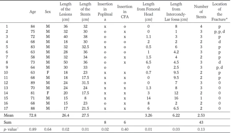
Table 1. Risk Factors of Stent Fracture
Length Length Insertion
Insertion Length Length
Number Location
Age Sex of the of the in
in from Femoral from
of of
Lesions Stents Popliteal
CFA Head Intercondy-
Stents the
(cm) (cm) a (cm) Lar fossa (cm) Fracture*
01 84 M 36 32 x o 0 8 4 p
02 75 M 32 30 o x 0 1 3 p, p, d
03 72 M 40 38 o x 1.1 3 3 p
04 68 M 18 30 o o 2 2 2 d
05 83 M 32 32.5 x o 0.5 6 3 p
06 63 M 28 36 o o 1 4.2 3 p
07 58 M 32 34 o x 1.5 4 2 p
08 73 M 50 36 o x 6.5 4.5 3 d
09 64 M 30 33 o 0 2.5 3 p, d
10 63 F 18 23 x x 0.7 9.5 2 p
11 68 M 18 17.5 x x 0 9.5 2 p
12 89 M 24 31.5 x o 0 7 3 0
13 70 M 24 24 x x 1.3 8 3 0
14 81 F 20 17.5 x x 3 12 2 0
15 71 M 15 8 x x 14 16 1 0
16 68 M 15 23 o x 8 2 2 0
17 88 M 17 21.5 x x 6 6.5 2 0
Mean 72.8 26.4 27.5 3.26 6.22 2.53
Sum 8 6 43
p- value� 0.89 0.64 0.02 0.01 0.02 0.40 0.01 0.03 0.13
Note.─* p: proximal 1/3 of SFA, d: distal 1/3 of SFA
�available, if less than 0.05
Fig. 4. Primary stent patency rates for fractured and non-frac- tured stents (p=0.440).
되고 있지만 이에 대하여 논란이 있다(11-13).
본 연구에서 사용된 Prote′ge′GPS 자가 확장성 니티놀 스텐 트는 intracoil 스텐트로 횡축으로는 방사 강도(radial strength)를 종축으로는 유연성을 가져 주변 근육에 의한 압 박, 구부림, 비틀림, 신장, 수축 등에 의한 스텐트의 변형을 최 소화할 수 있게 제작되었다. 실제로 Allie 등(7)에 의한 연구 에 따르면 intracoil (eV3, Plymouth, MN, USA) 스텐트에 서 골절이 발생하지 않았으며, 여러 스텐트를 이용한 Scheinert 등(11)에 의한 연구에서 병변의 길이가 8 cm에서 16 cm 미만의 병변에서 42.4%, 16 cm 이상에서 52.0%의 높 은 골절 발생률을 보였던 것과 달리 이 연구에서는 30.2%로 비교적 낮았다.
본 연구에서 스텐트 골절은 병변 및 스텐트의 길이가 길수록 빈번하게 발생하였으나 삽입된 개수가 많아 중첩될수록 축성 경직(axial stiffness)이 증가하여 골절이 잘 발생한다는 이전 에 제기되었던 연구 결과(11)와 달리 스텐트 숫자와의 관련성 은 없었다(p = 0.126). 실제로 14개의 스텐트 골절 중 스텐트 겹침 부위에서 골절은 두 개의 스텐트에서 발생하였고(Fig.
2) 재협착은 골절 부위에 관계없이 발생하였다(Fig. 3).
Scheinert 등(11)에 따르며 스텐트 골절이 천대퇴동맥에 고르게 발생하였으나 본 연구에서 13개의 골절 중 10개 (71.4%)가 상부 1/3에 분포하였으며 이는 1개를 제외한 나머 지 동맥에서 모두 상부 1/3에 스텐트가 삽입되었기 때문으로 생각한다. 관절과 가까이 스텐트가 삽입될수록 상부와 하부 1/3에 골절이 발생할 위험이 컸다. 또한, 오금동맥에 연장하여 스텐트가 삽입될수록 하부 1/3 골절의 발생 위험이 컸다. 총대 퇴동맥 내 스텐트 삽입 여부와 상부 1/3 골절의 발생 여부는 통계학적으로 유의한 상관관계를 보이지 않았다(p = 0.40).
골절과 재협착 간의 관계에 대하여 다양한 보고가 있지만 본 연구에서 Kaplan-Meier 방법을 통해 분석한 6개월 누적 개 통률은 골절이 없었던 환자에서 50%, 골절이 발생한 환자에 서 45%로 골절이 없으면 개통률이 비교적 높았으나 통계적으 로 유의하지 않았다(p = 0.440). 이는 본 연구에서 type III 이상의 심각한 골절이 없었으며 병변이 길어 골절에 관계없이 재협착 발생률이 높았기 때문으로 생각한다. 실제로 삽입된 스 텐트의 평균길이가 Schlager 등(8)은 13.9 ± 1.8 cm, Scheinert 등 (11)은 15.7 ± 5.9 cm였던 반면 본 연구에서 는 평균 27.5 cm로 길었고, 1명의 환자를 제외한 나머지 16명 의 환자에서 추적 기간에 재협착이 일어났다. 하지만, 본 연구 에서와같이 길게 삽입된 스텐트에서 스텐트의 숫자와 골절 발 생과의 연관성 및 골절과 재협착과의 관계에 대한 보고가 없어 이에 대한 추가적인 비교 연구가 필요할 것으로 생각한다.
본 연구는 17명의 작은 수의 환자를 연구 대상으로 하여 각 각의 요인들 간에 통계적으로 의의 있는 결과들을 도출해 내기
어려웠으며, 충분한 장기간의 추적 검사가 이뤄지지 못한 제한 점이 있다.
결론적으로 천대퇴동맥에 삽입했던 Prote′ge′GPS 스텐트는 추적 기간에 비교적 많은 골절이 발생했고, 발생빈도는 병변 및 스텐트의 길이와 유의한 상관관계를 보였으며 관절과 가까 이 스텐트가 삽입될수록 골절의 발생 빈도는 높아진다. 골절과 재협착 간의 유의한 상관관계는 보이지 않으나 삽입된 스텐트 의 길이에 따른 추가적인 연구조사가 필요할 것으로 생각한다.
참 고 문 헌
1. Chung SK, Kim JK, Yoon W, Kim J, Park JG, Kang HG, et al.
Metallic stent implantation in patients with iliac artery occlusion:
long term patency rate and factors related recurrence. J Korean Radiol Soc 2003;49:173-179
2. Lugmayr HF, Holzer H, Kastner M, Riedelsberger H, Auterith A.
Treatment of complex arteriosclerotic lesions with nitinol stents in the superficial femoral and popliteal arteries: a midterm follow-up.
Radiology 2002;222:37-43
3. Jahnke T, Voshage G, Muller-Hulsbeck S, Grimm J, Heller M, Brossmann J. Endovascular placement of self-expanding nitinol coil stents for the treatment of femoropopliteal obstructive disease.
J Vasc Interv Radiol 2002;13:257-266
4. Vogel TR, Shindelman LE, Nackman GB, Graham AM. Efficacious use of nitinol stents in the femoral and popliteal arteries. J Vasc Surg 2003;38:1178-1184
5. Ruef J, Hofmann M, Haase J. Endovascular interventions in iliac and infrainguinal occlusive artery disease. J Interven Cardiol 2004;
17:427-435
6. Dorrucci V. Treatment of superficial femoral artery occlusive dis- ease. J Cardiovasc Surg 2004;45:193-201
7. Allie DE, Hebert CJ, Walker CM, Nitinol stent fracture in the SFA.
Endovascular Today 2004;(7):22-34
8. Schlager O, Dick P, Sabeti S, Amighi J, Mlekusch W, Minar E, et al. Long-segment SFA stenting-the dark sides: in-stent restenosis, clinical deterioration, and stent fractures. J Endovasc Ther 2005;
12:676-684
9. Duda SH, Pusich B, Richter G, Landwehr P, Oliva VL, Tielbeek A, et al. Sirolimus-eluting stents for the treatment of obstructive su- perficial femoral artery disease: six-month results. Circulation 2002;106:1505-1509
10. Kim SK, Yang JS, Han JH, Chang NK, Yim NY, Yoon W, et al.
Endovascular stent placement for the treatment of superficial femoral artery stenoses and occlusions. J Korean Radiol Soc 2008;58:365-373
11. Scheinert D, Scheinert S, Sax J, Piorkowski C, Braunlich S, Ulrich M, et al. Prevalence and clinical impact of stent fractures after femoropopliteal stenting. J Am Coll Cardiol 2005;45:312-315 12. Ansel GM. Are stent fractures significant? Endovascular Today
2006;(11):45-52
13. Ansel GM. Clinical significance of stent fractures, Endovascular Today 2006;(5):37-40
J Korean Soc Radiol 2010;63:513-518
Address reprint requests to : Jae-Kyu Kim, M.D., Department of Radiology, Chonnam National University, School of Medicine, Chonnam University Hospital, 8 Hak-dong, Dong-gu, Gwangju 501-757, Korea.
Tel. 82-62-220-5746 Fax. 82-62-226-4380 E-mail: [email protected]
Risk Factors and Clinical Evaluation of Superficial Femoral Artery Stent Fracture: Prote ′ ge ′GPS Stent1
Daun Lee, M.D., Jae Kyu Kim, M.D., Hye doo Jung, M.D., Tae-Wook Huh, M.D., Nam Yeol Yim, M.D., Hyun-jun Oh, M.D., Nam Kyu Chang, M.D.2, Soo-Jin-Na Choi, M.D.3
1Department of Radiology, Chonnam National University Hospital, Chonnam National University, Medical School
2Department of Radiology, Chonnam National University Hwasun Hospital, Chonnam National University, Medical School
3Department of Surgery, Chonnam National University Hospital, Chonnam National University, Medical School
Purpose: To evaluate the occurrence of superficial femoral artery stent fractures, the risk factors of stent frac- ture, and the relationship between fractures and clinical findings.
Materials and Methods: Of the 38 patients who underwent treatment with Prote′ge′GPS stenting due to arterial occlusions on the superficial femoral artery, 17 also underwent a clinical analysis. Forty-three stents were in- serted in the 17 superficial femoral arteries, ranging between 15 and 50 cm in length, with a mean treated length of 26.4 cm (15-50 cm). A fracture was evaluated by taking a PA and lateral simple radiography, as well as a follow-up evaluation accompanied with a CT angiography, DSA, and a color Doppler sonography. The ex- amination involved the assessment of the difference between bone fractures due to length, placement, and fre- quency.
Results: Fractures occurred in 13 of 43 stents (30.2%). A total of 10 (71.4%) occurred in the upper third, com- pared to 4 (28.6%) in the lower third of the superficial femoral artery. In addition, 10 stents (71.4%) had a sin- gle strut fracture, whereas 4 (28.6%) had multiple strut fractures. A stent fracture occurred more frequently when the stents and lesions were longer (p=0.021, 0.012) and the stents were inserted near the joint.
However, there was no significant relationship between stent numbers and the fractures (p=0.126). When the stents were inserted along the popliteal artery, a stent fracture occurred more frequently in the lower third of the artery. The stent fractures did not significantly influence the patency rate of the stented artery (p=0.44) Conclusion: Prote′ge′GPS stents in the superficial femoral artery revealed a considerable number of fractures and the fracture frequency showed a significant relationship with the length of stents and lesions. The closer stent insertion was to the joints, the more frequently fractures occurred. There were no evident significant re- lationships between the presence of stent fractures and the patency of the stented arteries.
Index words :Stent fracture Stents and prostheses Peripheral Arterial Disease Femoral artery
Arteriosclerosis