서론
소아복부에서 사고에 의한 이물질은 삼키거나 외상에 의해 발생한다. 소아들은 모든 물체를 입으로 가져가는 버릇 때문에 이물질을 삼키는 사고가 흔하다. 미국의 AAPCC (American Association of Poison Control Centers)에서 2009년에 발표한 보고를 보면 전체 중독환자에서 3세 이하의 소아가 38.9%이 며, 6세 이하의 소아가 반 이상을 차지한다. 그리고 중독 중 이 물질 삼킴은 전체 환자의 4.3%로 다섯 번째로 흔한 원인을 차 지하며, 5세 이하의 소아에서는 7.0%로 네 번째로 흔한 원인 이다. 전체 중독환자의 82.4%에서 비고의적이며 이 중 65.6%가 5세 이하의 소아이다(1). 반면에 성인에서 사고에 의 한 이물질 삼킴은 정신지체나 정신질환자에서 일어나며 정상인 에서는 죄수나 자살을 목적으로 하는 고의적인 이물질 삼킴이 더 흔하다.
소아에서 이물질에 의한 외상은 부서진 나무의 파편, 바늘, 가시 등에 의한 천공 손상들이 대부분이며, 이물질이 복강 내 에 있는 경우는 드물다. 천공 손상에 의한 장기의 손상 여부와 정도를 보기 위해서는 초음파나 CT가 필요하다(2).
소아복부의 이물질에 관한 논문들은 삼킨 이물질의 원인이 나 치료에 관한 논문들이 대부분이며 영상에 관련된 논문들은
대부분이 증례 보고들이다. 또한, 복부의 이물질에 관한 영상 의학적 논문들은 성인들을 포함하여 같이 기술한 논문들이 대 부분이다(3). 이에 소아복부에서 비고의적으로 삼키거나 외상 에 의한 이물질로 인해 유발될 수 있는 합병증을 포함한 다양 한 영상소견에 대해 알아보고자 한다.
소아복부의 사고에 의한 이물질
소아에서 이물질을 삼키는 사고는 흔하며, 특히 3세 이하의 소아는 모든 물체를 입으로 가져가는 습관이 있어 삼킴 사고가 많다. 14세 이하의 소아에서 이물질을 삼킨 환아의 평균연령이 28개월이며 2세 이하의 어린 소아가 약 45%였다(4).
삼키는 이물질은 장소와 나라에 따라 다양하게 보고되고 있 다. 소아들은 뾰족한 물체보다 둥근 물체들을 많이 삼키는데 동전이 가장 흔하다고 하며 그 빈도는 27~89%로 다양하다 (4-6)(Fig. 1). 그 외 음식물 덩어리, 건전지, 장남감, 액세서 리, 핀, 생선뼈 혹은 닭뼈, 자석 등으로 다양하다. 생활의 다변 화로 다양한 물질들을 삼키는데 최근 들어 건전지와 자석이 합 병증을 많이 일으킴으로써 문제시 되고 있다. 삼킨 이물질의 종 류는 환자의 나이에 따라 다른데, 14세 이하의 소아에서는 동 전이 가장 흔하고, 15세에서 59세는 생선뼈가, 60세 이상의
J Korean Soc Radiol 2012;67(2):121-128
Received December 28, 2011; Accepted March 1, 2012 Corresponding author: Young Tong Kim, MD Department of Radiology, Soonchunhyang University Cheonan Hospital, Soonchunhyang University College of Medicine, 31 Suncheonhyang 6-gil, Dongnam-gu, Cheonan 330-721, Korea.
Tel. 82-41-570-3515 Fax. 82-41-579-9026 E-mail: [email protected]
This work was supported in part by the Soonchunhyang University Research Fund.
Copyrights © 2012 The Korean Society of Radiology
Foreign bodies in pediatric abdomens are caused by accidental ingestion or trauma.
The purpose of this article is to review the various findings of accidental foreign bodies in pediatric abdomens. Abdominal radiography, fluoroscopic examination, gastrointestinal contrast studies and CT may be useful in evaluating the location and type of foreign body, and for evaluating complications such as bowel perfora- tion and obstruction.
Index terms
Foreign Bodies Radiography Abdomen Child AccidentsAccidental Foreign Bodies in Pediatric Abdomens: A Pictorial Essay 소아복부의 사고에 의한 이물질: 임상화보
Chan Ho Park, MD, Young Tong Kim, MD
Department of Radiology, Soonchunhyang University Cheonan Hospital, Soonchunhyang University College of Medicine, Cheonan, Korea
10~20%는 내시경으로 제거되며 드물게 장천공이나 장폐색과 같은 합병증을 동반할 수 있기 때문에 1%에서는 수술적 치료 가 필요하다(6).
삼키는 이물질의 종류에 따라 각각의 특성을 보일 수 있는데 이물질의 독성, 모양과 크기에 따라 차이를 보일 수 있고 해부 학적 위치에 따라서도 차이를 보일 수 있다.
이물질의 종류
자석
자석물은 자석 특유의 자성으로 인하여 장폐색이나 천공을 유발할 수도 있고 자석 자체로 인한 인체의 유해성도 널리 알려 져 있다. 최근 들어 자석제품들이 더 강력해지고 가격도 싸져서 우리 주변에 많이 있기 때문에 삼키는 사고가 증가하고 있다.
자석은 한 개를 삼켰을 때는 거의 문제가 없지만 두 개 이상의 자석을 삼키거나 자석과 금속성 이물질을 같이 삼켰을 때, 궤 양, 장천공, 장폐색, 패혈증 등을 일으킬 수 있으며 자석을 삼 킨 환아의 사망사례가 보고되기도 하였다. 왜냐하면 두 개 이 상의 자석이나 자석과 금속성 이물질을 시간 간격을 두고 삼켰 을 때 다른 장관에 있는 자석끼리 혹은 자석과 금속성 이물질 이 장벽을 사이에 두고 강력하게 밀착해서 붙어서 궤양을 일으 키거나 장천공과 같은 합병증을 일으킬 수 있다(7-9). 그래서 자석은 하나를 삼켰을 때는 다른 이물질을 삼켰을 때와 같이 지켜볼 수 있지만 두 개 이상을 삼켰을 때는 자석이 위장관에 있을 때 내시경을 통해 제거해야 한다(Fig. 2). 한 개 이상의 성인은 치아보철과 음식물 덩어리가 가장 흔한 원인이다(5).
이물질을 삼킨 환아의 약 18%에서는 보는 이가 없는 상태에 서 사고가 일어나며 약 50~85%에서 증상이 없으며, 가장 흔 한 증상은 구토이다. 위장관에 있는 이물질은 거의 증상이 없 거나, 복통, 흑변(melena), 혈변(hematochezia) 등을 일으킬 수 있다. 약 9%에서 심한 연하곤란과 청색증을 보였다고 한다 (6). 이물질은 발견 당시 대부분이 위장이나 식도에 있었고 약 11%에서는 소장에 있었다고 한다(4-6).
삼킨 이물질의 80~90%는 위장관을 통과해서 배출되고,
Fig. 1. A coin in an 23-month-old child. Abdominal radiograph shows the coin in the stomach. Endoscopic retrieval was done because the coin was not passed through the gastric pylorus for four days.
Fig. 2. Two magnets removed from the stomach by endoscopic procedure in an 18-month-old child. He swallowed two magnetic blocks several hours ago.
A. Chest radiograph shows two metallic foreign bodies in the left upper abdomen.
B. Two educational magnet blocks are extracted from the stomach by endoscopy.
A B
독성이 없는 동전이나 금속성 이물질을 포함한 이물질 삼킴
식도에 있는 동전은 25~30%에서 자연 배출되기 때문에 8~16시간 정도 기다린다는 보고도 있고 즉시 제거하기도 한 다. 독성이 없는 이물질인 동전이 위장관에 있을 때는 수주 동 안 지켜보기도 한다.
이물질이 뾰족하거나 크기가 큰 경우 장이 좁아지는 부위에 서 걸릴 수도 있고 뾰족한 물질들은 장에 미만, 궤양을 일으킬 수 있으며 장천공을 유발할 수 있다. 이물질에 의한 장천공이 약 1% 미만으로 보고하고 있는데 뾰족한 이물질의 경우는 약 15~35%의 장천공률을 보인다고 한다. 하지만 이물질에 의한 합병증의 여부는 환아의 나이가 어릴수록 증가하며, 환자의 성 별, 이물질의 모양, 해부학적 위치, 증상과는 무관하다는 보고 도 있다(4). 성인에서 흔한 닭뼈나 생선뼈에 의한 장천공은 주 로 회장, 회맹부(ileocecal), 직장S상결장에 가장 흔하다. 이들 자석을 삼켰을 때 위장관에 있는 자석을 제거하고 난 후에 소
장에 있던 나머지 한 개의 자석이 자연배출 되었다는 보고도 있 다(7)(Fig. 3).
건전지
독성을 가진 이물질은 건전지가 대표적이며, 건전지를 이용 하는 장난감이나 물체들이 많아짐에 따라 소아에서 건전지를 삼키는 사고가 증가하고 있다. 대부분이 원형 건전지를 삼키는 데 건전지를 삼켜서 발생하는 손상은 건전지 자체에 의한 괴사 가 생길 수 있고 건전지에 포함되어 있는 물질에 의한 중독이 생길 수 있다. 예를 들어, 건전지는 수은, 카드뮴과 같은 중금 속과 알칼리성 부식성 물질을 포함하고 있어, 장기간 장에 있 을 때 장천공이나 수은중독을 유발할 수 있다. 이러한 이유 때 문에 건전지가 식도와 위장에 있을 때 제거해 준다(3).
Fig. 3. Two magnets in the stomach and the small bowel, attracting to each other in a 24-month-old boy. He swallowed the magnets three hours ago.
A, B. Abdominal supine (A) and lateral (B) radiographs reveal two small rod-shaped metallic foreign bodies in anterior portion of left upper ab- domen. There is some space (arrowhead) between the two magnets.
C. Endoscopic examination shows only one small magnet impacted on the gastric wall. It suggests that one magnet is in the stomach and the other in the small bowel, and two magnets are attracted through the intestinal walls to each other.
D. Endoscopic examination after the removal of one magnet from the stomach shows an ulceration (arrow) at the site of impacted foreign body.
E. Abdominal radiograph after endoscopic removal of one magnet from the stomach reveals the remaining magnet in the mid abdomen, sug- gesting a passage of the foreign body on serial abdominal radiographs three days later.
D A
E
B C
고, 이물질의 개수가 정확하지 않을 수 있다(6). 또한 이물질 삼킴과 무관한 증상으로 내원하였다가 위장관의 이물질이 우 연히 발견되기도 한다(4). 이물질을 삼킨 병력이 있는 환아에 서 경부, 흉부, 복부사진을 시행하여 이물질의 위치와 개수를 확인하는 동시에 흡인성 폐렴이나 종격동기종과 같은 합병증 을 확인하여야 한다. 또한 삼킨 이물질을 정확히 모를 때, 이물 질의 종류를 알아내는 데도 도움이 된다. 예를 들어, 동전이나 건전지는 전후 사진은 둥글지만 측면사진에서 차이를 보인다.
이물질의 정확한 위치를 파악하기 위해서는 상부 위장관조영 술, 소장 혹은 대장 조영술검사가 시행될 수 있다(Fig. 5D).
이물질은 종류에 따라 방사선투과성이 다른데 대표적인 방 사선비투과성 이물질에는 동전, 건전지, 동물뼈(생선뼈, 닭뼈), 유리 조각 등이 있으며 이물질의 삼킴 병력이 있는 환아에서 방 사선비투과성의 이물질이 약 64~86%이다(4, 6). 방사선투과 성 이물질에는 나무 조각, 플라스틱 조각 등이 있으며 방사선투 과성이 있는 이물질을 삼켰을 때 식도조영술을 시행할 수 있다.
하지만, 내시경을 시행할 때 방해를 줄 수 있고 흡인의 가능성 때문에 식도조영술보다 바로 내시경을 추천한다. 이물질의 위 치는 식도와 위장관에 많이 있고, 이물질이 한 개 이상 혹은 다 발성으로 있는 경우도 약 4%였다(4, 6). 이물질 삼킴에 의해 식도에 일으킬 수 있는 합병증으로 식도의 협착이나 천공, 또는 종격동염을 유발할 수 있으며, 드물게 기관식도루, 기종격증, 기도폐색을 일으킬 수 있는데 이러한 합병증은 성인에서 더 많 부위는 유동성이 있는 결장간막(mesocolon)과 비교적 움직임
이 적은 후복막강의 맹장과 직장 사이이다. 닭뼈나 생선뼈는 복부사진에서 보일 정도로 방사선비투과성이 높지 않기 때문 에 복부사진은 닭뼈나 생선뼈의 삼킴 여부나 위치 확인에는 도 움이 되지 않는다. 또한 장천공이 생겼을 때도 복부사진에서 기복을 확인할 수 없을 때가 흔히 있다. 이런 경우 CT가 이물 질의 위치를 확인하거나 장천공을 보는 데 도움이 된다(10).
이물질과 해부학적 위치와의 관계
삼킨 이물질이 식도를 지나면 장에 걸리거나 박히는 빈도가 아주 줄어든다. 대부분의 이물질들은 자연 배출되지만 길쭉하 거나 뾰족한 물체들, 예를 들면 바늘, 핀, 면도날 등은 장이 좁 아지는 부위에 걸릴 수가 있다. 해부학적으로 장이 좁아지는 부 위는 십이지장, 십이지장과 공장의 연결부위, 충수돌기, 회맹판 (ileocecal valve) 등이고 이전에 수술을 시행한 부위도 장이 좁 아지기 때문에 이물질이 잘 걸릴 수 있다. 이물질의 길이가 5 cm 이상인 경우는 십이지장을 지나 공장으로 넘어가기가 어렵 다. 특히 2세 이하의 소아에서는 더욱 더 어렵다(Fig. 1). 일단 이물질이 유문을 통과했을 경우는 이물질이 배출되기를 기다려 야 하는데 서너 달까지 배출이 지연되기도 한다(4)(Fig. 4).
이물질 삼킴의 영상소견
이물질을 삼킨 환아의 약 18%는 삼킴을 지켜보는 이가 없었
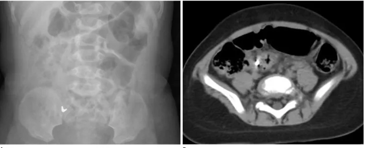
Fig. 4. Delayed passage of a small metallic foreign body five months after ingestion. A 11-month-old girl swallowed a part of cell phone chain.
A. Abdominal radiograph shows a small V-shaped metallic foreign body in the right lower abdomen. Foreign body was not found in the colon by colonoscopy.
B. Abdominal CT scan reveals a metallic foreign body in the right lower abdomen.
There is no extraluminal gas around the foreign body or fluid collection in the abdominal cavity. It was confirmed by the passage of the foreign body 5 months after ingestion of foreign body through serial abdominal radiographs.
A B
Fig. 5. Intestinal perforation of the colon and small bowel by ingestion of a magnet and metals. A 16-month-old boy swallowed one small steel marble three days ago and the other rod-shaped magnet on that day.
A. Initial abdominal radiograph shows a mass of magnet (M), steel marble (S) and tiny metals (black arrowhead) in the midline abdomen. Also note some tiny metallic foreign bodies (open arrows) in the midline abdomen.
B. Abdominal radiograph 9 hours later reveals an attached marble and magnet in the left abdomen, and multiple tiny metals are in left upper and right mid abdomen.
C. Abdominal radiograph one day later shows that attached marble and magnet are not progressed and some tiny metals are attached to the magnet.
D. Colon study reveals a mass of marble, magnet and metals in the descending colon, but some metals (arrow) seem to be outside of intralumi- nal gas.
E. Colonoscopic examination shows a mass of marble, rod shaped magnet (open arrow) and some small metals (open arrowheads).
F. Colonoscopic examination after removal of foreign bodies from the descending colon shows multiple small ulcerations (arrowheads) at the site of impacted foreign body.
G. Abdominal radiograph obtained two hours after the removal of foreign bodies shows free air in the abdominal cavity. Surgery confirmed the perforation of the sigmoid colon and small bowel.
A
E
B
F
C
G
D
Fig. 6. Multiple metallic foreign bodies (sewing needles and metallic chains) in a 16-year-old boy.
A. Abdominal radiograph shows multiple different kinds of metallic foreign bodies in the abdominal cavity.
B. Follow-up abdominal radiograph reveals a remaining sewing needle in the left lower abdomen.
C. Gross specimen photograph demonstrates impaction of a sewing needle (arrow), and ulceration and perforation (open arrowheads) at the im- pacted site of the ileum.
A B C
복통을 유발할 수 있는 다른 여러 질환들을 제외시킬 수 있다 는 장점이 있다(10)(Figs. 4B, 7).
자석이 두 개 이상이면 합병증을 유발할 수 있기 때문에 자 석이 하나인지 두 개인지 여부를 보기 위해서는 복부사진의 전 후와 측면사진이 도움이 되며, 또한 투시검사에서 각도를 달리 해서 자석의 개수를 살펴봄이 도움이 될 수 있다(Fig. 3). 또한 자석 사이의 간격이 있으면 자석 사이에 장벽이 있음을 짐작할 수 있다고 한다(8)(Fig. 3B). CT는 삼킨 이물질이 금속이나 자석, 건전지 같은 방사선비투과성 물질이면 이물질에 의한 beam hardening artifact에 의해 이물질 주변의 변화를 알기가 어렵다(Fig. 8). 자석에 의한 소장 천공의 복부사진에서 복강 내 유리가스가 보일 수도 있지만 복강 내 유리가스가 보이지 않았다는 보고들도 있다. 또한 수술에서 소장간의 다발성 루를 형성하거나 천공이 있었던 여러 증례 보고에서 복강 내 유리가 스의 소견이 보이지 않았다고 한다. 이는 다른 장에 있는 두 개 의 자석끼리 붙어서 자석 사이에 끼여 있는 장벽에 괴사를 유발 하면서 장천공을 일으키지만 공기가 복강 내로 다량으로 새어 나가지는 못하는 상태이기 때문에 복부사진에서 복강 내 유리 가스가 보이지 않는다(9)(Fig. 5). 하지만 장벽으로부터 자석 을 제거하면 복강 내로 공기가 새어나가면서 복부사진에서 복 강 내 유리가스가 보일 수 있다(Fig. 5G). 그래서 자석에 의한 장천공을 시사하는 주요 소견은 복강 내의 자석들이 시간의 흐 름에 따라 진행하지 않고 같은 위치에 있는 소견으로(Figs. 5, 8), 이는 다른 장에 있는 자석들이 서로 붙어서 더 진행하지 않 음을 시사한다(8, 9). 또한 장폐색의 소견이 있으며 이미 장천 공이 되어 있을 가능성이 높다.
이 발생한다.
이물질에 의한 장천공은 자석이나 뾰족한 이물질에 의해 보 고되고 있다. 뾰족한 이물질에 의한 장천공에서 복부사진에서 복강 내 유리가스가 잘 보이지 않는데 이는 뾰족한 이물질이 장벽에 서서히 박히면서 피브린과 주위 장벽으로 싸이기 때문 에 복강 내로 다량의 액체나 기체가 나가는 것을 막기 때문이 다(Fig. 6). 이물질이 방사선투과성이거나 비투과성이 약할 때 복부사진에 이물질을 확인할 수 없다. 이럴 때 CT는 이물질의 위치를 파악하는 데 도움이 될 수 있으며 또한 이물질에 의한 합병증을 보는 데도 도움이 된다. 삼킨 이물질에 의한 소장천 공의 CT소견은 두꺼워진 장, 국한된 복강 내 유리가스, 주변 의 지방침윤, 동반된 장폐색 등이다. 또한 CT의 장점은 급성
Fig. 7. A foreign body (a plum seed) in the terminal ileum in a 9-year- old girl. She had a history of ileocecectomy due to ileal atresia during neonatal period. Abdominal CT scan shows a 2 cm sized intraluminal foreign body (arrow) in the terminal ileum. Preoperative diagnosis was partial small bowel obstruction used by a foreign body. Foreign body was removed through ileoileal anastomosis.
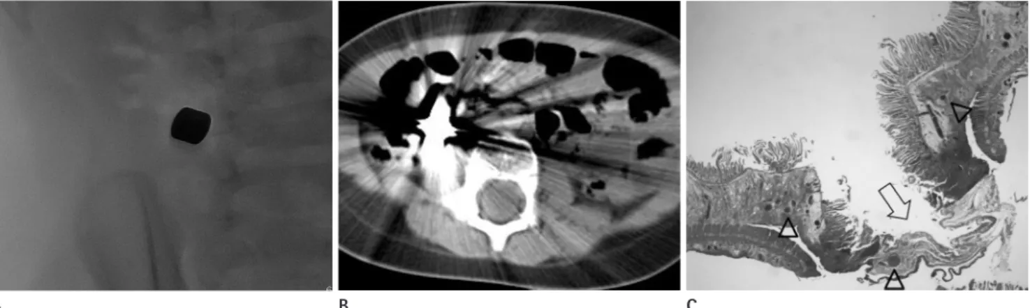
Fig. 8. Multiple small bowel perforation by abutting magnets. A 17-month-old boy presented vomiting about 5 hours ago. Initial abdominal ra- diograph (not shown) demonstrated two abutting magnets in the right mid abdomen.
A. On spot view of foreign body through fluoroscopic examination, two magnets are attracted tightly without any gap.
B. Abdominal CT scan shows severe beam hardening artifact caused by magnets, so CT is not useful for the evaluation of extraluminal gas or flu- id collection. Serial abdominal radiograph (not shown) revealed that the magnets were fixed for three days. At surgery, multiple small perfora- tions were on ileum, and segmental resection of ileum about 10 cm was done.
C. Microscopically, the small intestine demonstrates ischemic necrosis of the mucosal and submucosal layers and fragmented muscle layers (ar- row) (H&E, × 40). Vascular congestion (open arrowheads) is also seen.
A B C
주위의 지방침윤도 장 손상의 간접적인 증거가 될 수 있다. 복 강 내 액체도 둔상에서 보일 때처럼 뚜렷한 소견은 아니지만 장 손상을 의심해 볼 수 있다(5)(Fig. 9).
참고문헌
1. Bronstein AC, Spyker DA, Cantilena LR Jr, Green JL, Ru- mack BH, Giffin SL. 2009 Annual Report of the American Association of Poison Control Centers' National Poison Data System (NPDS): 27th Annual Report. Clin Toxicol (Phila) 2010;48:979-1178
2. Anderson SW, Soto JA. Anorectal trauma: the use of com- 외상에 의한 이물질
대부분이 부서진 나무의 파편, 바늘, 가시 등에 의한 천공 손 상들이 대부분이다. 외상에 의한 장기들의 손상 여부와 정도를 확인할 때 CT가 유용하다. 외상에 의한 장 손상의 가장 민감 한 소견은 이물질이 지나간 자리가 장까지 확장된 것을 보이는 것이다. 입이나 항문으로 넣은 조영제가 장외로의 유출은 장 손 상의 직접적이고 가장 특이적인 소견이며, 직장이나 항문의 전 층의 벽 소실을 보일 때 의심할 수 있다. 둔상에 의한 것과는 달리 장외 공기는 장 손상의 지표가 되지 못하지만, 장외 공기 가 국소적이고 비대칭적으로 있을 때 장 손상의 가능성을 생각 해 볼 수 있다. 그 외에도 드물기는 하지만 급성 출혈이 보일 경 우에도 장 손상을 생각해 볼 수 있다. 장벽의 비후, 직장, 항문
Fig. 9. Traumatic perineal injury from bamboo sticks in a 10-year-old girl.
A. The CT scan at the level of the ischial tuberosity shows an irregular shaped air containing structure (*) in the levator ani muscle and perianal area. The anal mucosa seems to be intact.
B. The CT scan at the level of pelvic cavity shows an irregular shaped air containing structure (*) without sigmoid colon wall thickening or adja- cent fat stranding. There is no transmural defect in the wall of the sigmoid colon.
C. Coronal CT with wide window-width image reveals a tubular air containing structure (*) in the pelvic cavity. The outer margin (open arrow- heads) of tubular air containing structure shows low attenuation, which is higher than air and lower than adjacent fat. It is suggestive of a bam- boo foreign body itself. Also note multiple air shadows (open arrow) in the left sided retroperitoneum.
D. Removed specimen by operation shows multiple fragmented bamboo sticks.
C A
D B
view of the literature. Eur J Pediatr 2001;160:468-472 7. Kim MJ, Kwak AJ, Choi KH. Gastric ulcer due to three
magnets ingestion in a 37-month-old girl. Korean J Pedi- atr Gastroenterol Nutr 2002;5:68-72
8. Kircher MF, Milla S, Callahan MJ. Ingestion of magnetic foreign bodies causing multiple bowel perforations. Pedi- atr Radiol 2007;37:933-936
9. Sahin C, Alver D, Gulcin N, Kurt G, Celayir AC. A rare cause of intestinal perforation: ingestion of magnet. World J Pe- diatr 2010;6:369-371
10. Coulier B, Tancredi MH, Ramboux A. Spiral CT and multi- detector-row CT diagnosis of perforation of the small in- testine caused by ingested foreign bodies. Eur Radiol 2004;14:1918-1925
puted tomography scan in diagnosis. Semin Ultrasound CT MR 2008;29:472-482
3. Gayer G, Petrovitch I, Jeffrey RB. Foreign objects encoun- tered in the abdominal cavity at CT. Radiographics 2011;
31:409-428
4. Choi SW, Kim DK. Ingested gastrointestinal foreign body in children: retrospective review in a pediatric emergency department. J Korean Soc Emerg Med 2010;21:88-93 5. Li ZS, Sun ZX, Zou DW, Xu GM, Wu RP, Liao Z. Endoscopic
management of foreign bodies in the upper-GI tract: ex- perience with 1088 cases in China. Gastrointest Endosc 2006;64:485-492
6. Arana A, Hauser B, Hachimi-Idrissi S, Vandenplas Y. Man- agement of ingested foreign bodies in childhood and re-
소아복부의 사고에 의한 이물질: 임상화보
박찬호 · 김영통
소아복부의 사고에 의한 이물질은 삼킴이나 외상에 의해 유발된다. 이 논문은 소아복부에서 사고에 의한 이물질의 다양 한 영상소견에 대해 기술하고자 한다. 복부사진, 투시검사, 위장관 조영검사와 CT는 이물질의 위치와 종류, 그리고 장천 공과 폐색을 포함한 합병증을 평가하는 데 도움이 된다.
순천향대학교 의과대학 천안병원 영상의학과