Anatomical Observation on Components Related to Foot Gworeum Meridian Muscle in Human
Kyoung-Sik Park
Dept. of Anatomy, College of Oriental Medicine, Sangji University Original Article
⋅Received:23 November 2010 ⋅Revised:7 March 2011 ⋅Accepted:9 March 2011
⋅Correspondence to:Kyoung-Sik Park
Dept. of Anatomy, College of Oriental Medicine, Sangji University, Woosan-dong 660, Wonju, Gangwon-do, Korea Tel:+82-33-730-0667, Fax:+82-33-730-0653, Email:[email protected]
Objectives: This study was carried out to observe the foot gworeum meridian muscle from a viewpoint of human anatomy on the assumption that the meridian muscle system is basically matched to the meridian vessel system as a part of the meridian system, and further to support the accurate application of acupuncture in clinical practice.
Methods: Meridian points corresponding to the foot gworeum meridian muscle at the body surface were labeled with latex, being based on Korean standard acupuncture point locations. In order to expose components related to the foot gworeum meridian muscle, the cadaver was then dissected, being respectively divided into superficial, middle, and deep layers while entering more deeply.
Results: Anatomical components related to the foot gworeum meridian muscle in human are composed of muscles, fascia, ligament, nerves, etc. The anatomical components of the foot gworeum meridian muscle in cadaver are as follows:
1. Muscle: Dorsal pedis fascia, crural fascia, flexor digitorum (digit.) longus muscle (m.), soleus m., sartorius m., adductor longus m., and external abdominal oblique m. aponeurosis at the superficial layer, dorsal interosseous m.
tendon (tend.), extensor (ext.) hallucis brevis m. tend., ext. hallucis longus m. tend., tibialis anterior m. tend., flexor digit. longus m., and internal abdominal oblique m. at the middle layer, and finally posterior tibialis m., gracilis m. tend., semitendinosus m. tend., semimembranosus m. tend., gastrocnemius m., adductor magnus m. tend., vastus medialis m., adductor brevis m., and intercostal m. at the deep layer.
2. Nerve: Dorsal digital branch (br.) of the deep peroneal nerve (n.), dorsal br. of the proper plantar digital n., medial br. of the deep peroneal n., saphenous n., infrapatellar br. of the saphenous n., cutaneous (cut.) br. of the obturator n., femoral br. of the genitofemoral n., anterior (ant.) cut. br. of the femoral n., ant. cut. br. of the iliohypogastric n., lateral cut. br. of the intercostal n. (T11), and lateral cut. br. of the intercostal n. (T6) at the superficial layer, saphenous n., ant. division of the obturator n., post. division of the obturator n., obturator n., ant. cut. br. of the intercostal n. (T11), and ant. cut. br. of the intercostal n. (T6) at the middle layer, and finally tibialis n. and articular br. of tibial n. at the deep layer.
Conclusion: The meridian muscle system seemed to be closely matched to the meridian vessel system as a part of the meridian system. This study shows comparative differences from established studies on anatomical components related to the foot gworeum meridian muscle, and also from the methodical aspect of the analytic process. In addition, the human foot gworeum meridian muscle is composed of the proper muscles, and also may include the relevant nerves, but it is as questionable as ever, and we can guess that there are somewhat conceptual differences between terms (that is, nerves which control muscles in the foot gworeum meridian muscle and those which pass nearby) in human anatomy.
Key Words : Foot gworeum meridian muscle, LR1-14, muscle, tendon, ligament, fascia, nerve.
Introduction
This paper follows a series of research papers which
I have reported in various journals with relation to the
twelve meridian muscles, most recently On Foot
Taeyang Meridian Muscle, J. Korean. Oriental Med.,
2009.
With relation to the study of meridian muscles, it is thought most important, even elementary to elucidate what the components related to the meridian muscle are, since the concept of meridian muscle (MM) has been shown in Ling Shu (Miraculous Pivot) of Huandi Neijing. For all divergent views it is a common view that MM is closely connected with the twelve (12) main meridians (TMM). Where the meridian muscle system is basically matched to the meridian vessel system as a part of the meridian system, it is different in that TMM are internally connected to
one
1,2,3)of the twelve organs (Zang Fu) but MM is
not so. Meridian muscle is a general term of the muscular system distributed among the twelve (12) main meridians, and is composed of muscular components
1,2)such as muscle (including tendon), fascia, ligament and so on, where gi (life energy) in TMM is collected or concluded or translated
1,4)to the muscular system. According to these opinions, there are three Yin & Yang channels on each arm, and three on each
5,6)leg
1,. Whereas the liver meridian is a negative meridian with energy running from the foot to the chest and this meridian is composed of fourteen bilateral acupoints, the foot gworeum meridian muscle is thought to be a colligated conception of body muscles distributed among this meridian.
Acupuncture theory, especially the theory of TMM or MM, although supported by numerous empirical studies, has received various criticisms.
There is not yet a generally-accepted anatomical and histological basis for their existence or hypothesis. It also seems that the anatomical constituents of individual MM are wrongly known to the academic world of oriental medicine
1,7). This may bring about a mistaken clinical application or a wrong diagnosis as well as misunderstanding of the mode of action of acupuncture, though the term of MM means a lot to myology, arthrology, rehabilitation, and the other clinics.
On the other hand, Korean standard acupuncture
point locations were established on February 11
th2006
8)and STRICTA was revised on August 17
th2010
9). It is necessary for such situations to be considered when the meridian muscle is studied.
This study was carried out in order to investigate correct constituents related to the foot gworeum meridian muscle and to theoretically support the meridianology or the clinics of oriental medicine, following Lung MM
10), pericardium MM
11), and foot Taeyang MM
12).
Materials and Methods
1. Preservative preparations and injection
1) The preparation of a preservative
1 kg of Phenol was dissolved in 1 ℓ of methyl alcohol (The 1st solution). 500ml of glycerin was dissolved in 2 ℓ of methyl alcohol and thereafter an additional 500 ㎖ of glycerin was dissolved in this solution (the 2nd solution). The 1st and 2nd solutions were mixed well, and warmed (30min, 20
℃). 1 ℓof methyl alcohol was added to this mixed solution and stirred for 10 minutes. Finally 1.5 ℓ of formalin was added to the mixed solution.
2) Injection to cadaver
The sheath of the femoral artery & vein was exposed by vertical incision at the medial third of the inguinal ligament, and the femoral artery was carefully separated from the femoral vein. A preservative was injected into the femoral artery at a speed of 150 ㎖ per minute. After 6 ℓ of preservative was injected, the needle-inserted part was ligated, and subsequently an injector needle was inserted downwards for the preservation of the leg.
2. Embalmment of cadaver
1) Cadaver was pending in the embalmment system for 40 hrs at 40℃.
2) Cadaver was exposed for 1hr at room
temperature, and after that, kept in refrigerated storage (3 ℃, 30% humidity).
3. Experimental procedure
1) The foot gworeum meridian in the body surface was labeled with latex at the surface of the cadaver, subsequently being photographed.
2) Meridian points were based on standard Korean acupuncture point locations
8).
3) Pores were made by a drill in the vertical direction at each meridian point.
4) Skin and superficial fascia were removed in order and the exposed deep fascia surface was thereafter labeled by latex, and photographed again.
5) Deep fascia was also removed.
6) Subsequently muscle, tendon, and nerve were investigated and photographed, being grouped into three layers (superficial, middle, and deep layers), herein first-exposed muscle layer was named superficial layer, while entering more deeply, named middle and deep layer in order.
Results
The foot gworeum meridian was labeled at the surface of the cadaver, and also relevant constituents were dissected with groupings in the order of the depth of the three layers (superficial, middle, deep layers).
The results were identified as follows:
1. Daedon (LR
1)
This point located at 0.1 chon lateral posterior to the angle of the toenail of the hallux. The dorsal pedis fascia is extended up to this place. Here are distributed the dorsal digital (digit.) branch (br.) of the deep peroneal nerve (n.) and the dorsal br. of the proper plantar digital n. (Fig. 1).
2. Haenggan (LR
2)
This point is located at the dorsal surface between the 1st and 2nd toes. At the superficial layer there is the dorsal pedis fascia, and right under it the dorsal interosseous muscle (m.) tendon (tend.). At the superficial layer there is the dorsal digital br. of the deep peroneal n. (Fig. 1)
3. Taechung (LR
3)
This point is located at the dorsal surface between the 1st and 2nd metatarsal bones. There are the dorsal pedis fascia at the superficial layer, the extensor (ext.) hallucis brevis m. tend. and the lateral margin of the ext. hallucis longus m. tend. The medial br. of the deep peroneal n. is distributed (Fig.
1).
4. Jungbong (LR
4)
This point is located at the recession medial to the tibialis anterior m. tend. There is the dorsal pedis fascia and medial margin of the tibialis anterior m.
tend. in the superficial layer. Subcutaneously is situated the fascia saphenous n. (Fig. 1)
5. Yeogu (LR
5)
This point is located at 5 chon upward to the tip of the medial malleorus. At the superficial layer there are crural fascia and flexor (flex.) digit. longus m. and at the deep layer the posterior tibialis muscle.
At the superficial layer the saphenous n. is situated, and at the deep layer the tibialis n. (Fig. 1, 2). The crural fascia, flexor (flex.) digit. longus m. and saphenous n. are situated in the superficial layer. The posterior tibialis m. and tibialis n. are situated in the deep layer.
6. Jungdo (LR
6)
This point is located at 7 chon upward to the tip
of the medial malleorus. The superficial layer of
Fig. 1. Photograph shows the foot gworeum meridian points (LR1-LR5) at the level of the deep fascia of the leg (● : Meridian points, A: LR1, B: LR2, C: LR3, D: LR4, E: LR6, F: LR5, △: Muscles, a: tibialis ant. m. tend., b: ext. hallucis longus m. tend., c: ext. digit. longus m. tend., d: calcaneal (Achilles) tend., e: gastrocnemius m., f: dorsalis pedis fascia, g: dorsal interosseous m. tend., h: ext. hallucis longus m. tend., i: ext. hallucis brevis m. tend., j: crural fascia,
▲: other surface markings, k: tibia).
Fig. 2. Photograph shows the foot gworeum meridian points (LR5-LR9) at the level of deep fascia and inner layer of leg (●: Meridian points, A: LR5, B: LR6, C: LR7, D: LR8, E: LR9, △: Muscles, a: flex. digit. longus m., b: soleus m., c: gastrocnemius m., d: sartorius m., e: semimembranosus m., f: semitendinosus m., g: rectus femoris m., i: crural fascia, j: gracilis m. tend. and semitendinosus m. tend., k: add. magnus m. tend., l: vastus medialis m., ▲: other surface markings, h: tibia).
Jungdo corresponds to the space between the flex.
digit. longus m., and the soleus m.; the posterior tibialis muscle is situated in the deep layer. The relevant nerve is very alike to Yeogu in this respect (Fig. 2).
7. Seulgwan (LR
7)
This point is located below the medial tibia condyle, 1 chon posterior to Eumneungchon (SP
9).
Sartorius m. and saphenous n. are situated in the superficial layer, and the inner layer corresponds to the anterior space to tendons of gracilis m., semitendinosus m., and semimembranosus m. (or gastrocnemius with aforementioned muscles in case of inclined acupuncture); tibialis n. are situated in the deep layer (Fig. 2).
8. Gokcheon (LR
8)
This point is located at the extremity of the crease formed by the knee flexure. The relevant muscle is most alike to Seulgwan in this respect but at the inner layer there is the tip of the adductor magnus m. tend. The relevant nerve is the infrapatellar br. of saphenous n. in the superficial layer and the articular br. of tibial n. in the deep layer (Fig. 2).
9. Eumpo (LR
9)
This point is located at 4 chon above the medial
condyle of the femur. According to the direction of
the acupuncture needle (upright or inclined position)
the relevant muscles or nerves differ. Muscle in the
superficial layer corresponds to sartorius m. The
deep layer corresponds to the space between the
adductor magnus m. and the medial margin of the
Fig. 3. Photograph shows the foot gworeum meridian points (LR10-LR14) at the level of the deep fascia of the abdomen
& upper thigh, body surface of thorax (●: Meridian points, A: LR10, B: LR11, C: LR12, D: LR13, E: LR14, F: ST30,
△: Muscles, a: gracilis m., b: sartorius m., c: adductor longus m., d: external abdominal oblique m., e: internal abdominal oblique m., f: inguinal lig., g: rectus abdominis m., ▲: other surface markings, h: ant. sup. iliac spine, i: fossa ovalis (great saphenous v.), j: umbilicus, k: xiphisternal joint, l: 7th intercostal space, m: tip of 11th rib.
Fig. 4. Photograph shows the foot gworeum meridian points (LR10-LR12) and Gichung (ST30) at the level of deep fascia and inner layer of lower abdomen& upper thigh (●: Meridian points, A: LR10, B: LR11, C: LR12, D: ST30, △:
Muscles, a: adductor longus m., b: pectineus m., c: adductor brevis m., d: adductor magnus m., e: gracilis m., f:
sartorius m., g: rectus femoris m., i: fascia of ext. oblique m., ▲: other surface markings, h: supf. circumflex iliac vessels, j: symphysis pubis, k: inguinal lig., l: fossa ovalis, m: femoral artery and vein, n: penis spongy).
vastus medialis m. The relevant nerves are the saphenous n. in the middle layer and the cutaneous (cut.) br. of the obturator n. in the superficial layer (Fig. 2).
10. Jogori (LR
10)
This point is located at 3 chon at a right angle to Gichung (ST
30) situated at 2 chon lateral to Gokgol (CV
2) on the median line of the abdomen. The relevant muscles are adductor longus m. in the
superficial layer and the adductor brevis m. and adductor magnus m. in the deep layer. The relevant nerves are the femoral br. of genitofemoral n. and the anterior (ant.) cut. br. of the femoral n. at the outer layer, ant. division of the obturator n. in the middle layer, and lastly post. division of the obturator n. (Fig. 3, 4).
11. Eumnyeom (LR
11)
This point is located at 2 chon at a right angle
downward to Gichung (ST
30). There are the adductor longus m. in the superficial layer and the adductor brevis m. and adductor magnus m. in the deep layer.
Relevant nerves are the obturator n. in the middle layer, the femoral br. of the genitofemoral n., the ant. cut. br. of the femoral n., and the ant. cut. br. of iliohypogastric n. in the superficial layer (Fig. 3, 4).
12. Geummaek (LR
12)
This point is located at 2.5 chon lateral to Gokgol (CV
2) and 0.5 chon lateral to Gichung (ST
30). In the superficial layer there is the aponeurotic part of the external abdominal oblique m., and the internal oblique m. inside this muscle. The relevant nerves are the obturator n. at the middle layer, the femoral br. of the genitofemoral n., the ant. cut. br. of the femoral n., the ant. cut. br. of the iliohypogastric n.
in the superficial layer (Fig. 3, 4).
13. Jangmun (LR
13)
This point is located at the lower edge of the inner extremity of the 11th rib. In the superficial layer there is the external abdominal oblique m., and the internal oblique m. inside this muscle. The relevant nerves are the lateral cut. br. of the intercostal n. (T11) in the superficial and the ant.
cut. br. of the intercostal n. (T11) at the deeper layer (Fig. 3).
14. Gimun (LR
14)
This point is located at a position crossing with the mammillary line on the 6th intercostal space.
There are the external abdominal oblique m. in the superficial layer and the intercostal muscles in the deep layer. The relevant nerves are the lateral cut.
br. of the intercostal n. (T6) in the superficial layer and the ant. cut. br. of the intercostal n. (T6) at a deeper layer (Fig. 3).
Discussion
Foot gworeum MM originates at Daedon (LR
1) in the lateral posterior to the angle of the hallux toenail, goes upward to the medial part of the lower limb passing by the dorsum of the foot, and reaches Gimun (LR
14) in the lower chest via the pubis. So muscles or nerves relevant to this meridian muscle are chiefly situated at the medial parts of the lower limb and lower chest.
As mentioned above, anatomical knowledge of the
meridian muscle is essentially required for the
clinical application of MM. At the same time such
knowledge must be exact for effective application of
MM to clinics
13-20). But it is criticizable on muscles
correspondent to the meridian muscle. We
investigated muscles or components correspondent to
the meridian muscle according to a common view
that basically MM is closely connected with the
meridian vessel as a part of the meridian, though we
admit the necessity of the strict interpretation of
Ling Shu of HUANDI NEIJING
1,2,3,21). Study carried
out from this viewpoint shows some differences from
already established data
1,7)on MM (refer to Table 1
and 2); that is, anatomical constituents of MM such
as muscle, fascia, ligament, nerve, and furthermore,
different analytical methods from established
procedure. But it is necessary to study further
whether nerves are included among meridian muscle,
considering flow pathways of MM, disease
symptoms in relation with MM, the interpretation of
Ling Shu of HUANDI NEIJING, etc. Above all the
components of each meridian point investigated in
this study were divided into three layers according to
their depth from the body surface but on the other
hand we came across that it might be wide
differences in opinion according to the disparity of
actual meridian points or the application angle of
9,22)acupuncture. The present study was performed with
upright acupuncture position for a basis. As we have
already brought out several times, the direction of
Meridian
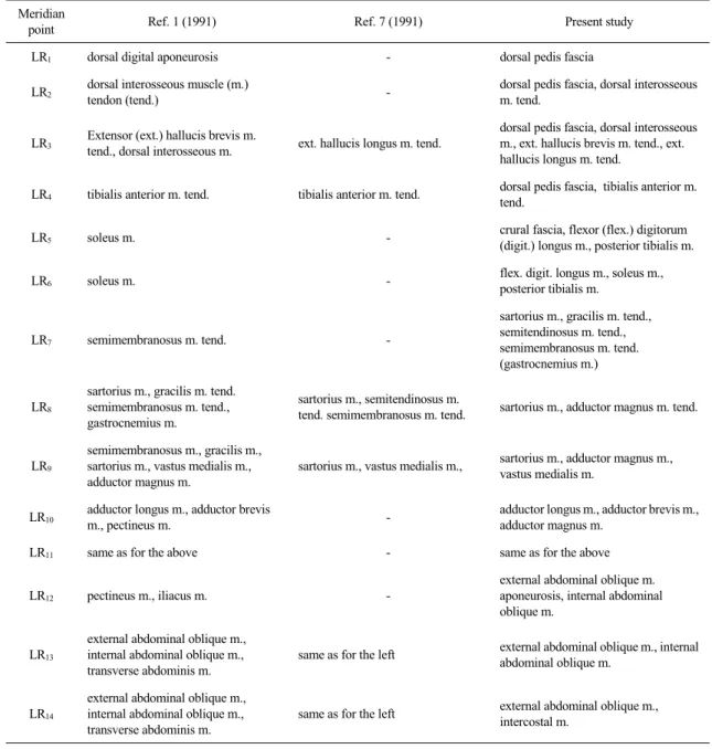
point Ref. 1 (1991) Ref. 7 (1991) Present study
LR1 dorsal digital aponeurosis - dorsal pedis fascia
LR2 dorsal interosseous muscle (m.)
tendon (tend.) - dorsal pedis fascia, dorsal interosseous
m. tend.
LR3 Extensor (ext.) hallucis brevis m.
tend., dorsal interosseous m. ext. hallucis longus m. tend.
dorsal pedis fascia, dorsal interosseous m., ext. hallucis brevis m. tend., ext.
hallucis longus m. tend.
LR4 tibialis anterior m. tend. tibialis anterior m. tend. dorsal pedis fascia, tibialis anterior m.
tend.
LR5 soleus m. - crural fascia, flexor (flex.) digitorum
(digit.) longus m., posterior tibialis m.
LR6 soleus m. - flex. digit. longus m., soleus m.,
posterior tibialis m.
LR7 semimembranosus m. tend. -
sartorius m., gracilis m. tend., semitendinosus m. tend., semimembranosus m. tend.
(gastrocnemius m.)
LR8
sartorius m., gracilis m. tend.
semimembranosus m. tend., gastrocnemius m.
sartorius m., semitendinosus m.
tend. semimembranosus m. tend. sartorius m., adductor magnus m. tend.
LR9
semimembranosus m., gracilis m., sartorius m., vastus medialis m., adductor magnus m.
sartorius m., vastus medialis m., sartorius m., adductor magnus m., vastus medialis m.
LR10 adductor longus m., adductor brevis
m., pectineus m. - adductor longus m., adductor brevis m.,
adductor magnus m.
LR11 same as for the above - same as for the above
LR12 pectineus m., iliacus m. -
external abdominal oblique m.
aponeurosis, internal abdominal oblique m.
LR13
external abdominal oblique m., internal abdominal oblique m., transverse abdominis m.
same as for the left external abdominal oblique m., internal abdominal oblique m.
LR14
external abdominal oblique m., internal abdominal oblique m., transverse abdominis m.
same as for the left external abdominal oblique m., intercostal m.
Table 1. Differences from Established Studies from a Viewpoint of the Muscular Components of the Foot Gworeum Meridian Muscle in Human.
the acupuncture needle in oriental medicine is generally composed of 3 types, the upright position inserted vertical to the skin, the down or transverse position, and the inclined position inserted at the angle of 45 degrees to the skin. The upright position
is required for the application of the needle to LR
1,5,7-13(according to Ahan, Ref. 7), or LR
1-14(according to Ref. 1), inclined or transverse position for LR
2-6,13,14(according to Ahan, Ref. 7) or LR
1-14(according to Ref. 1). Speaking for the last time, the
Meridian
point Ref. 1 (1991) Ref. 7 (1991) Present study
LR1 dorsal digital branch (br.) of deep
peroneal nerve (n.). - dorsal digital br. of deep peroneal n.,
dorsal br. of proper plantar digital n.
LR2 dorsal digital br. of peroneal n. dorsal digital br. of deep peroneal n. dorsal digital br. of deep peroneal n.
LR3 deep peroneal n. deep peroneal n. med. br. of deep peroneal n.
LR4 med. crural cutaneous (cut.) br. of
saphenous n. med. crural cut. br. of saphenous n. saphenous n.
LR5 same as for the above ant. br. of saphenous n. saphenous n., tibial n.
LR6 same as for the above - same as for the above
LR7 saphenous n., tibial n. - same as for the above
LR8 same as for the above saphenous n. infrapatellar br. of saphenous n., articular br. of tibial n.
LR9 ant. cut. br. of femoral n., saphenous n. - cut. br. of obturator n., saphenous n.
LR10 obturator n. obturator n.,
femoral br. of genitofemoral n., ant. cut.
br. of femoral n., ant. division of obturator n., post. division of obturator n.
LR11 obturator n., -
femoral br. of genitofemoral n., ant. cut.
br. of femoral n., ant. cut. br. of iliohypogastric n., obturator n.
LR12 obturator n., femoral br. of
ilioinguinal n. - femoral br. of genitofemoral n., ant. cut.
br. of iliohypogastric n., obturator n.
Table 2. Differences from Established Studies from a Viewpoint of the Nervous Components of the Foot Gworeum Meridian Muscle in Human.