online©ML Comm
125 臨床耳鼻:第18卷 第1號 2007
• • • • • • • • • • • • • • • • • • • • • • • • • • • • • • • • • • • • • • • • • • • • • • • • • • • • • • • • • • • • • • • • • • • • • • • • • • • • • • • • • • • • • • • • • • • • • • • • • • • • • • • • • • • • • • • • • • • • • • • • • • • • • • • • • • • • • • • • • • • • • • • • • • • • • • • • • • • • • • • • • • • • • • • • • • • • • • • • • • • • • • • • • • •
J Clinical Otolaryngol 2007;18:125-127 □ 증 례□
이수에 국한된 선천성 이개 누공
인제대학교 의과대학 이비인후-두경부외과학교실
허 경 욱
Congenital Auricular Fistula Confined to an Ear Lobule
Kyung Wook Heo, MD
Department of Otorhinolaryngology-Head and Neck Surgery, College of Medicine, Inje University, Busan, Korea
- ABSTRACT -
Congenital anomalies of the external ear are rare, and can have negative effects on children due to unaesthetic appearance and repeated infection. Surgical management is required to correct the deformity. Of the external ear deformities, congenital fistulae are rare and manifest as blindly ending narrow tubes, sometimes mere pits or dimples. The fistular opening can be located in various regions of the periauricular area. The present case involved 3-year-old girl with two congenital openings at the intertragic incisura and the middle of the free border of ear lobule, which caused frequent odorous discharge. A fistulogram showed that the two openings were interconnected via a fistular tract. The patient underwent fistulectomy under general anesthesia. The author reports this case with review of a literature. (J Clinical Otolaryngol 2007;18:125-127)
KEY WORDS:External Ear·Fistula·Congenital.
서 론
이개는 태생 4주에 제 1새열을 둘러싸고 있는 제 1, 2새궁에서 발생한 6개의 이개융기에 의해 형성되기 시 작한다. 이개 융기들은 태생 5~6주에 걸쳐 농축되고 융 합하여 이개를 형성한다.1)2) 태생 50일경 원시적인 형태 의 이개가 완성되어 태아의 성장에 따라 크기가 커지고 모 양이 바뀌게 되며, 태생 20주에는 그 성장이 완성된다.1) 태생초기에 출현한 이개는 하악 부위의 미측에 위치하지 만 하악골이 성장함에 따라 이개가 두부쪽으로 이동하여,
태생 20주에는 신생아 이개의 모양과 위치를 가지게 된 다. 이러한 과정 중에 다양한 내적 및 외적인 요인들에 의 해 이개의 기형이 발생하며, 기형의 대다수가 이개의 상 측에 위치한다.2) 기형이 이수에 국한되는 것은 매우 드물 며2)3) 이수에 국한된 선천성 누공은 전세계적으로 거의 보고가 없다. 이에 저자는 3세 여아에서 이수에 국한되 어 발생한 선천성 이개 누공을 치험하였기에 문헌고찰과 함께 보고하는 바이다.
증 례
3세 여아가 좌측 이개에 존재하는 두개의 누공으로부 터 잦은 농성 분비물을 주소로 본원 이비인후과 외래를 방문했다. 병력청취상 환아는 정상분만으로 출생했고 영 아기에 특별한 문제는 없었다. 좌측 이개의 누공은 출생 당시부터 존재했으며, 출생 후 약 6개월 경부터 누공에 논문접수일:2007년 4월 3일
심사완료일:2007년 5월 7일
교신저자:허경욱, 614-735 부산광역시 부산진구 개금1동 633-165 인제대학교 의과대학 이비인후-두경부외과학교실 전화:(051) 890-6375·전송:(051) 892-3831 E-mail:heokw96@kornet.net
J Clinical Otolaryngol 2007;18: 125-127
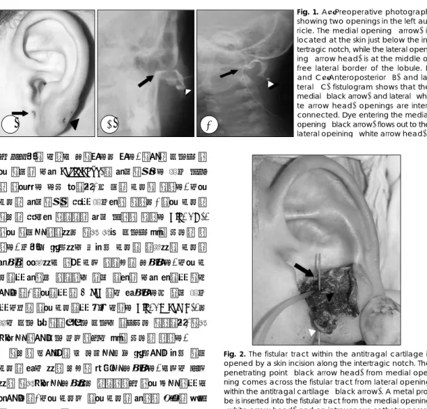
126 서 분비물이 나오기 시작했다고 했다. 이학적 검사상 좌 측 이개의 대주(antitragus)와 주간절흔보다 아래 부분 은 우측보다 다소 컸으며, 두 개의 누공이 있었다. 내측 누공은 주간절흔의 바로 아래쪽에 있었고, 외측 누공은 이수의 바깥쪽 자유연의 중간 부위에 있었다(Fig. 1A).
좌측 이개를 제외하고 전신 신체 검사상 특이 소견은 없 었다. 드물게 발생하는 선천성 누공을 의심하여 누공의 주행을 파악하기 위해 누관 조영술을 시행했다. 내측 누 공으로 주입된 조영제는 약간 윗쪽의 대주 쪽으로 이동 한 다음, 외측으로 약 2 cm 정도 진행했다가 약간 아래 로 내려와 외측 누공으로 흘러 나왔다(Fig. 1B and C). 고 실도 검사 및 이음향방사 검사는 정상소견 이었으며, 전신 마취를 위한 검사 결과에서도 특이 소견은 없었다.
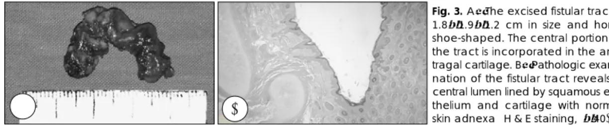
이수에 국한되어 그 경계를 따라 발생한 선천성 이개 누공의 진단 하에 수술적 치료를 시행했다. 기관내 삽관 하에 전신마취를 시행하고 앙와위에서 좌측 귀를 위로 노 출한 다음, 내측 누공과 외측 누공의 주위에 환상의 피부 절개를 시행하고 양측 누공루를 따라 박리를 시행했다. 박 리 중에 양측의 누공으로 삽입한 탐침자들은 대주연골에 서 만나고 있었다(Fig. 2). 누공루를 따라 내측 누공과 대주연골 사이에 2 cm 정도의 피부절개를 가한 다음, 교 차부위의 대주연골과 전체 누공루를 일괄하여(en bloc) 절제하였다. 누공루는 비대칭적인 말발굽 모양이었고 1.8
×1.9×1.2 cm 크기였으며, 상처는 일차봉합을 시행했 다(Fig. 3A). 병리조직검사상 누공루는 정상적인 피부부 속기를 가진 중층 편평상피로 덮여 있었고 정상적인 연 골과 접하고 있어 새열 기형에 합당한 소견이었다(Fig.
3B). 수술후 좌측 이개의 아래 부분은 우측과 같은 크기 로 줄었으며 환자는 수술후 5일째 피부의 봉합사를 제
거하고 특별한 문제 없이 퇴원하였다. 술 후 26개월이 경과한 현재 특이 소견 없이 외래 추적관찰 중이다.
고 찰
이개 기형의 빈도는 잘 알려져 있지 않지만, 인종간에 차이가 있는 것으로 알려지고 있다.4) 소이증이나 무이 증 등의 드문 이개 기형은 만 명당 0.8~2.4명의 빈도로 발생하고, 아시아계와 라틴아메리카계에 흔하며 유럽계 나 아프리카계에는 드물게 발생한다.4)5) 대부분의 이개 기
A B C
Fig. 1. A:Preoperative photograph showing two openings in the left au- ricle. The medial opening (arrow) is located at the skin just below the in- tertragic notch, while the lateral open- ing (arrow head) is at the middle of free lateral border of the lobule. B and C:Anteroposterior (B) and la- teral (C) fistulogram shows that the medial (black arrow) and lateral (whi- te arrow head) openings are inter- connected. Dye entering the medial opening (black arrow) flows out to the lateral opeining (white arrow head).
Fig. 2. The fistular tract within the antitragal cartilage is opened by a skin incision along the intertragic notch. The penetrating point (black arrow head) from medial ope- ning comes across the fistular tract from lateral opening within the antitragal cartilage (black arrow). A metal pro- be is inserted into the fistular tract from the medial opening (white arrow head) and an intravenous catheter passes through the fistular tract from the lateral opening.
허경욱:이수에 국한된 선천성 이개 누공
127 형은 산발적으로 발생하지만 드물게 상염색체 우성 또는 열성 유전, 성염색체 관련 유전의 경우가 알려져 있다.6)
복외측 경부의 상당부분을 형성하는 새궁이 태생기 6주부터 이개의 형성에 관여하게 된다. 제 2새궁과 제 3새궁의 배측 부위를 덮는 외배엽 조직은 중배엽 조직에 서 콜라겐의 침착과 연골세포 분화가 일어나도록 자극하 여 태생기 7주에는 중배엽에서 연골 전 단계의 여섯 개의 이개융기가 만들어진다.7) 이 융기들은 중배엽 성장의 강 력한 중심이 되지만 이개융기들이 어떤 방식으로 증식 과 융합을 일으켜 이개의 특정 부분을 형성하는 지는 정 립되어 있지 않으며, 다양한 이개기형이 이개 발생의 단 서를 제공하게 된다.7)
이개의 각 구조물들의 기원에 대해서는 여러 연구자들 이 다양하게 기술해 왔다.8-10) 최근 Park3)은 동맥의 분 포범위에 따라 이개의 기원을 추정했는데, 천측두동맥에 의해 공급받는 지역이 제 2새궁에 의해 형성되는 부분과 일치하고 후이개 동맥에 의해 공급받는 지역이 제 3새궁 에 의해 형성되는 부분과 일치함을 보고 하였다. 이를 근 거로 Park은 제 1, 6이개 융기가 이수를, 제 4, 5이개융 기가 이륜 또는 대이륜을, 제 2이개 융기가 이주를, 그리 고 제 3이개융기가 상행 이륜각을 형성한다고 제시했다.3)
본 증례에서 누공루의 주행이 주간 절흔에서 이수의 자 유연으로 이어지고 있어 Park3)과 Davis10)의 제안을 고 려해 볼 때, 제 1과 제 2이개 융기가 불완전 결합되어 내 측의 누공루를 형성하고, 제 5와 제 6이개 융기가 불완전 결합되어 외측의 누공루를 형성한 것으로 추정할 수 있을 것이다. 좀 더 많은 이개 기형의 증례를 모아서 분석한다 면 각 이개 융기가 이개의 어느 부위를 형성하는 지를 좀
더 정확히 알 수 있을 것으로 사료된다. 또, 본 증례와 같 이 모양이 특수하거나 위치가 비전형적인 누공이 존재할 경우 누관 조영술 등의 수술전 검사를 반드시 시행해서 수 술의 계획시 기초자료로 사용해야 할 것이다.
저자는 이수에 국한되어 선천적으로 발생한 이개 누 공을 성공적으로 치험하였기에 문헌고찰과 함께 보고하 는 바이다.
중심 단어:외이·누공·선천성.
REFERENCES
1) Altmann F. Normal development of the ear and its mecha- nics. AMA Arch Otolaryngol 1950;52:725-66.
2) Kenna MA, Hirose K. Embryology and Developmental anatomy. In: Charles DB, Sylvan ES, Cuneyt MA, Ellis MA, Margaretha LC, Joseph ED, editors. Pediatric Otolaryn- gology, 4th ed. Philadelphia, PA: Saunders;2003. p.129-46.
3) Park C. Lower auricular malformations: Their represen- tataion, correction, and embryologic correlation. Plast Re- constr Surg 1999;104:29-40.
4) Karmody CS, Annino DJ. Embryology and anomalies of the external ear. Facial Plast Surg 1995;11:251-6.
5) Harris J, Kallen B, Robert E. The epidemiology of anotia and microtia. J Med Genet 1996;33:809-13.
6) Oliveira CA, Pinheiro LC, Gomes MR. External and middle ear malformations: autosomal dominant genetic transmi- ssion. Ann Otol Rhinol Laryngol 1989;98:772-6.
7) Porter CJ, Tan ST. Congenital auricular anomalies: Topo- graphic Anatomy, Embryology, Classification, and Treatment Strategies. Plast Reconstr Surg 2005;115:1701-12.
8) Streeter GL. Development of the auricle in the human em- bryo. Contrib Embryol 1922;69:111-39.
9) Wood-Jones F, Wen IC. The development of the external ear.
J Anat 1933;68:525-33.
10) Davis J. Surgical embryology. In: Davis J, editor. Aesthetic and Reconstructive Otoplasty. 1st ed. New York, NY: Sprin- ger-Verlag;1987. p.93-125.
Fig. 3. A:The excised fistular tract is 1.8×1.9×1.2 cm in size and horse shoe-shaped. The central portion of the tract is incorporated in the anti- tragal cartilage. B:Pathologic exami- nation of the fistular tract reveals a central lumen lined by squamous epi- thelium and cartilage with normal skin adnexa (H & E staining, ×40).
A B