Primary Isolated Bone Marrow Diffuse Large B-cell Lymphoma with Hemolytic Anemia as the First Manifestation
Ja Young Lee, M.D.1, Jae Hyun Kim, M.D.1, Jeong Hwan Shin, M.D.1, Hye Ran Kim, M.D.1, Jeong Nyeo Lee, M.D.1, Ki Hyang Kim, M.D.2, Won Sik Lee, M.D.2,
Young Don Joo, M.D.2, Chang Hak Sohn, M.D.2 and Chan-Hwan Kim, M.D.3 Departments of 1Laboratory Medicine, 2Hemato-oncology and 3Pathology,
Busan Paik Hospital, College of Medicine, Inje University, Busan, Korea
Primary isolated bone marrow disease as a presenting feature of diffuse large B-cell lymphoma is very rare. We describe the first Korean case of isolated bone marrow diffuse large B-cell lymphoma with hemolytic anemia as the first manifestation. A32-year-old man was admitted to our hospital presenting with fever and hematuria. He had severe anemia and high lactate dehydrogenase activity. His peripheral blood smear and laboratory findings were suggestive of intravascular hemolytic anemia. The bone marrow biopsy revealed involvement with diffuse large B-cell lymphoma. A computed tomographic scan showed splenomegaly, but no lymphadenopathy. Our case shared some clinical features with the Asian variant of intravascular B-cell lymphoma, but there was infrequent involvement of the sinusoids of lymphoma cells and no hemophagocytosis. Our patient was treated with R-CHOP regimen for six cycles and he is in remission after autologous peripheral blood stem cell transplantation. (Korean J Hematol 2008;
43:48-52.)
Key Words: Diffuse large B-cell lymphoma, Bone marrow, Hemolytic anemia
48 접수:2007년 12월 26일, 수정:2008년 2월 5일
승인:2008년 2월 15일
교신저자:이정녀, 부산시 부산진구 개금동 633-165
614-735, 인제대학교 의과대학 부산백병원 진단검사의학과
Tel: 051-890-6862, Fax: 051-893-1562 E-mail: [email protected]
Correspondence to:Jeong Nyeo Lee, M.D.
Department of Laboratory Medicine, Busan Paik Hospital, College of Medicine, Inje University
633-165, Gaegeum-dong, Busanjin-gu, Busan 614-735, Korea Tel: +82-51-890-6862, Fax: +82-51-893-1562
E-mail: [email protected] INTRODUCTION
The most frequent type of non-Hodgkin's lym- phoma (NHL) is diffuse large B-cell lymphoma (DLBL). Patients may present with nodal or ex- tranodal disease. Extranodal involvement is most- ly considered as stage IV, and portends a poorer prognosis. The most common extranodal site is the gastrointestinal tract, but virtually any loca- tion has been reported as a primary site. Al- though bone marrow (BM) involvement in DLBL is relatively infrequent at presentation and is gen-
erally believed to represent systemic dissem- ination of disease, some cases may arise primarily in BM.1-6) Primary BM involvement in NHL may be more frequent in cases of T-cell lymphoma, as only eight cases in 32 previous reports were DLBL in English literature.3,5,6) But primary iso- lated BM DLBL was not reported in Korea.
We describe a patient with primary isolated bone marrow DLBL (IBMDLBL) who presented with hemolytic anemia as the first manifestation.
Hemolytic anemia is a relatively common compli- cation in patients with NHL; however, there have been only one report of autoimmune hemolytic
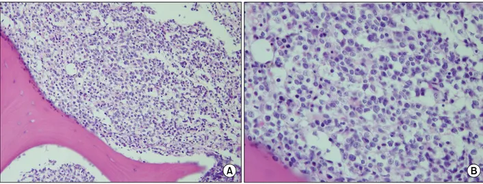
Fig. 1. Histologic features of diffuse large B cell lymphoma in the bone marrow. (A) Atypical large lymphoma cells are diffusely infiltrated (H&E stain, original magnification ×200). (B) Higher power view shows a diffuse lymphoid infiltrate with typical morphologic features of diffuse large B-cell lymphoma (H&E stain, original magnification ×400).
anemia (AIHA) in association with IBMDLBL.7) This is the first case of IBMDLBL with hemo- lytic anemia as the first manifestation.
CASE REPORT
A 32-year-old man was hospitalized with hema- turia and a febrile sensation for 2 weeks. Physical examination revealed hepatosplenomegaly but no overt lymphadenopathy. No specific previous me- dical history and family history were observed.
He had no medication history up to recently.
Laboratory studies revealed a hemoglobin level 3.8g/dL, a hematocrit 12.4%, a white blood cell count 5.4×10⁹/L with 54% granulocytes, 37%
lymphocytes, 9% monocytes, a platelet count 254
×109/L, and reticulocytes 7.4%. Erythrocyte sed- imentation rate was 24mm/hr. Total protein was 5.7g/dL, albumin 3.0g/dL, total bilirubin 2.0mg/
dL, AST/ALT 44/28U/L and Lactate dehydroge- nase (LDH) 728IU/L (reference range, 232∼410 IU/L). The blood urea nitrogen was elevated (24mg/dL), however the levels of serum crea- tinine and electrolytes were normal. On uri- nalysis, urobilinogen and hemosiderin were pos- itive, urine myoglobin was negative. Serum hap- toglobin was 220.8mg/dL (reference range, 30∼
200mg/dL) and C reactive protein was 8.11mg/
dL. An extensive work-up for hemolytic anemia including direct/indirect Coombs' test, autohemo- lysis test, acidified serum lysis test, and sucrose hemolysis test were all negative except the os- motic fragility test, which was slightly increased.
A peripheral blood smear showed moderate ani- sopoikilocytosis with spherocytosis, polychroma- sia, and nucleated red blood cells (4/100 WBCs).
Thus it was suggestive of intravascular hemolytic anemia although haptoglobin was not decreased.
Repeated blood and urinary cultures were ne- gative. Tests for viruses including HCV, HBs, CMV, and HIV were negative. The results of se- rologic testing for Epstein-Barr virus were con- sistent with past infection. Antinuclear antibodies and rheumatoid factor were negative or normal.
Initial broad-spectrum antibiotics with third gen- eration cephalosporin and aminoglycosides and blood transfusions were administered. Hemoglo- bin level was elevated to 8.0g/dL, however high fever continued and a platelet count dropped to 88×109/L.
Thus BM aspiration and biopsy were per- formed. The specimen was markedly hypercel- lular (80∼90%) and diffusely infiltrated by atyp- ical large lymphoid cells with round nuclear con- tours and scanty cytoplasm (Fig. 1). Immunohis- tochemical studies revealed that the neoplastic
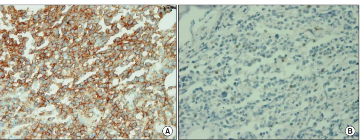
Fig. 2. Immunohistochemical stain of the bone marrow. (A) Lymphoma cells are positive for CD20 (original magnification
×400). (B) Lymphoma cells are negative for CD3 (original magnification ×400).
cells were positive for CD20 and CD45 and neg- ative for TdT, CD45RO, CD3, CD43, CD5, and CD79a (Fig. 2). The morphologic features, to- gether with the immunohistochemical results, were consistent with diffuse large B-cell lymph- oma. A whole-body staging computed tomog- raphy (CT) scan showed no nodal involvement except a mild enlarged spleen. The whole-body bone scan was normal. On 18F-fluorodeoxyglucose- positron emission tomography, BM hyperplasia was observed.
Primary isolated BMDLBL was diagnosed, and the patient was treated with cyclophosphamide, doxorubicin, vincristine, and prednisone (CHOP) plus rituximab for six cycles. After the first che- motherapy, fever and hematuria were subsided and the laboratory finding of the patient was promptly normalized except neutropenia because of BM depression. After 2 months, the BM speci- men was negative for lymphoma cells, and he subsequently received an autologous peripheral blood stem cell (PBSC) transplantation. At pres- ent, he is in remission.
DISCUSSION
Non-Hodgkin's lymphoma restricted to the BM is extremely rare, with only 32 fully documented cases in the English-language literature,1-6) of
which 8 were IBMDLBL.3,5,6) These patients con- sisted of four men and four women with an age range from 42 to 86 years. The most frequent clinical findings were weakness, fatigue, and B symptoms of NHL according to the Ann Arbor staging system. Most patients had neither hep- atosplenomegaly nor lymphadenopathy. Only one patient had hepatosplenomegaly. Six patients had bicytopenia or pancytopenia and elevated serum LDH. Peripheral blood smears in five patients showed a leukoerythroblastic reaction.
Our patient also presented fever and general weakness. He had mild hepatosplenomegaly and the similar laboratory findings in previous cases of IBMDLBL. But his complaint of hematuria is unusual. He had severely decreased hemoglobin (3.8g/dL). Laboratory findings, including a pe- ripheral blood smear were compatible with the diagnosis of intravascular hemolytic anemia ex- cept elevated haptoglobin. Serum haptoglobin level was generally decreased or absent in hemo- lytic anemia. But our patient presented with sus- tained high fever and elevated CRP at diagnosis of hemolytic anemia, it presumed that he had acute inflammation and haptoglobin was elevated as an acute phase reactant.
Hemolytic anemia was rarely found in previous cases of IBMDLBL and only one case was re- ported about IBMDLBL complicated with AIHA
in Japan.7) Hemolytic anemia is often among the clinical manifestations of intravascular B-cell lym- phoma (IVL). This rare type of large-cell lympho- ma is associated with hematologic abnormalities, including autoimmune hemolytic anemia, pan- cytopenia, and disseminated intravascular coa- gulation.8) It is well known that IVL often in- volves the nervous system, the skin, or both but rarely the BM, lymph nodes, liver, or spleen. Our patient did not have skin lesions or neurologic signs, and his BM did not demonstrate intra- vascular infiltration of lymphoma cells. So this case is different from typical IVL observed in Western countries but rather is similar to Japan- ese cases recently reported by Murase and Naka- mura9) and called the Asian variant of IVL (AIVL). To determine whether AIVL was present, we reassessed the BM aspirate and biopsy but found neither hemophagocytosis nor sinusoidal involvement with neoplastic large B cells, making the diagnosis of AIVL strongly indicated from clinical viewpoint. This case met all three clinical and laboratory criteria but only one of the three histopathologic criteria. So this case was not con- firmed AIVL. It was confirmed to IBMDLBL with hemolytic anemia as the first unusual clin- ical manifestation. This case demonstrates that hemolytic anemia could be accompanied by the course of IBMDLBL and underlines the im- portance of BM examination as a diagnostic tool in the lymphoid malignancy combined with the unusual hematologic abnormalities.
Various chemotherapeutic protocols were used in the eight patients in previous reports of IBMDLBL.3,5,6) Except for one patient, all pa- tients manifested initial transient improvement or complete remission. Five of them showed a good response to CHOP or CHOP with ritux- imab, but no standard protocol seems to offer a definitive treatment for IBMDLBL.4) In our case, the patient was treated with CHOP plus ritux- imab for six cycles, and subsequent BM examina- tion showed complete remission.
The prognosis of IBMDLBL generally is poor
because most patients are older than 60 years, elevated LDH is frequently found, and BM in- volvement is defined as stage Ⅳ disease. Among the previously reported eight patients with IBMDLBL, four died either of complications of chemotherapy or of relapse after treatment. Their survival after diagnosis ranged from 10 days to 17 months. Two younger patients, who were 42 and 45 year olds, survived for more than 4 years, however. One had received BM transplantation, and the other showed complete remission in re- sponse to chemotherapy after relapse. Our patient was younger than other patients in previous re- ports, and his IPI score was 2. He received autol- ogous PBSC transplantation after complete re- mission in response to treatment with CHOP plus rituximab. At present, he has survived for more than one year.
In conclusion, isolated BM disease in DLBL is rare. Clinical characteristics, therapy, and prog- noses are diverse and not well defined. We report a unique case of IBMDLBL with hemolytic ane- mia as the initial clinical manifestation in Korea and describe details of the clinicopathologic fea- tures of IBMDLBL from a review of the related literature.
요 약
원발성 골수 침범 미만성 대세포 B 림프종은 매우 드물게 보고되고 있다. 저자들은 용혈성 빈혈의 임상 증상을 보인 환자에서 원발성 골수 침범 미만성 대세 포 B 림프종 1예를 경험하였기에 보고하는 바이다. 32 세 남자 환자가 발열과 혈뇨를 주소로 내원하였다. 말 초혈액검사에서 심한 혈색소 감소와 LDH 증가를 보였 다. 말초혈액 도말검사상 용혈성 빈혈 소견이 있어 실 시한 골수 검사에서 미만성 대세포 B 림프종의 골수 침범이 관찰되었다. 그러나 전신 전산화단층촬영술에 서 경도의 비장비대 외에 다른 림프절 비대는 나타나 지 않았다. 본 증례에서 환자가 보인 임상증상은 아시 아 변형 혈관 내 림프종과 유사하였으나 골수도말에서 아시아 변형 혈관 내 림프종에 특징적인 림프종의 si- nusoid 침범 및 혈구포식증이 관찰되지 않았다. 환자는 6차의 R-CHOP 항암치료를 받은 이후 자가조혈모세포
이식을 받았으며 현재 1년 이상 완전 관해상태를 유지 하고 있다.
REFERENCES
1) Barton JC, Conrad ME, Vogler LB, Parmley RT.
Isolated marrow lymphoma: an entity of possible T-cell derivation. Cancer 1980;46:1767-74.
2) Hassan K, Nagi AH, Hayee A. Non-Hodgkin's lym- phoma of bone marrow-an unusual presentation. J Pak Med Assoc 1982;32:230-6.
3) Ponzoni M, Li CY. Isolated bone marrow non- Hodgkin's lymphoma: a clinicopathologic study.
Mayo Clin Proc 1994;69:37-43.
4) Chachashvili S, Almoznino-Sarafian D, Yona R, et al.
Isolated bone marrow non-Hodgkin's lymphoma.
Report of two cases and literature review. Eur J Intern Med 2003;14:116-9.
5) Strauchen JA. Primary bone marrow B-cell lympho- ma: report of four cases. Mt Sinai J Med 2003;
70:133-8.
6) Alvares CL, Matutes E, Scully MA, et al. Isolated bone marrow involvement in diffuse large B cell lymphoma: a report of three cases with review of morphological, immunophenotypic and cytogenetic findings. Leuk Lymphoma 2004;45:769-75.
7) Sumi M, Ichikawa N, Shimizu I, Yotsumoto M, Ueno M, Kobayashi H. Primary diffuse large B-cell lymphoma of the bone marrow complicated with au- toimmune hemolytic anemia and erythroid hypo- plasia. Rinsho Ketsueki 2007;48:571-5.
8) Harris NL, Jaffe ES, Diebold J, et al. World Health Organization classification of neoplastic diseases of the hematopoietic and lymphoid tissues: report of the Clinical Advisory Committee meeting-Airlie House, Virginia, November 1997. J Clin Oncol 1999;
17:3835-49.
9) Murase T, Nakamura S. An Asian variant of intra- vascular lymphomatosis: an updated review of malig- nant histiocytosis-like B-cell lymphoma. Leuk Lym- phoma 1999;33:459-73.