lial dysplasia ranging from mild to severe. The likelihood of developing an invasive carcinoma increases with the severity of dysplasia. The frequency of carcinomatous changes in oral leukoplakia varies from 6.6% to 36%2.
Immunohistochemical (IHC) studies have investigated OSCC to better understand the biology, diagnosis, progno- sis, and treatment3. Rapid progress has been made in the last several years to elucidate the underlying cell cycle regulation and other molecular mechanisms in mammalian cells. Cyclin D1, a 45 kDa protein, is part of the molecular system that regulates the cell cycle G1 to S transition4. Dysregulation of the cell cycle machinery is a fundamental hallmark of cancer, and this is emerging as a central theme in oral carcinogenesis.
Therefore, the genes involved in cell cycle control represent targets for oncogenic abnormalities, and cyclin D1 could prove to be a worthwhile target for treatment approaches3.
p63 protein has been regarded as a novel basal cell IHC marker. A dual role of p63 protein has been reported5,6. p63 protein is expressed in the proliferative layer of cells near
I. Introduction
Oral cancer is the sixth most common malignancy and is a major cause of cancer morbidity and mortality worldwide1. More than 90% of tumors that originate in the head and neck area are squamous cell carcinoma (SCC), a tumor of epithe- lial origin. Oral squamous cell carcinoma (OSCC) can be preceded by clinically evident premalignant lesions. Among the premalignant lesions, leukoplakia is the most commonly encountered lesion and can show varying degrees of epithe- Bhari S. Manjunatha
Department of Oral Biology, Basic Dental Sciences, Faculty of Dentistry, Al-Huwaiyah, Taif University, Taif 21944, Kingdom of Saudi Arabia TEL: +966-012-7272020 FAX: +966-012-7274299
E-mail: [email protected]
ORCID: http://orcid.org/0000-0002-4415-2538
This is an open-access article distributed under the terms of the Creative Commons Attribution Non-Commercial License (http://creativecommons.org/
licenses/by-nc/4.0/), which permits unrestricted non-commercial use, distribution, and reproduction in any medium, provided the original work is properly cited.
CC
Immunohistochemical evaluation of p63 and cyclin D1 in oral squamous cell carcinoma and leukoplakia
Sunit B. Patel1, Bhari S. Manjunatha2, Vandana Shah3, Nishit Soni4, Rakesh Sutariya5
1Department of Oral Pathology, Ahmedabad Dental College, Ahmedabad, India,
2Department of Oral Biology, Basic Dental Sciences, Faculty of Dentistry, Al-Huwaiyah, Taif University, Taif, Kingdom of Saudi Arabia,
3Department of Oral Pathology, K.M.Shah Dental College, Vadodara,
4Department of Oral Pathology, Karnavati School of Dentistry, Gandhinagar,
5Department of Oral Pathology, Vaidik Dental College, Daman, India
Abstract(J Korean Assoc Oral Maxillofac Surg 2017;43:324-330)
Objectives: There are only a limited number of studies on cyclin D1 and p63 expression in oral squamous cell carcinoma (OSCC) and leukoplakia.
This study compared cyclin D1 and p63 expression in leukoplakia and OSCC to investigate the possible correlation of both markers with grade of dys- plasia and histological grade of OSCC.
Materials and Methods: The study included a total of 60 cases, of which 30 were diagnosed with OSCC and 30 with leukoplakia, that were evalu- ated immunohistochemically for p63 and cyclin D1 expression. Protein expression was correlated based on grades of dysplasia and OSCC.
Results: Out of 30 cases of OSCC, 23 cases (76.7%) were cyclin D1 positive and 30 cases (100%) were p63 positive. Out of 30 cases of leukoplakia, 21 cases (70.0%) were cyclin D1 positive and 30 (100%) were p63 positive (P<0.05).
Conclusion: The overall expression of cyclin D1 and p63 correlated with tumor differentiation, and increases were correlated with poor histological grades, from well-differentiated to poorly-differentiated SCC. Increased cyclin D1 and p63 expression was associated with the severity of leukoplakia.
Based on these results cyclin D1 and p63 products can be a useful tool for improved leukoplakia prognosis.
Key words: Cyclin D1, Immunohistochemistry, Leukoplakia, Oral squamous cell carcinoma, p63
[paper submitted 2017. 1. 3 / revised 2017. 4. 30 / accepted 2017. 7. 5]
Copyright © 2017 The Korean Association of Oral and Maxillofacial Surgeons. All rights reserved.
paraffinized and rehydrated through changes of xylene and descending grades of alcohol. Antigen retrieval was carried out in citrate buffer with a pressure cooker for 5 minutes. The pressure cooker was allowed to cool to room temperature with the slides remaining in the buffer for 15 to 20 minutes.
The sections were incubated with peroxidase blocking re- agent for 15 minutes, followed by incubation with ready to use monoclonal p63 and cyclin D1 antibody (Dako EnVision FLEX system; Dako, Glostrup, Denmark) for 1 hour at room temperature.
After additional incubation with secondary antibody for 45 minutes, visualization was performed using freshly prepared substrate chromogen solution for 10 minutes. The slides were then counterstained with Harri’s hematoxylin for 2 minutes.
The presence of brown colored end product at the site of target antigen was indicative of positive immunoreactivity.
The absence of staining in the negative control tissue demon- strated specificity.
The positive results were further assessed for intensity of staining, which was graded for statistical analyses. In cases with staining heterogeneity, the expression was grouped ac- cording to the predominant staining intensity as (+) mild staining, (++) moderate staining, and (+++) intense staining based on a study by Castle et al.8. All observations were per- formed by two more observers in order to eliminate interob- server bias.
2. Statistical analyses
A chi-square test was used for comparison and correla- tion between leukoplakia and OSCC and between grades of dysplasia and OSCC. Statistical significance was defined as P<0.05. The data collected were analysed statistically using SPSS Statistics ver. 17 (SPSS Inc., Chicago, IL, USA).
III. Results
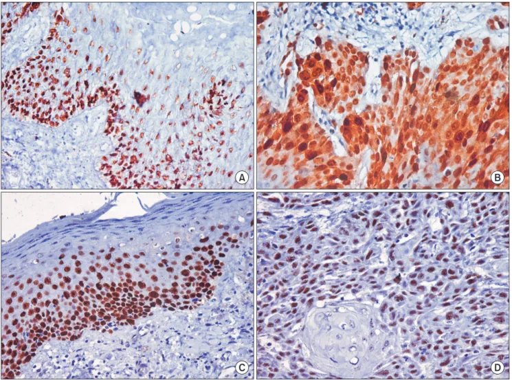
Cyclin D1 positivity was seen in 21 cases (70.0%) of leu- koplakia in Fig. 1. A and 23 cases (76.7%) of OSCC in Fig. 1.
B. Mild dysplasia (37.5%), moderate dysplasia (66.7%), and severe dysplasia (92.3%) were identified, and OSCC grades included WDSCC (66.7%), MDSCC (87.5%), and PDSCC (85.7%). These dysplasia and OSCC grades showed (Table 1) p63 positivity in all cases of leukoplakia in Fig. 1. C and OSCC in Fig. 1. D.(Table 2) Statistical significance at P<0.05 was observed for cyclin D1 and p63 expression between 2 categories of dysplasia and OSCC.(Tables 3, 4) Higher levels the basement membrane of the normal oral mucosa, where
it likely prevents basal cells from differentiating and thereby helps to maintain their basal cell status. However, upon dys- plastic change (i.e., transition from normal oral mucosa to epithelial dysplasia), dysplastic keratinocytes above the basal layer can shift to a status similar to the embryogenesis condi- tion and are still able to express p63 protein, which produces an antidifferentiation effect as well as a proliferative capacity of dysplastic cells in the oral dysplastic mucosa. p63 can con- tribute to the development of epithelial dysplasia by altering stem cell function in the basal layer, resulting in an increased number of proliferating cells, and this can contribute to the altered distribution in basal and suprabasal layers within oral epithelial dysplasia4,7.
This study of cyclin D1 as well as p63 expression indicated that these gene products can be useful for a more precise di- agnosis of leukoplakia and OSCC.
II. Materials and Methods
This was a laboratory-based study that involved using 10%
neutral buffered fixed formalin and representative paraffin- embedded histopathologically-diagnosed tissue cases of OSCC and leukoplakia, which were retrieved from the De- partment of Oral and Maxillofacial Pathology, K.M.Shah Dental College and Hospital (Vadodara, India). The samples were retrieved after the study was approved by the ethical committee of the institution, K.M.Shah Dental College and Hospital.
A total of 60 cases, comprised of leukoplakia and OSCC, were evaluated for p63 and cyclin D1 expression. The study included 30 cases of OSCC and 30 leukoplakia cases. OSCC was grouped into 3 categories based on Broder’s histopatho- logical grading: well-differentiated (WDSCC), moderately- differentiated (MDSCC), and poorly-differentiated (PDSCC) carcinoma.
Leukoplakia tissues were subdivided according to the World Health Organization classification from 2005, using the architecture and cytology criteria of mild dysplasia, mod- erate dysplasia, and severe dysplasia.
1. Immunohistochemistry
Two to three serial sections, 4 μm thick, were placed on silanized slides for p63 and cyclin D1 IHC staining. The protocols for both markers were performed according to the manufacturer’s recommendations. The sections were de-
notable in poorly-differentiated SCC.(Table 6) Comparison of both cyclin D1 and p63 expression in different grades of dysplasia of leukoplakia cases showed no statistically signifi- cant differences. Cyclin D1 expression was highly significant compared to severe and mild grades of dysplasia.(Table 7) and increased expression of cyclin D1 and p63 were found in
different grades of dysplasia in leukoplakia cases, as shown in Table 5. Similarly, the expression of both cyclin D1 and p63 was pronounced in well-differentiated SCC. However, the intensity of both cyclin D1 and p63 expression was also
Fig. 1. A. Cyclin D1 expression in leukoplakia (×400). B. Cyclin D1 expression in oral squamous cell carcinoma (×400). C. p63 expression in leukoplakia (×400). D. p63 expression in oral squamous cell carcinoma (×400).
Sunit B. Patel et al: Immunohistochemical evaluation of p63 and cyclin D1 in oral squamous cell carcinoma and leukoplakia. J Korean Assoc Oral Maxillofac Surg 2017
A B
C D
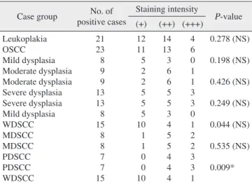
Table 1. Dysplasia and OSCC grading intensity: cyclin D1 positive cases
Case group No. of total cases No. of positive cases (%) Staining intensity
(+) (++) (+++)
Leukoplakia Mild dysplasia Moderate dysplasia Severe dysplasia OSCC
Well-differentiated Moderately-differentiated Poorly-differentiated
30 8 9 13 30 15 8 7
21 (70.0) 3 (37.5) 6 (66.7) 12 (92.3) 23 (76.7) 10 (66.7) 7 (87.5) 6 (85.7)
11 3 2 6 6 5 1 0
7 0 4 3 10 4 4 2
3 0 0 3 7 1 2 4 (OSCC: oral squamous cell carcinoma)
Sunit B. Patel et al: Immunohistochemical evaluation of p63 and cyclin D1 in oral squamous cell carcinoma and leukoplakia. J Korean Assoc Oral Maxillofac Surg 2017
Both cyclin D1 and p63 expression was statistically signifi- cant compared with poor grades of SCC, as shown in Table 8.
IV. Discussion
Many studies have assessed SCCs, with a primary objec- tive to understand the biology and determine the severity and prognosis of oral cancers with diverse behavior8. The cell cycle impacts the complexity of growth, renewal, and repair in all organisms9 and can be visualized as a relay race con- sisting of G1, S, G2, M, and G0 phases. Each of these laps
Table 4. Statistical analysis of dysplasia and OSCC in p63 Case group No. of
positive cases
Staining intensity
P-value (+) (++) (+++)
Leukoplakia OSCC Mild dysplasia Moderate dysplasia Moderate dysplasia Severe dysplasia Severe dysplasia Mild dysplasia WDSCC MDSCC MDSCC PDSCC PDSCC WDSCC
21 23 8 9 9 13 13 8 15 8 8 7 7 15
12 11 5 2 2 5 5 5 10 1 1 0 0 10
14 13 3 6 6 5 5 3 4 5 5 4 4 4
4 6 0 1 1 3 3 0 1 2 2 3 3 1
0.278 (NS) 0.198 (NS) 0.426 (NS) 0.249 (NS) 0.044 (NS) 0.535 (NS) 0.009*
(OSCC: oral squamous cell carcinoma, WDSCC: well-differentiated OSCC, MDSCC: moderately-differentiated OSCC, PDSCC: poorly- differentiated OSCC, NS: not significant)
*P<0.05.
Sunit B. Patel et al: Immunohistochemical evaluation of p63 and cyclin D1 in oral squamous cell carcinoma and leukoplakia. J Korean Assoc Oral Maxillofac Surg 2017 Table 2. Dysplasia and OSCC grading intensity: p63 positive
cases
Case group No. of total cases
Staining intensity (+) (++) (+++) Leukoplakia
Mild dysplasia Moderate dysplasia Severe dysplasia OSCC
Well-differentiated Moderately-differentiated Poorly-differentiated
30 8 9 13 30 15 8 7
12 5 2 5 11 10 1 0
14 3 6 5 13 4 5 4
4 0 1 3 6 1 2 3 (OSCC: oral squamous cell carcinoma)
Sunit B. Patel et al: Immunohistochemical evaluation of p63 and cyclin D1 in oral squamous cell carcinoma and leukoplakia. J Korean Assoc Oral Maxillofac Surg 2017
Table 3. Statistical analysis of dysplasia and OSCC in cyclin D1 Case group No. of
positive cases
Staining intensity
P-value (+) (++) (+++)
Leukoplakia OSCC Mild dysplasia Moderate dysplasia Moderate dysplasia Severe dysplasia Severe dysplasia Mild dysplasia WDSCC MDSCC MDSCC PDSCC PDSCC WDSCC
21 23 3 6 6 12 12 3 10 7 7 6 6 10
11 6 3 2 2 6 6 3 5 1 1 0 0 5
7 10 0 4 4 3 3 0 4 4 4 2 2 4
3 7 0 0 0 3 3 0 1 2 2 4 4 1
0.278 (NS) 0.097 (NS) 0.133 (NS) 0.029*
0.273 (NS) 0.517 (NS) 0.04*
(OSCC: oral squamous cell carcinoma, WDSCC: well-differentiated OSCC, MDSCC: moderately-differentiated OSCC, PDSCC: poorly- differentiated OSCC, NS: not significant)
*P<0.05.
Sunit B. Patel et al: Immunohistochemical evaluation of p63 and cyclin D1 in oral squamous cell carcinoma and leukoplakia. J Korean Assoc Oral Maxillofac Surg 2017
Table 5. Cyclin D1 and p63 expression based on intensity and grade of dysplasia
Case Cyclin D1 intensity p63 intensity
Total (+) (++) (+++) Total (+) (++) (+++)
Mild dysplasia Moderate dysplasia Severe dysplasia
3 (37.5) 6 (66.7) 12 (92.3)
3 (37.5) 2 (22.2) 6 (46.2)
0 4 (44.4) 3 (23.1)
0 0 3 (23.1)
8 (100) 9 (100) 13 (100)
5 (62.5) 2 (22.2) 5 (38.5)
3 (37.5) 6 (66.7) 5 (38.5)
0 1 (11.1) 3 (23.1) Values are presented as number (%).
Sunit B. Patel et al: Immunohistochemical evaluation of p63 and cyclin D1 in oral squamous cell carcinoma and leukoplakia. J Korean Assoc Oral Maxillofac Surg 2017
Table 6. Cyclin D1 and p63 expression based on intensity and grade of OSCC
Case Cyclin D1 intensity p63 intensity
Total (+) (++) (+++) Total (+) (++) (+++)
Well-differentiated Moderately-differentiated Poorly-differentiated
10 (66.7) 7 (87.5) 6 (85.7)
5 (33.3) 1 (12.5)
0
4 (26.7) 4 (50.0) 2 (28.6)
1 (6.7) 2 (25.0) 4 (57.1)
15 (100) 8 (100) 7 (100)
10 (6.7) 1 (12.5)
0
4 (26.7) 5 (62.5) 4 (57.1)
1 (6.7) 2 (25.0) 3 (42.9) (OSCC: oral squamous cell carcinoma)
Values are presented as number (%).
Sunit B. Patel et al: Immunohistochemical evaluation of p63 and cyclin D1 in oral squamous cell carcinoma and leukoplakia. J Korean Assoc Oral Maxillofac Surg 2017
with Angadi and Krishnapillai13 (88.90%). In addition, Goto et al.15 (100%) indicated that cyclin D1 expression increases and is correlated with poor histological grade. The difference in expression was statistically significant (P<0.05). More- over, cyclin D1 overexpression has been reported in various diverse histogenesis tumors, such as breast carcinomas16, hepatocellular carcinomas17, endometrial carcinoma18, colon carcinomas16, esophageal carcinoma19,20, and lung carcino- ma12.
p63 is a p53 homologue that plays a distinctive role in the physiology of the epithelium. TAp63, like p53, exhibits tumor-suppressive properties, while up-regulation of ΔNp63 isoforms is common and a distinctive feature of tumorigen- esis21. The role of p63 in oral dysplasia and tumors has been the focus of numerous studies, although the results remain conflicting. It is widely accepted that normal and non-dys- plastic mucosa show nuclear expression of p63 in the basal and parabasal keratinocytes of the epithelium, with the major functioning isoform being ΔNp63. As dysplasia increased in severity, the percentage of p63 positive cells increases and extends into the upper epithelial layers. In addition, over- expression of p63, especially ΔNp63, is frequently seen in HNSCC tumors and HNSCC-derived cell lines22-25. Addition- ally, oral leukoplakia with increased ΔNp63 expression and inflammatory cell infiltration exhibits a higher rate of cancer development and worse prognosis26.
In our study, all cases of leukoplakia showed p63 expres- sion, which is similar to studies by Takeda et al.27, Chen et al.24, Vered et al.28, Bortoluzzi et al.23, and Choi et al.29. In the present study, 62% cases of mild dysplasia showed mild expression, while 66.54% cases of severe dysplasia showed moderate to intense expression. Thus, p63 expres- sion increased parallel to the severity of dysplasia, similar to findings of Takeda et al.27. In addition, all cases of OSCC showed positive p63 expression. This finding is correlated with reports from other studies of Sakiz et al.30, Moergel et is regulated by a distinct set of “cyclins” that activate their
CDK partners and propel the cell forward in its proliferative pool. Negative control over the cell cycle is exerted by CDK inhibitors10.
Cancer cell escape from the cell cycle machinery reflects a fundamental hallmark of cancer progression and is emerging as a central theme in oral carcinogenesis3. Indeed, the stron- gest connection between cyclins and oncogenesis was shown in studies on cyclin D111. The underlying mechanisms for cyclin D1 overexpression include gene amplification, chro- mosomal translocation, and other post translational mecha- nisms3,12.
This study utilized the cyclin D1 monoclonal antibody for OSCC to elucidate the role of this key cell cycle regulator in this neoplasia. In our study, 21 cases (70.0%) of leukopla- kia showed cyclin D1 positive expression, which is slightly lower than the findings reported by Castle et al.8, which found 84.62% positive expression. Eleven cases (52.38%) showed mild, 7 cases (33.33%) showed moderate, and 3 cases (14.29%) showed intense cyclin D1 expression. These findings were similar to Castle et al.8 who found that 45.45%
of patients showed mild, 40.90% showed moderate, and 13.63% showed intense cyclin D1 expression. All positive mild dysplasia cases showed mild expression, while 50% of severe dysplasia cases showed moderate to intense expres- sion, which shows that the cyclin D1 expression increased in parallel with the severity of dysplasia.
Twenty-three cases (76.7%) of OSCC showed positive cyclin D1 expression, which is slightly higher than studies re- ported by Angadi and Krishnapillai13, Lam et al.14, and Goto et al.15, which found 70.73%, 63%, and 65.9% positive cyclin D1 expression, respectively. In this study, 60% of WDSCC cases showed mild to moderate expression that was similar to results reported by Angadi and Krishnapillai13 (46.9%) and Goto et al.15 (67.74%). All PDSCC cases showed moderate to intense cyclin D1 expression, which is also in concordance
Table 7. Cyclin D1 and p63 expression in different grades of leu- koplakia (dysplasia)
Dysplasia Cyclin D1 intensity p63 intensity Chi-square P-value Chi-square P-value Mild vs moderate
Moderate vs severe Severe vs mild
4.657 5.601 8.986
0.097 (NS) 0.133 (NS) 0.029*
3.238 1.706 2.448
0.198 (NS) 0.426 (NS) 0.294 (NS) (NS: not significant)
*P<0.05.
Sunit B. Patel et al: Immunohistochemical evaluation of p63 and cyclin D1 in oral squamous cell carcinoma and leukoplakia. J Korean Assoc Oral Maxillofac Surg 2017
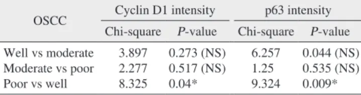
Table 8. Cyclin D1 and p63 expression in different grades of OSCC
OSCC Cyclin D1 intensity p63 intensity Chi-square P-value Chi-square P-value Well vs moderate
Moderate vs poor Poor vs well
3.897 2.277 8.325
0.273 (NS) 0.517 (NS) 0.04*
6.257 1.25 9.324
0.044 (NS) 0.535 (NS) 0.009*
(OSCC: oral squamous cell carcinoma, NS: not significant)
*P<0.05.
Sunit B. Patel et al: Immunohistochemical evaluation of p63 and cyclin D1 in oral squamous cell carcinoma and leukoplakia. J Korean Assoc Oral Maxillofac Surg 2017
between increased cyclin D1 expression in mild and severe dysplasia in leukoplakia cases, but not with other groups (mild vs moderate and moderate vs severe).(Table 7) Hence, the results indicate that the severity of dysplasia increases with increased expression of cyclin D1 and can indicate a high risk for malignant transformation. Cyclin D1 and p63 expression was significantly higher than that in well-differentiated SCC and poorly-differentiated SCC, respectively.(Table 8) IHC demonstration of these gene products can be a useful tool for a more precise prognosis of leukoplakia and OSCC. Similar studies with a larger sample size in different areas of the oral cavity and that assess all stages of SCC will provide addi- tional insight into oral carcinogenesis and the roles of cyclin D1 and p63.
Conflict of Interest
No potential conflict of interest relevant to this article was reported.
ORCID
Sunit B. Patel, http://orcid.org/0000-0003-4701-2466 Bhari S. Manjunatha, http://orcid.org/0000-0002-4415- 2538
Vandana Shah, http://orcid.org/0000-0001-9049-3942 Nishit Soni, http://orcid.org/0000-0001-6548-9255 Rakesh Sutariya, http://orcid.org/0000-0002-0302-4468
References
1. Fregonesi PA, Teresa DB, Duarte RA, Neto CB, de Oliveira MR, Soares CP. p16(INK4A) immunohistochemical overexpression in premalignant and malignant oral lesions infected with human pap- illomavirus. J Histochem Cytochem 2003;51:1291-7.
2. Peghini PE, Fehr J. Analysis of cyclin D1 expression by quan- titative real-time reverse transcription-polymerase chain reac- tion in the diagnosis of mantle cell lymphoma. Am J Clin Pathol 2002;117:237-45.
3. Todd R, Hinds PW, Munger K, Rustgi AK, Opitz OG, Suliman Y, et al. Cell cycle dysregulation in oral cancer. Crit Rev Oral Biol Med 2002;13:51-61.
4. Hosokawa Y, Arnold A. Cyclin D1/PRAD1 as a central target in oncogenesis. J Lab Clin Med 1996;127:246-52.
5. Fracchiolla NS, Pruneri G, Pignataro L, Carboni N, Capaccio P, Boletini A, et al. Molecular and immunohistochemical analysis of the bcl-1/cyclin D1 gene in laryngeal squamous cell carcinomas:
correlation of protein expression with lymph node metastases and advanced clinical stage. Cancer 1997;79:1114-21.
6. Hu H, Xia SH, Li AD, Xu X, Cai Y, Han YL, et al. Elevated ex- pression of p63 protein in human esophageal squamous cell carci- nomas. Int J Cancer 2002;102:580-3.
7. Shah RB, Zhou M, LeBlanc M, Snyder M, Rubin MA. Compari- son of the basal cell-specific markers, 34betaE12 and p63, in the
al.31, Chen et al.24, Bortoluzzi et al.23, Chen et al.32, Faridoni- Laurens et al.33, and Choi et al.29, who reported 100% positive p63 expression.
Out of 30 OSCC cases, 11 cases (36.7%) showed mild, 13 cases (43.3%) showed moderate, and 6 cases (20.0%) showed intense p63 expression. This is similar the results of a study by de Oliveira et al.25, who found 31.1% mild, 50% moder- ate, and 18.9% intense p63 expression. The results indicated that 93.33% cases of WDSCC showed mild to moderate, 62.5% cases of MDSCC showed moderate, and 100% cases of PDSCC showed intense p63 expression, indicating that p63 expression increased with poor histological grade. This result was statistically significant (P<0.05).
A literature search indicated that very few studies have re- ported isolated examination of either leukoplakia or OSCC.
This study involved both cyclin D1 and p63 expression in leukoplakia as well as all grades of OSCC. The expression of both cyclin D1 and p63 increased as the stage of dysplasia in leukoplakia cases increased from mild to severe.(Table 5) The total cyclin D1 and p63 expression was more general with well-differentiated SCC. In contrast, the intensity of OSCC in poorly-differentiated cases was greater than that in well- and moderately-differentiated SCC.(Table 6) This indicates that the cancer prognosis is evident from increased expression as dysplasia increases, which is in accordance with Todd et al.3.
Overexpression of p63 has been reported in various tumors, including adenocarcinoma and SCC34,35, adenoid carcinoma, polymorphous low-grade adenocarcinoma and basal cell and canalicular adenomas36, esophageal carcinoma6, and prostate cancer7. p63 expression was mainly noted in the peripheral cells of tumor nests in the well-differentiated tumor area, similar to a report by Chen et al.32.
The overall expression of cyclin D1 and p63 correlates with poor histological grades of OSCC and leukoplakia. The increased cyclin D1 and p63 expression parallel with the severity of lesions likely reflects the intense proliferative ac- tivity and invasiveness of these lesions. In general, cyclin D1 and p63 expression increased with increased grade of dyspla- sia in leukoplakia and OSCC.
V. Conclusion
In this study, we conclude that cyclin D1 and p63 are in- volved in carcinogenesis, which is associated with changes from early dysplasia to poorly-differentiated carcinoma, and these changes are noted with higher expression patterns in leukoplakia and OSCC. We found a significant association
diagnosis of prostate cancer. Am J Surg Pathol 2002;26:1161-8.
8. Castle JT, Cardinali M, Kratochvil FJ, Abbondanzo SL, Kessler HP, Auclair PL, et al. P53 and cyclin D1 staining patterns of malig- nant and premalignant oral lesions in age-dependent populations.
Oral Surg Oral Med Oral Pathol Oral Radiol Endod 1999;88:326- 9. Poon RYC. Cell cycle control. Encyclopedia Cancer 1997;1:246-32.
10. Kumar V, Cotran RS, Robbins SL. Robbins basic pathology. 7th 55.
ed. Philadelphia: Saunders; 2003.
11. Weinstein IB, Zhou P. Cell cycle control gene defects and human cancer. Encyclopedia Cancer 1997;1:256-67.
12. Mate JL, Ariza A, Aracil C, López D, Isamat M, Pérez-Piteira J, et al. Cyclin D1 overexpression in non-small cell lung carcinoma:
correlation with Ki67 labelling index and poor cytoplasmic differ- entiation. J Pathol 1996;180:395-9.
13. Angadi PV, Krishnapillai R. Cyclin D1 expression in oral squa- mous cell carcinoma and verrucous carcinoma: correlation with histological differentiation. Oral Surg Oral Med Oral Pathol Oral Radiol Endod 2007;103:e30-5.
14. Lam KY, Ng IO, Yuen AP, Kwong DL, Wei W. Cyclin D1 ex- pression in oral squamous cell carcinomas: clinicopathological relevance and correlation with p53 expression. J Oral Pathol Med 2000;29:167-72.
15. Goto H, Kawano K, Kobayashi I, Sakai H, Yanagisawa S. Expres- sion of cyclin D1 and GSK-3beta and their predictive value of prognosis in squamous cell carcinomas of the tongue. Oral Oncol 2002;38:549-56.
16. Bartkova J, Lukas J, Strauss M, Bartek J. Cell cycle-related varia- tion and tissue-restricted expression of human cyclin D1 protein. J Pathol 1994;172:237-45.
17. Zhang YJ, Jiang W, Chen CJ, Lee CS, Kahn SM, Santella RM, et al. Amplification and overexpression of cyclin D1 in hu- man hepatocellular carcinoma. Biochem Biophys Res Commun 1993;196:1010-6.
18. Ruhul Quddus M, Latkovich P, Castellani WJ, James Sung C, Steinhoff MM, Briggs RC, et al. Expression of cyclin D1 in nor- mal, metaplastic, hyperplastic endometrium and endometrioid car- cinoma suggests a role in endometrial carcinogenesis. Arch Pathol Lab Med 2002;126:459-63.
19. Tsuruta H, Sakamoto H, Onda M, Terada M. Amplification and overexpression of EXP1 and EXP2/Cyclin D1 genes in hu- man esophageal carcinomas. Biochem Biophys Res Commun 1993;196:1529-36.
20. Naitoh H, Shibata J, Kawaguchi A, Kodama M, Hattori T. Over- expression and localization of cyclin D1 mRNA and antigen in esophageal cancer. Am J Pathol 1995;146:1161-9.
21. Mills AA. p63: oncogene or tumor suppressor? Curr Opin Genet Dev 2006;16:38-44.
22. Foschini MP, Gaiba A, Cocchi R, Pennesi MG, Gatto MR, Frezza GP, et al. Pattern of p63 expression in squamous cell carcinoma of
the oral cavity. Virchows Arch 2004;444:332-9.
23. Bortoluzzi MC, Yurgel LS, Dekker NP, Jordan RC, Regezi JA. As- sessment of p63 expression in oral squamous cell carcinomas and dysplasias. Oral Surg Oral Med Oral Pathol Oral Radiol Endod 2004;98:698-704.
24. Chen YK, Hsue SS, Lin LM. Expression of p63 protein and mRNA in oral epithelial dysplasia. J Oral Pathol Med 2005;34:232-9.
25. de Oliveira LR, Ribeiro-Silva A, Zucoloto S. Prognostic impact of p53 and p63 immunoexpression in oral squamous cell carcinoma. J Oral Pathol Med 2007;36:191-7.
26. Saintigny P, El-Naggar AK, Papadimitrakopoulou V, Ren H, Fan YH, Feng L, et al. DeltaNp63 overexpression, alone and in combi- nation with other biomarkers, predicts the development of oral can- cer in patients with leukoplakia. Clin Cancer Res 2009;15:6284-91.
27. Takeda T, Sugihara K, Hirayama Y, Hirano M, Tanuma JI, Semba I. Immunohistological evaluation of Ki-67, p63, CK19 and p53 expression in oral epithelial dysplasias. J Oral Pathol Med 2006;35:369-75.
28. Vered M, Allon I, Dayan D. Maspin, p53, p63, and Ki-67 in epi- thelial lesions of the tongue: from hyperplasia through dysplasia to carcinoma. J Oral Pathol Med 2009;38:314-20.
29. Choi HR, Batsakis JG, Zhan F, Sturgis E, Luna MA, El-Naggar AK. Differential expression of p53 gene family members p63 and p73 in head and neck squamous tumorigenesis. Hum Pathol 2002;33:158-64.
30. Sakiz D, Turkmenoglu TT, Kabukcuoglu F. The expression of p63 and p53 in keratoacanthoma and intraepidermal and invasive neo- plasms of the skin. Pathol Res Pract 2009;205:589-94.
31. Moergel M, Abt E, Stockinger M, Kunkel M. Overexpression of p63 is associated with radiation resistance and prognosis in oral squamous cell carcinoma. Oral Oncol 2010;46:667-71.
32. Chen YK, Hsue SS, Lin LM. Immunohistochemical demonstration of p63 in DMBA-induced hamster buccal pouch squamous cell carcinogenesis. Oral Dis 2003;9:235-40.
33. Faridoni-Laurens L, Bosq J, Janot F, Vayssade M, Le Bihan ML, Kaghad M, et al. P73 expression in basal layers of head and neck squamous epithelium: a role in differentiation and carcinogenesis in concert with p53 and p63? Oncogene 2001;20:5302-12.
34. Shirendeb U, Hishikawa Y, Moriyama S, Win N, Thu MM, Mar KS, et al. Human papillomavirus infection and its possible correla- tion with p63 expression in cervical cancer in Japan, Mongolia, and Myanmar. Acta Histochem Cytochem 2009;42:181-90.
35. Di Como CJ, Urist MJ, Babayan I, Drobnjak M, Hedvat CV, Ter- uya-Feldstein J, et al. p63 expression profiles in human normal and tumor tissues. Clin Cancer Res 2002;8:494-501.
36. Edwards PC, Bhuiya T, Kelsch RD. Assessment of p63 expression in the salivary gland neoplasms adenoid cystic carcinoma, poly- morphous low-grade adenocarcinoma, and basal cell and canalicu- lar adenomas. Oral Surg Oral Med Oral Pathol Oral Radiol Endod 2004;97:613-9.