Introduction
There has been increasing interest in placement of a dental implant into a fresh extraction socket and many authors have tried to improve clinical efficacy on immediate implant in human clinical studies1-15). The advantages of immediate implantation are as fol- lows: total treatment time can be shortened; the hori- zontal and vertical levels of the residual socket wall
can be more easily preserved than with the delayed implantation; positioning of the implant is optimized;
the need for additional surgeries like bone augmenta- tion is lessened; and the healing potential of residual periodontal ligament cells is helpful for successful os- seointegration2,3,8,9). On the other hand, coronal gaps made around the implants placed immediately into fresh extraction sockets and the lack of soft tissue that makes it difficult to maintain a primary closure of the surgical site can be problematic6,10-12).
Several studies have been published the relationship between gap width and healing pattern around imme- diate implants16-24). Carlsson et al.16) used titanium implants with initial gap widths of 0.00, 0.35, and 0.85mm to find out that there was no osseointegration
The evaluation of healing patterns in surgically created circumferential gap defects around dental implants according to implant surface, defect width and defect morphology
Se-Ung Im
1, Ji-Youn Hong
1, Gyung-Joon Chae
1, Ui-Won Jung
1, Chang-Sung Kim
1, Yong-Keun Lee
2, Kyoo-Sung Cho
1, Chong-Kwan Kim
1, Seong-Ho Choi
1*1. Department of Periodontology, Research Institute for Periodontal Regeneration, College of Dentistry, Yonsei University 2. Department and Research Institute of Dental Biomaterials and Bioengineering, College of Dentistry, Yonsei University
ABSTRACT
Purpose: The aim of this study was to evaluate the factors affecting healing patterns of surgically created circumferential gap defects around implants in dogs.
Materials and Methods: In four mongrel dogs, all mandibular premolars were extracted. After 8 weeks of healing periods, implants were submerged. According to the surface treatment, turned surface was designated as a group A and rough surface as a group B. In each dog, surgical defects on the left side were made with a customized tapered step drill and on the right with a customized paralleled drill. Groups were also divided according to the width of the coronal gaps: 1.0mm, 1.5mm, or 2.0mm. The dogs were sacrificed following 8 weeks and the specimens were analyzed histologically and histomorphometrically.
Results: During the postoperative period, healing was uneventful and implants were well-maintained. As the size of the coronal gap was increased, the amount of bone-to-implant contact was decreased. The bone healing was greater in rough surface implants compared to the turned ones. About the defect morphology, tapered shape showed much bone healing and direct bone to implant contact even in the smooth surface implants.
Conclusion: Healing of the circumferential defect around dental implant is influenced by the implant surface, defect width and the morphology of the defect. When using rough surface implants, circumferential gap defects within 2 mm do not need any kinds of regenerative procedures and the healing appeared to be faster in the tapered defect morphology than the paralleled one. (J Korean Acad Periodontol 2008;38:385-394)
KEY WORDS: defect width; defect morphology; implant surface; gap.
Correspondence : Seong-Ho Choi
Department of Periodontology, College of Dentistry, Yonsei University, Shinchon-Dong 134, Seodaemoon-gu, Seoul, 120-752, Korea.
e-mail: [email protected], Tel: 82-2-2228-3189, Fax: 82-2-392-0398
* This study was supported by a grant of the Medical Science and Engineering Research Center (No.R13-2003-013-02001-0) from the Ministry of Education, Science Technology.
Received: May 26, 2008; Accepted: Jun 30, 2008
Akimoto et al.19) studied a smooth surface implant placed in the surgically created bone defect sites after tooth extraction in dog experimental model. Bone re- generation was found in gap width more than 0.5mm clinically, but histologically there was no direct con- tact between bone and implant.
Botticelli et al.20,21) suggested that implant surface characteristics can affect the healing pattern of gap defect around implant. They used rough surface im- plant (SLA) in dogs by creating bone defects with 1 to 1.25mm gaps and barrier membranes were used to cover the coronal defects. They suggested that the gaps were healed by appositional bone growth from the lateral and apical bone walls of the defects. More recently, they also compared bone healing around the implants with turned or rough surface topographies placed in self-contained defects using either a sub- merged or non-submerged technique and suggested that the rough surface showed superior healing pat- tern than the turned one22).
The actual shape of the fresh extraction socket is ap- proximately conical, but the paralleled defects made sur-
Materials and methods
1. Animals
Four male mongrel dogs with 18 to 24 months old and weighing about 30kg were chosen. They had intact dentitions and healthy periodontium. Animal selection, management, preparation and surgical protocol were followed by the routine procedures approved by the Animal Care and Use Committee, Yonsei Medical Center, Seoul, Korea.
2. Experimental Design
Animals were divided into the group A those with turned surface implants and the group B, rough (resorbable blast media ; RBM) surface implants.
The defects in the left side were made_surgically with a customized tapered step drill and those in right with customized paralleled drill. Groups were also divided according to the width of the coronal gaps:
1.0mm, 1.5mm, 2.0mm (Fig. 1, Fig. 2a, 2b).
2mm defect
1.5mm defect 1mm defect Paralleled
defect
Tapered defect
Paralleled defect 1mm defect 2mm defect
1.5mm defect
Tapered defect
3. Surgical protocol
Teeth were extracted under general anesthesia un- der sterile conditions in an operating room using Atropine 0.05 mg/kg SQ, xylazine (Rompun®, Bayer Korea, Seoul, Korea) 2 mg/kg, and ketamine hydro- chloride (Ketalar®, Yuhan Co., Seoul, Korea) of 10 mg/kg IV. Dogs were placed on a heating pad, in- tubated, administered 2% enflurane, and monitored with an electrocardiogram. After disinfecting the sur- gical site, 2% lidocaine HCl with epinephrine 1:100,000 (Kwangmyung Pharm., Seoul, Korea) was administered by infiltration. Crevicular incisions were made and all premolars were carefully extracted. Prior to ex- traction, P2-P4 were sectioned to avoid tooth fracture. Flap was sutured with 5-0 resorbable suture material (Polyglactin 910 braided absorbable suture, Ethicon, Johnson & Johnson Int., Edinburgh, UK) by the vertical mattress suture technique. On the day of surgery, the dogs received 10 mg/kg IV of the anti- biotics Cefazoline.
The implants (Restore®, Lifecore, USA) were placed after a healing period of 8 weeks under the same sur- gical conditions as those for tooth extraction. A cres- tal incision was made to preserve keratinized tissue and mucoperiosteal flap was carefully reflected on the buccal and lingual aspect. The edentulous ridge was carefully flattened with a surgical bur and irrigated with sterile saline. Implant osteotomy was performed at 800 rpm under chilled saline irrigation and circum- ferential defects of 1.0mm, 1.5mm and 2.0mm gaps with 5mm defect depth were created surgically with a customized paralleled step drill, and same procedure was done on the left side of mandible using a cus- tomized tapered step drill. Implant placement was made without tapping to obtain good initial stability.
Turned surface implants in Group A and rough RBM surface implants in Group B were used. Three sub- merged type implants (3.5mm diameter, 10.0mm length) were placed on the right side of the mandible (Fig. 3).
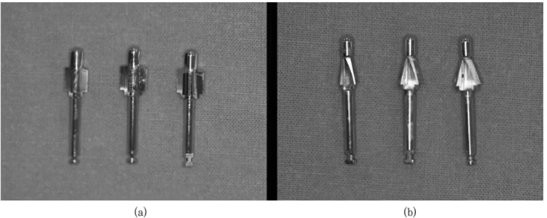
Figure 2. Customized drills
a. Stepped paralleled drills, from the left, a 5.5-mm diameter drill for the 1.0-mm gap defect, a 6.5-mm diameter drill for the 1.5-mm gap defect and a 7.5-mm diameter drill for the 2.0-mm gap defect are represented, respectively. The length of the drill is 5mm.
b. Stepped tapered drills, from the left, a 5.5-mm diameter drill for the 1.0-mm gap defect, a 6.5-mm diameter drill for the 1.5-mm gap defect and a 7.5-mm diameter drill for the 2.0-mm gap defect are represented, respectively. The length of the drill is 5mm.
(a) (b)
Flaps were closed with 5-0 resorbable suture mate- rials and implants were submerged. Post-operative care was similar as that for tooth extraction. Sutures were removed after 7 to 10 days and a soft diet was provided throughout the study period.
Dogs were sacrificed 8 weeks after surgery.
Euthanasia was performed by anesthesia drug overdose. Block sections including segments with im- plants were preserved and fixed in 10% neutral buf- fered formalin.
The specimens were dehydrated in ethanol, em- bedded in methacrylate, and sectioned in the me- sio-distal plane using a diamond saw (Exakt®, Apparatebau, Norderstedt, Germany). From each im- plant site, the central section was reduced to a final thickness of about 20μm by microgrinding and polish- ing with a cutting-grinding device (Exakt®, Apparatebau, Norderstedt, Germany). The sections were stained in hematoxyline-eosin.
4. Histologic analysis
General histologic findings were observed with a stereoscope (LEICA MZFLIII, LEICA, WETZLAR, Germany) and microscope. After conventional micro- scopic examinations, computer-assisted histometric
Cybernetics, Silverspring M.D., USA) coupled with a video camera mounted on a light microscope (LEICA DM-LB, LEICA, WETZLAR, Germany). The measuring points were as follows.
1) distance (mm) from the implant margin to the most coronal level of contact between bone and implant
2) bone to implant contact percentage (BIC%) in the coronal 5mm of the implant
Results
1. Clinical findings
During the postoperative periods, healing was un- eventful and implants were well-maintained. There were no signs of inflammation observed in the mucosa adjacent to the implants.
2. Histologic findings
1) Implant surface
The healing of rough surface implants was superior to smooth surface implants. Wedge shaped defect in coronal portion was found in the 2mm width of paral- leled defect in group A, when there was good Figure 3. Clinical photograph representing the experimental design; from the left, 1.0mm, 1.5mm and
2.0mm gaps were prepared, respectively.
2) Defect width
More remaining area which was not filled with bone was found at the larger width of the defect around the implant. There was a remaining wedge shaped de- fect in 2mm tapered gap in group A (Fig. 5a, 5b).
3) Defect morphology
Most of the tapered defects showed good bone fill- ing compared to the paralleled defects. In 1.0mm gap of Group A, good bone fill was found in the tapered defect when there was no direct bone-to-implant contact in paralleled one (Fig. 6a, 6b).
(a) (b)
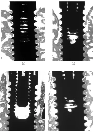
Figure 5. Histologic view of group A (turned surface) in tapered defect with gap defect width of 1mm (a) and with gap defect width of 2mm (b), H-E stain, magnification X8.
Figure 6. Histologic view of group A (turned surface) in 1 mm gap defect with tapered de- fect morphology (a) and with paralleled defect morphology (b), H-E stain, magnification X8.
(b) (a)
(b) (a)
3. Histomorphometric analysis
1) Distance (mm) from the implant margin to the most coronal level of contact between bone and implant
With increasing size of the coronal gap, the dis- tance tended to be greater. Regarding the implant surface characteristics, greater distance was shown in the turned implant than the rough one. In defect morphology, paralleled shape demonstrated greater distance than the tapered one (Table 1).
2) Bone-to implant contact percentage (BIC%) in the coronal 5mm of the implant
With decreasing size of coronal gap, bone to im- plant contact tended to be greater. Regarding the im- plant surface characteristics, rough surface showed greater bone to implant contact than turned surface implant. In defect morphology, tapered shape showed
Discussion
The immediate implant placement technique was in- troduced to shorten the rehabilitation periods and re- searches have been carried out to provide the theo- retical backgrounds. There are many methods in- troduced to overcome the coronal gap associated with immediate implant3,5,6,10-12,15,23-25) however, the critical size of defect allowing spontaneous healing has yet to be determined. To make the treatment procedure more simplified and save the time for treatment benefiting both patient and practitioner, effort to figure out the critical size is of importance. Besides defect width, implant surface and defect morphology can influence the healing of circumferential gap defect around implants.
About defect width, Akimoto et al.19) used dog model to evaluate the bone fill that occurred in defects ad- jacent to implants designed with machined surface.
Implants were placed in simulated extraction sockets Table 2. Bone to implant contact percentage (BIC%) in the coronal 5mm of the implant (N=2)
defect group
Paralleled defect Tapered defect
1.0mm 1.5mm 2.0mm 1.0mm 1.5mm 2.0mm
Group A 8.9 5.2 2.6 34.2 28.7 10.5
Group B 28.7 25.2 10.7 42.7 41.5 27.4
Mean values are shown in this table.
N : number of the dogs used in each group (Group A, Group B) Group A : turned implant surface
Group B : rough implant surface
and the bone. A clinical examination was performed after 12 weeks of healing showing that all the defects, independent of size, had healed properly. Histologic evaluations in biopsies obtained from the different defect sites, however, revealed that there were con- sistent certain distances between the marginal borders of the implants and the most coronal levels of bone-to-implant contact. Further, this distance var- ied with the initial size of the defect, so that when the defect was wider the distance between the rim of the implant and the level of bone to implant contact became longer. In the present study, the 2mm defect width tended to make more wide and deep wedge shaped coronal defect than 1mm defect width. It sug- gests that the greater defect width will need more healing time to fill the bone defect.
To mention the implant surface characteristics in the present study, rough surface implants with re- sorbable blast media (RBM) were used. To obtain RBM surface, a machined titanium implant was blasted with calcium phosphate ceramic and then passivated to completely remove the residual media. The surface roughness ranged from 3.09±0.38 microns and mi- cro-pit diameter ranged from 5 to 10 microns.
Osteoblasts may lay down bone on the old bone surface or on the implant surface itself. Davies26) suggested that there are two different phenomena by which bone can become juxtaposed to an implant sur- face: distance and contact osteogenesis. Distance os- teogenesis is that in which new bone is formed on the surfaces of bone in the peri-implant site through ap- positional growth and contact osteogenesis or osteo- conduction is that in which de novo bone formation occurs directly on the implant surface. He suggested that implant with roughened surface, as opposed to implant with smooth surface, may promote osteo- conduction by both increasing available surface area for fibrin attachment and providing surface features with which fibrin could become entangled.
More recently, Davies27) also explained that the im-
plant surface design will play an important role in the fibrin retention. Fibrin retention is so critical to os- teogenic cell migration to the implant surface. Bone cells will reach the implant surface by migration through fibrin, and these cells will then be available to synthesize de novo bone on the implant surface itself.
Akimoto19) studied marginal bone defects of varying dimensions that occurred following placement of im- plants with turned surface failed to heal with proper osseointegration. In contrast, similar experiments21) was done with rough surface implants demonstrated that marginal bone defects were resolved by de novo formation of hard tissue. Botticelli et al.22) compared bone healing at implants with turned or rough surface in self-contained defects using dogs. After 4 months of healing, the marginal defects around rough surface implants exhibited substantial bone fill and a high degree of osseointegration, but healing at turned im- plants was characterized by incomplete bone fill and the presence of a connective tissue zone between the implant and the newly formed bone.
Present study showed that bone healing was superi- or in bone defects adjacent to implants with a rough compared to smooth surface implants, and it is similar to a previous study22). The reason can be explained that the defect healing of rough surface implants is occurred by combination of contact osteogenesis and distance osteogenesis, but healing of smooth surface implants is done only by distance osteogenesis.
Therefore, the remodeling of defect will be faster in the rough than smooth surface implants.
In defect morphology, several studies have been published the relationship between gap width and healing pattern around implants in immediate implantation. Most of these studies used a paralleled defect model. However a shape of fresh extraction socket is a conical, so this study used a tapered step- ped drill to reproduce an actual extraction socket. In this study, most of tapered defect were found good
a lateral wall at defect base is closer in tapered defect than paralleled defect. That means appositional bone growth occurred faster in tapered defect than paral- leled defect.
Botticelli et al.21) explained bone-to-implant contact was first established in the apical portion of the gap.
This new bone tissue was in the coronal direction continuous with a dense, non-mineralized implant- attached soft tissue which, over time, also became mineralized and, hence, the height of the zone of bone-to-implant contact was increased.
Therefore, it can be concluded that healing of cir- cumferential gap defects around implants is influenced by the implant surface, defect width, defect morphology. If using rough surface implants, circum- ferential gap defect within 2mm does not need any kind of regenerative procedure, and tapered defect morphology showed faster healing than paralleled de- fect morphology.
References
1. Wilson T, Weber H. Classification of and therapy for areas of deficient bony housing prior to dental implant placement. Int J Periodont Rest Dent 1993;13:451–459.
2. Lazzara RJ. Immediate implant placement into extraction sites: Surgical and restorative advantages. Int J Periodont Rest Dent 1989;9:333-343.
3. Werbitt M, Goldberg P. The immediate implant: Bone preservation and bone regeneration. Int J Periodont Rest Dent 1992;12:206-217.
7. Becker BE, Becker W, Ricci A, Geurs N. A prospective clinical trial of endosseous screw-shaped implants placed at the time of tooth extraction without augmentation. J Periodontol 1998;69:920-926.
8. Watzek G, Haider R, Mensdorff-Pouilly N, Haas R.
Immediate implants and delayed implantation for complete restoration of the jaw following extraction of all residual teeth: A retrospective study comparing different types of serial immediate implantation. Int J Oral Maxillofac Implants 1995;10:561-567.
9. Denissen HW, Kalk W, Veldhuis HA, van Waas MA.
Anatomic consideration for preventive implantation. Int J Oral Maxillofac Implants 1993;82:191-196.
10. Becker W, Becker BE. Guided tissue regeneration for im- plants placed into extraction sockets and for implant de- hiscences: surgical techniques and case report. Int J Periodont Rest Dent 1990;10:376-391.
11. Gotfredsen K, Nimb L, Buser D, Hjörting-Hansen E.
Evaluation of guided bone generation around implants placed into fresh extraction sockets: An experimental study in dogs. J Oral Maxillofac Sur 1993;51:879-884.
12. Goldstein M, Boyan BD, Schwartz Z. The palatal advanced flap: a pedicle flap for primary coverage of immediately placed implants. Clin Oral Imp Res 2002;13:644-650.
13. Rosenquist B, Grenthe B. Immediate placement of implants into extraction sockets: Implant survival. Int J Oral Maxillofac Implants 1996;11:205-209.
14. Schwarz AD, Chaushu G. Placement of implants into fresh extraction sites: 4-7 years retrospective evaluation of 95 immediate implants. J Periodontol 1997;68:1110-1116.
15. Schwartz AD, Grossman Y, Chaushu G. The clinical effec- tiveness of implants placed immediately into fresh extraction sites of molar teeth. J Periodontol 2000;71:839-844.
17. Knox R, Caudill R, Meffert R. Histologic evaluation of dental endosseous implants placed in surgically created ex- traction defects. Int J Periodont Rest Dent 1991;11:364-375.
18. Thomas G, Wilson Jr., Schenk R, Buser D. Implants placed in immediate extraction sites: A report of histologic and histomorphometric analyses of human biopsies. Int J Oral Maxillofacial implants 1998;13:333-341.
19. Akimoto K, Becker W, Persson R et al. Evaluation of tita- nium implants placed into simulated extraction sockets: A study in dogs. Int J Oral Maxillofac Implants 1999;14:351-360.
20. Botticelli D, Berglundh T, Buser D, Lindhe J. Hard-tissue alterations following immediate implant placement in ex- traction sites. J Clin Periodontol 2004;31:820-828.
21. Botticelli D, Berglundh T, Buser D, Lindhe J. Appositional bone formation in marginal defects at implants. Clin Oral Imp Res 2003;14:1-9.
22. Botticelli D, Berglundh T, Persson LG, Lindhe J. Bone re- generation at implants with turned or rough surfaces in self-contained defects: An experimental study in the dog. J
Clin Periodontol 2005;32:448–455.
23. Botticelli D, Berglundh T, Buser D, Lindhe J. Resolution of bone defects of varying dimension and configuration in the marginal portion of the peri-implant bone: An ex- perimental study in the dog. J Clin Periodontol 2004;31:
309-317.
24. Botticelli D, Berglundh T, Buser D, Lindhe J. The influ- ence of a biomaterial on the closure of marginal hard tis- sue defect adjacent to implants: An experimental study in the dog. Clin Oral Implants Res 2004;15:285-292.
25. Caudill RF, Meffert RM. Histologic analysis of the os- seointegration of endosseous implants in simulated ex- traction sockets with and without e-PTFE barriers. Part I.
Preliminary findings. Int J Periodont Rest Dent 1991;11:
207-215.
26. Davies JE. Mechanisms of endosseous integration. Int J Prosthodont 1998;11:391-401.
27. Davies JE. Understanding peri-implant endosseous healing.
J Dent Edu 2003;8:932-949.