서 론
미 만성 간질성 폐질환(diffuse interstitial lung di- sease)은 폐포보다는 폐간질을 주로 침범하는 다양한 정도의 염증과 섬유화 소견을 보이는 만성 질환으로 결체조 직질환이나 약물 등 원인이 밝혀진 경우와 특발성인 경우로 나눌 수 있다. 특발성 간질성 폐렴의 분류는 2002년
American Thoracic Society와 European Respiratory So- ciety의 합의를 통한 임상적, 영상의학적 그리고 병리학적 소견을 종합한 다각적 접근에 따른 분류 체계가 널리 받아 들여 지고 있다(1, 2). 이에 따르면 특발성 간질성 폐렴은 상용 간질성 폐렴(idiopathic pulmonary fibrosis/usual interstitial pneumonia, IPF/UIP), 비특이적 간질성 폐렴 (nonspecific interstitial pneumonia, NSIP), 특발성 기질화
간질성 폐질환의 영상의학적 소견
Radiologic Diagnosis of Interstitial Lung Diseases
이 진 아·이 경 수 | 성균관의대 삼성서울병원 영상의학과 | Chin A Yi, MD·Kyung Soo Lee, MD Department of Radiology, Samsung Medical Center, Sungkyunkwan University School of Medicine E - mail : kyungs.lee@samsung.com
J Korean Med Assoc 2009; 52(1): 14 -21
D
iffuse interstitial lung diseases (DILD) are a group of chronic disorders showing varying degrees of active inflammation and established fibrosis mainly involving the interstitium of the lungs. DILD can be classified into two groups in an etiologic aspect; idiopathic and DILD with known-cause, such as connective tissue diseases associated with DILD. Although there had been complexity and variability in the classification of idiopathic interstitial pneumonia (IIP), an international standard was established for the classification of IIPs including seven clinical- radiologic-pathologic entities; idiopathic pulmonary fibrosis (IPF), nonspecific interstitial pneu- monia (NSIP), cryptogenic organizing pneumonia (COP), acute interstitial pneumonia (AIP), respiratory bronchiolitis-associated interstitial lung disease (RB-ILD), desquamative interstitial pneumonia (DIP), and lymphoid interstitial pneumonia (LIP). The prognosis of fibrotic NSIP and IPF is much poorer compared to those of other spectrum of IIPs, such as COP, RB-ILD, DIP, and LIP. Therefore, fibrotic NSIP and IPF can be considered separately as a group of fibrotic IIPs. Trying to predict the prognosis of IIPs, there has been an effort to differentiate inflammation- predominant lesions from fibrosis-predominant lesions in patients with fibrotic IIPs. Radiologic features of low fibrotic scores at high-resolution CT and early enhancement patterns at dynamic enhancement of MRI can be useful prognostic determinants for the prediction of disease improvement in patients with fibrotic IIPs.Keywords: Diffuse interstitial lung disease; Idiopathic interstitial pneumonia; CT·idiopathic interstitial pneumonia; MR·idiopathic interstitial pneumonia; Prognosis
핵 심 용 어: 미만성 간질성 폐질환; 특발성 간질성 폐렴; CT·특발성 간질성 폐렴; MR·특발성 간질성 폐렴; 예후
Abstract
폐렴(cryptogenic organizing pneumonia, COP), 급성 간질성 폐렴(acute interstitial pneumonia, AIP), 세기관 지 간질성 폐렴(respiratory bronchiolitis-associated in- terstitial lung disease, RB-ILD), 박리성 간질성 폐렴 (desquamative interstitial pneumonia, DIP) 그리고 림 프양 간질성 폐렴(lymphoid interstitial pneumonia, LIP) 의 7가지 질환으로 나눌 수 있으며, 이들 중 IPF와 NSIP는 전체 특발성 간질성 폐렴의 2/3 가량을 차지한다(1). 특발 성 간질성 폐렴의 정확한 진단은 환자의 예후와 치료 방침 을 결정하는 데 있어 필수적인데, 5년 생존율의 경우 IPF는 43%, 섬유화성 NSIP는 86%으로 차이가 나고 나머지 특발 성 간질성 폐렴들은 거의 100%로 큰 차이를 보인다. 예후
결정에 있어서의 병리조직학적 접근은 정확한 진단을 가능 하게 한다는 면에 있어서 매우 중요하지만(3~5), 수술적으로 얻어지는 극히 일부분의 조직을 이용한 병리조직학적 소견 이 불균질한 전체 폐 병변을 반영하지 못한다는 한계가 크다.
반면, 고해상도 단층 촬영(high-resolution computed to- mography, HRCT)을 통하여 폐 전체에 걸친 병변의 형태 학적 특성을 관찰한 결과는 질병의 특성과 예후 예측 인자 로서의 역할이 클 것으로 생각된다. 실제로 특발성 간질성 폐렴 환자의 병리조직학적 소견은 병변의 위치에 따라서 차 이가 많다고 알려져 있으며(6, 7), 병리 의사의 경험 정도에 따라 진단 결과의 차이가 큰 것으로 나타났다(8). 그러므로 특발성 간질성 폐렴 환자의 예후 예측에 기여하는 흉부
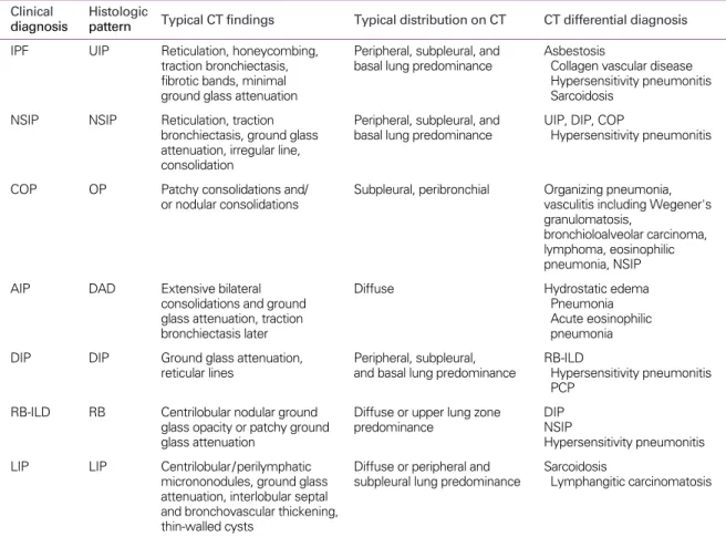
Table 1. Typical CT findings and differential diagnosis of idiopathic interstitial pneumonias (1) Clinical Histologic
Typical CT findings Typical distribution on CT CT differential diagnosis diagnosis pattern
IPF UIP Reticulation, honeycombing, Peripheral, subpleural, and Asbestosis
traction bronchiectasis, basal lung predominance Collagen vascular disease
fibrotic bands, minimal Hypersensitivity pneumonitis
ground glass attenuation Sarcoidosis
NSIP NSIP Reticulation, traction Peripheral, subpleural, and UIP, DIP, COP
bronchiectasis, ground glass basal lung predominance Hypersensitivity pneumonitis attenuation, irregular line,
consolidation
COP OP Patchy consolidations and/ Subpleural, peribronchial Organizing pneumonia,
or nodular consolidations vasculitis including Wegener's
granulomatosis,
bronchioloalveolar carcinoma, lymphoma, eosinophilic pneumonia, NSIP
AIP DAD Extensive bilateral Diffuse Hydrostatic edema
consolidations and ground Pneumonia
glass attenuation, traction Acute eosinophilic
bronchiectasis later pneumonia
DIP DIP Ground glass attenuation, Peripheral, subpleural, RB-ILD
reticular lines and basal lung predominance Hypersensitivity pneumonitis PCP
RB-ILD RB Centrilobular nodular ground Diffuse or upper lung zone DIP glass opacity or patchy ground predominance NSIP
glass attenuation Hypersensitivity pneumonitis
LIP LIP Centrilobular/perilymphatic Diffuse or peripheral and Sarcoidosis
micrononodules, ground glass subpleural lung predominance Lymphangitic carcinomatosis attenuation, interlobular septal
and bronchovascular thickening, thin-walled cysts
HRCT의 소견 및 염증성 병변과 섬유화성 병변의 감별에 도움 이 되는 자기공명영상(MRI)의 영상의학적 특징을 살펴본다.
특발성 간질성 폐렴의 HRCT 소견
특발성 간질성 폐렴(idiopathic interstitial pneumo- nia)의 주요한 HRCT 소견은 양측성으로 나타나는 흉막 하 망상 혹은 간유리 음영이며, RB-ILD를 제외하고는 많은 경 우 폐 기저부를 주로 이환하여 나타난다. 각각의 특발성 간 질성 폐렴은 특징적인 HRCT의 소견을 나타내지만 많은 부 분 중복되는 소견이 보일 수 있으므로 CT 소견의 특징적인 정도에 따라 함께 감별해야 할 질환들이 있다(Table 1). 폐 기저부에 나타나는 벌집모양 음영(honeycombing)과 폐 상부에 나타날 수 있는 흉막 하 망상음영(reticulation)은 병리학적 진단인 상용 간질성 폐렴(UIP)과 밀접하게 관련 되는 HRCT 소견으로 보고되고 있다(9). 이와 같은 UIP를 시사하는 전형적인 HRCT 소견이 보일 경우에는 조직학적 검사 없이도 IPF를 진단할 수 있다. 하지만 많은 경우 IPF 와 NSIP의 CT 및 임상 양상 뿐만 아니라 병리조직학적 소 견도 중복되어 나타나므로 감별이 쉽지 않으며(10), 전형 적인 IPF를 제외하고는 수술적 방법을 이용한 병리조직학 적 방법을 통하여 확진할 것을 권고하고 있다(1). HRCT 소 견을 이용하여 NSIP와 UIP를 감별 진단하는 후향적 분석 연구에서 CT를 이용한 NSIP 진단의 정확도(accuracy)는 66% 가량(sensitivity; 70%, specificity; 63%)으로 이전에 보고된 결과들에 비해서는 높았으나 중등도의 진단 정확도 를 보였다. UIP와 비교시 NSIP가 나타내는 특징적인 HRCT 소견은 간유리음영(ground glass attenuation)이 더 많고 망상음영(reticulation)이 더 섬세하게 나타난다는 것이다(10).
특발성 간질성 폐렴의 예후를 예측할 수 있는 HRCT 소견
폐 전체를 영상화한 HRCT 소견들을 통하여 치료에 대한 반응이나 예후를 예측하고자 하는 시도들이 이루어지고 있
는데, 이는 특발성 간질성 폐렴의 예후가 그 복잡한 분류체 계에서 알 수 있듯이 폐의 극히 일부분을 절제한 병리 소견 에만 근거하여서는 전체적인 질환의 상태를 이해하기 어렵 기 때문이다. 그러므로 간질성 폐렴의 주요한 HRCT 소견 인 간질성폐음영(ground glass opacity), 망상음영(reticu- lation), 경화성음영(consolidation), 그리고 벌집양음영 (honeycombing)의 정도를 숙련된 영상의학과 의사들이 정량화하고 이를 폐 기능 검사 결과나 추적검사로 시행한 HRCT에서의 호전 혹은 진행 여부와 상호 연관시켜 보는 시 도들이 이루어지고 있으며 더 나아가 환자의 생존율에 영향 을 미치는 독립인자로서의 HRCT 소견을 분석하여 보고하 고 있다.
1992년에 Lee 등은 fibrosing alveolitis 환자를 대상으
로 하여 스테로이드 치료에 후 폐 기능 수치들의 호전을 예
측하게 하는 HRCT 소견을 분석하였다(11). Ground glass
attenuation의 면적을 정량화 하였을 때, 보다 광범위한
ground glass attenuation을 보일수록 치료에 대한 반응 효
과가 크게 나타나는 양의 상관관계를 보여, HRCT상 나타나
는 ground glass attenuation이 치료에 반응할 것임을 나타
내는 예측 인자가 될 수 있음을 보고하였다(11). 이후
Flaherty 등은 UIP와 NSIP 환자들을 대상으로 한 연구에서
조직병리 결과 UIP로 진단되는 환자가 가장 높은 mortality
를 보이는 것으로 보고하였고, UIP를 시사하는 특징적인
HRCT 소견인 honeycombing을 수술적 조직 검사가 어려
운 환자들의 나쁜 예후를 예측할 수 있는 대리 인자가 되는
것으로 보고하였다(4). 비슷한 시기에 UIP를 시사하는 특징
적인 HRCT 소견의 특징들에 대해 다변수분석(multiva-
riate logistic regression)을 시행하였는데, 이 결과 폐 기저
부의 honeycombing 뿐만 아니라 폐 상부의 reticulation
또한 UIP의 예측에 유의한 HRCT 소견으로 나타났다. 그러
므로 이 결과를 통해 HRCT에서 나타나는 honeycombing
과 reticulation의 합한 fibrotic score라는 지표를 통해 환자
의 예후를 예측해 보고자 하는 연구가 시도되었다. Shin 등
은 임상-영상-병리 결과를 토대로 하여 진단된 114명의
UIP 및 fibrotic NSIP 환자들을 대상으로 하여 폐기능검사
결과와 bronchoalveolar lavage (BAL) 결과 그리고 HRCT
소견들에 대한 정량화된 데이터를 수집하였다. 이들 값들을 이용하여 환자의 mortality에 대한 multivariate logistic regression을 후향적으로 시행하였을 때, 낮은 DLco (diffu- sion capacity of the lung for carbon dioxide)와 높은 fibrotic score (summation of honeycombing and
reticulation)만이 환자의 mortality를 증가시키는 유의한 지표로 나타났다(Figure 1, 2)(12). 재미있는 점은 이 연구 에서 병리적으로 UIP를 진단하는 요인은 단변수분석에서는 유의하게 mortality를 증가시키는 요인으로 나타났으나 다 변수분석에서는 그 유의성이 증명되지 않아, 이제까지 UIP
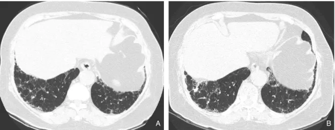
Figure 1. Stable UIP (concordant diagnosis in all three pathologists) in a 67- year- old woman.
(A) Transverse thin-section (1.0-mm section thickness) CT scan obtained at the level of liver dome shows bilateral subpleural distribution of reticulation and ground glass attenuation. Total extent of parenchymal abnormalities on CT scans was 15%
and fibrotic score (reticulation plus HC) was 10%.
(B) Seven-year follow-up CT scan obtained at the similar level shows no progression of disease (total extent of parenchymal abnormalities; 15%, fibrotic score; 10%).
A B
Figure 2. Progressive UIP (concordant diagnosis in all three pathologists) in a 63- year- old man.
(A) Transverse thin-section (1.0-mm section thickness) CT scan obtained at the level of liver dome shows bilateral subpleural distribution of reticulation, honeycombing, and ground glass attenuation. Total extent of parenchymal abnormalities on CT scans was 35% and fibrotic score (reticulation plus HC) was 30%.
(B) One-year follow-up CT scan obtained at the similar level shows progression of disease (total extent of parenchymal abnor- malities; 50%, fibrotic score; 35%).
A B
가 mortality 증가를 예측하게 하는 유의한 인자라는 보고들 과 상충한다는 점이다(4, 13). 이러한 결과는 병리적으로 UIP 혹은 NSIP를 진단하는 것보다 HRCT상 특징적인 UIP 의 feature를 보이는 경우가 예후와 더 밀접한 관계가 있을 가능성을 시사한다. 실제로 HRCT상 확실한 UIP의 소견을 보이는 그룹의 예후가 UIP를 시사하는 중등도의 HRCT 소 견을 보이거나 NSIP의 HRCT 소견을 보이는 그룹에 비하여 유의하게 나쁜 것으로 나타났으며, 병리적으로 UIP의 진단 을 받은 환자들 중에서도 비전형적인 UIP의 HRCT 소견을 보이는 그룹이 전형적인 UIP의 HRCT 소견을 보이는 그룹 에 비하여 예후가 좋은 것으로 나타났다(14). 병리적으로
NSIP를 진단받은 환자들의 HRCT 소견을 분석하였을 때, 예상과 달리 UIP pattern의 HRCT 소견을 보이는 경우를 포 함하여 78%의 NSIP 환자들이 이제까지 NSIP의 알려진 HRCT 소견과 다른 양상을 나타내어, 병리적 NSIP를 진단 받은 환자들의 HRCT 소견이 다양할 수 있음을 알게 되었다 (15). 그러므로 이상의 연구 결과들을 토대로 생각해 볼 때, 각각의 환자가 가지는 병리적 혹은 영상의학적 근거에 바탕 을 둔 UIP 혹은 NSIP라는 진단명 자체 보다는 전체적으로 폐를 이환하고 있는 섬유화의 정도를 반영하는 HRCT상의 fibrotic score나 PFT상의 DLco 값들이 환자의 예후를 보다 잘 예측할 수 있는 지표가 될 수 있다.
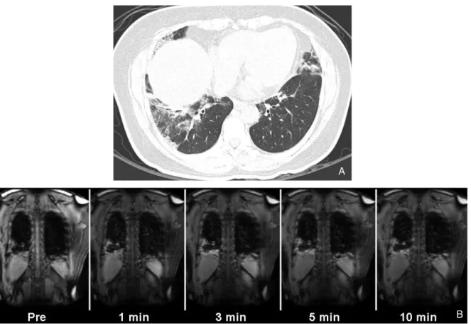
Figure 3. A 55-year-old woman with nonspecific interstitial pneumonia (group 1, cellular) exhibiting early enhancement.
(A) Transverse high-resolution CT scan (1.25-mm section thickness) at level of liver dome shows subpleural patchy parenchymal opacities in both lungs.
(B) Serial dynamic MR images show early enhancement with peak enhancement at 3 minutes. Biopsy was performed on lesions in left upper and lower lobes. Pathologic specimens disclosed inflammation-predominant nonspecific interstitial pneumonia (group 1, cellular).
A
B
미만성 간질성 폐렴의 예후를 예측할 수 있는 MRI 소견
미만성 간질성 폐렴의 치료에 대한 반응과 예후를 예측하 기 위한 방법으로서 MRI를 이용한 시도들도 이루어 지고 있 다. 폐 영역의 MR 기법은 다른 신체 부위와 달리 대부분의
조직이 공기로 채워져 있는 해부학적 특성 때문에 발생되는 MR signal이 적고, 공기와 조직 사이에 발생할 수 있는 susceptibility artifact가 커서 영상화 하기 어려운 단점이 있다. 하지만 최근 고자장 MR 기기의 발전과 susceptibility artifact를 최소화는 빠른 영상 기법들의 도입됨에 따라 특 발성 간질성 폐질환 환자에서 inflammation-predom-
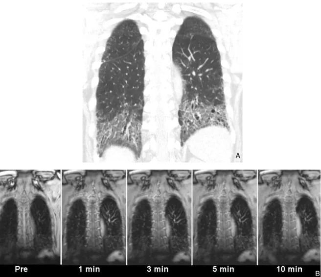
Figure 4. A 78- year- old man with interstitial pneumonia (group 3, fibrotic) exhibiting slight enhancement.
(A) Coronal thin- section (2.5-mm section thickness) CT scan shows subpleural reticulation and traction bronchiectasis in both lungs.
(B) Serial dynamic MR images show no significant enhancement. Biopsy was performed on lesions in left lower lobe. Pathologic specimens disclosed fibrosis-predominant nonspecific interstitial pneumonia (group 3).
A
B
inant 병변과 fibrosis-predominant 병변을 감별하는 데 MR 영상을 이용할 수 있다고 보고되고 있다.
2000년 Gaeta 등은 처음으로 다양한 chronic infiltrative lung disease 환자를 대상으로 조영증강 MRI를 시행하였 으며 활동성 질환을 보인 17명의 환자 중 14명의 환자에서 조영 증강되는 폐병변이 보인다고 하였고 조영 증강되는 폐 병변을 가지고 있던 14명의 환자는 모두 활동성 질환을 가 진 환자였다고 보고하였다(16). 초창기 MR 영상의 유용성 을 제시한 이러한 보고는 대상이 된 환자의 진단명이 매우 다양하였으며, 널리 받아들여지는 활동성 질환의 정의에 대 한 일치된 의견이 부족하고, 섬유성 조직에서 MR 조영제의 재분포(redistribution)가 일어나는 3분 이후의 조영 증강 양상에 대한 분석이 없다는 점들에서 한계가 있다. 최근에 3T의 고자장 MRI를 이용하여 UIP의 typical HRCT 소견을 보인 환자들을 제외하고, 수술적 조직 검사가 필요했던 33 명의 UIP와 fibrotic NSIP 환자들을 대상으로 하여 얻은 T2 강조영상과 1, 3, 5, 10분 지연영상을 포함하는 dynamic enhanced MR 영상을 분석한 연구가 보고되었다(17). 이 연구에서 조직학적으로 염증 소견이 주로 나타나는 병변들 은 섬유화 소견이 많이 나타나는 병변에 비하여 T2 강조영 상에서 밝게 나타나며(9/17 lesion sites, 53%), 조영 증강 시 3분 이내에 최고 조영 증가되는 early enhancement pattern을 보이는 것으로 나타났다(14/17 lesion sites, 82%)(Figure 3). 반면 섬유화 소견이 주가 되는 병변들은 전혀 조영 증강되지 않거나(19/35 lesion sites, 54%), 5분 이후에 늦게 조영 증강되는 양상(15/35 lesion sites, 43%) 의 dynamic enhancement pattern 을 보였다(Figure 4).
결 론
결론적으로, 각각의 환자가 가지는 병리적 혹은 영상의학 적 근거에 바탕을 둔 UIP 혹은 NSIP라는 진단명 자체 보다 는 전체적으로 폐를 이환하고 있는 섬유화의 정도를 반영하 는 HRCT상의 fibrotic score나 PFT상의 DLco 값들이 환자 의 예후를 보다 잘 예측할 수 있는 지표가 될 수 있다고 생각 된다. 또한 방사선 노출의 위험없이 다중 조영 증강 영상을
얻을 수 있는 MRI 기법은 염증이 심한 부위와 섬유화가 심 한 부위를 효과적으로 구별해 내어 HRCT 소견에 필적하는 예후 예측 인자로서의 역할을 할 수 있을 것으로 기대된다.
참고문헌
11. American Thoracic Society/European Respiratory Society International Multidisciplinary Consensus Classification of the Idiopathic Interstitial Pneumonias. This joint statement of the American Thoracic Society (ATS), and the European Respi- ratory Society (ERS) was adopted by the ATS board of direc- tors, June 2001 and by the ERS Executive Committee, June 2001. American journal of respiratory and critical care medi- cine 2002; 165: 277-304.
12. Demedts M, Costabel U. ATS/ERS international multidis- ciplinary consensus classification of the idiopathic interstitial pneumonias. Eur Respir J 2002; 19: 794 -796.
13. Monaghan H, Wells AU, Colby TV, du Bois RM, Hansell DM, Nicholson AG. Prognostic implications of histologic patterns in multiple surgical lung biopsies from patients with idiopathic interstitial pneumonias. Chest 2004; 125: 522-526.
14. Flaherty KR, Toews GB, Travis WD, Colby TV, Kazerooni EA, Gross BH, Jain A, Strawderman RL, 3rd, Paine R, Flint A, Lynch JP, 3rd, Martinez FJ. Clinical significance of histological classification of idiopathic interstitial pneumonia. Eur Respir J 2002; 19: 275-283.
15. Bjoraker JA, Ryu JH, Edwin MK, Myers JL, Tazelaar HD, Schroeder DR, Offord KP. Prognostic significance of histo- pathologic subsets in idiopathic pulmonary fibrosis. American journal of respiratory and critical care medicine 1998; 157:
199-203.
16. Flaherty KR, Travis WD, Colby TV, Toews GB, Kazerooni EA, Gross BH, Jain A, Strawderman RL, Flint A, Lynch JP, Mar- tinez FJ. Histopathologic variability in usual and nonspecific interstitial pneumonias. American journal of respiratory and critical care medicine 2001; 164: 1722-1727.
17. Qureshi RA, Ahmed TA, Grayson AD, Soorae AS, Drakeley MJ, Page RD. Does lung biopsy help patients with interstitial lung disease? Eur J Cardiothorac Surg 2002; 21: 621- 626;
discussion 626.
18. Lettieri CJ, Veerappan GR, Parker JM, Franks TJ, Hayden D, Travis WD, Shorr AF. Discordance between general and pulmonary pathologists in the diagnosis of interstitial lung disease. Respiratory medicine 2005; 99: 1425 -1430.
19. Hunninghake GW, Lynch DA, Galvin JR, Gross BH, Muller N, Schwartz DA, King TE Jr, Lynch JP, 3rd, Hegele R, Waldron J, Colby TV, Hogg JC. Radiologic findings are strongly asso-
ciated with a pathologic diagnosis of usual interstitial pneu- monia. Chest 2003; 124: 1215 -1223.
10. MacDonald SL, Rubens MB, Hansell DM, Copley SJ, Desai SR, du Bois RM, Nicholson AG, Colby TV, Wells AU. Non- specific interstitial pneumonia and usual interstitial pneumonia:
comparative appearances at and diagnostic accuracy of thin- section CT. Radiology 2001; 221: 600 - 605.
11. Lee JS, Im JG, Ahn JM, Kim YM, Han MC. Fibrosing alveolitis:
prognostic implication of ground-glass attenuation at high- resolution CT. Radiology 1992; 184: 451- 454.
12. Shin KM, Lee KS, Chung MP, Han J, Bae YA, Kim TS, Chung MJ. Prognostic determinants among clinical, thin-section CT, and histopathologic findings for fibrotic idiopathic interstitial pneumonias: tertiary hospital study. Radiology 2008; 249:
328 - 337.
13. Riha RL, Duhig EE, Clarke BE, Steele RH, Slaughter RE, Zimmerman PV. Survival of patients with biopsy-proven usual interstitial pneumonia and nonspecific interstitial pneumonia.
Eur Respir J 2002; 19: 1114 -1118.
14. Flaherty KR, Thwaite EL, Kazerooni EA, Gross BH, Toews GB, Colby TV, Travis WD, Mumford JA, Murray S, Flint A, Lynch JP, 3rd, Martinez FJ. Radiological versus histological diagnosis in UIP and NSIP: survival implications. Thorax 2003; 58: 143 - 148.
15. Hartman TE, Swensen SJ, Hansell DM, Colby TV, Myers JL, Tazelaar HD, Nicholson AG, Wells AU, Ryu JH, Midthun DE, du Bois RM, Muller NL. Nonspecific interstitial pneumonia:
variable appearance at high-resolution chest CT. Radiology 2000; 217: 701-705.
16. Gaeta M, Blandino A, Scribano E, Minutoli F, Barone M, Ando F, Pandolfo I. Chronic infiltrative lung diseases: value of ga- dolinium-enhanced MRI in the evaluation of disease activity- early report. Chest 2000; 117: 1173 -1178.
17. Yi CA, Lee KS, Han J, Chung MP, Chung MJ, Shin KM. 3-T MRI for differentiating inflammation- and fibrosis-predominant lesions of usual and nonspecific interstitial pneumonia: com- parison study with pathologic correlation. Ajr 2008; 190: 878 - 885.
Peer Reviewers Commentary
이 논문은 간질성 폐질환의 최근 분류와 UIP 및 NSIP의 HRCT 소견, 그리고 최근 임상적 중요성이 강조되고 있는 특발 성 간질성 폐렴의 예후를 예측할 수 있는 인자에 대해 기술하고 있다. 간질성 폐질환에서 MRI 소견과 역동적 조영기법 은 염증 부위와 섬유화 부위를 효과적으로 구별할 수 있어 임상적 유용성이 크지만 고자장(3.0 Tesla)을 이용한 기기라 는 한계점을 고려하여야 하겠다. HRCT상 fibrotic score는 전체적으로 진행되고 있는 섬유화의 정도를 잘 반영하여 환 자의 예후 예측인자로 유용하게 이용될 수 있으며 이는 병리학적 혹은 영상의학적 근거에 의한 간질성 폐질환의 진단 보 다 실제 임상적으로 더 큰 의미가 있다고 할 수 있다. 향후 영상의학적 접근을 통하여 현재 섬유화의 시간적 또는 공간적 침범 정도 뿐만 아니라 병태생리에 관여하는 여러 다른 인자들과의 연관성에 대한 연구가 필요하며 필자들이 사진 설명 에서 언급한 바와 같이 병변의 진행 속도에 따른 분류와 이에 중요한 역할을 할 HRCT 소견에 대한 연구가 필요할 것으 로 사료된다.
[정리:편집위원회]