www.krspine.org
Conservative Treatment of Lumbar Disc Herniation
- A Prospective Study of Disc Herniation Encroaching More than One-third of Spinal Canal -
Byung-Joon Shin, M.D., Jae Chul Lee, M.D., Ho-Hyoung Lee, M.D., Hae-Dong Jang, M.D.
J Korean Soc Spine Surg 2011 Sep;18(3):123-131.
Originally published online September 30, 2011;
http://dx.doi.org/10.4184/jkss.2011.18.3.123
Korean Society of Spine Surgery
Department of Orthopedic Surgery, Inha University School of Medicine
#7-206, 3rd ST. Sinheung-Dong, Jung-Gu, Incheon, 400-711, Korea Tel: 82-32-890-3044 Fax: 82-32-890-3467
©Copyright 2011 Korean Society of Spine Surgery pISSN 2093-4378 eISSN 2093-4386
The online version of this article, along with updated information and services, is located on the World Wide Web at:
http://www.krspine.org/DOIx.php?id=10.4184/jkss.2011.18.3.123
This is an Open Access article distributed under the terms of the Creative Commons Attribution Non-Commercial License (http://
creativecommons.org/licenses/by-nc/3.0) which permits unrestricted non-commercial use, distribution, and reproduction in any medium, provided the original work is properly cited.
Journal of Korean Society of
Spine Surgery
Received: January 17, 2011 Revised: July 21, 2011 Accepted: July 21, 2011
Published Online: September 30, 2011 Corresponding author: Jae Chul Lee, M.D.
Department of Orthopaedic Surgery, Soonchunhyang University College of Medicine, 657, Hannam-dong, Yongsan-gu, Seoul, Korea
TEL: 82-2-709-9808, FAX: 82-2-794-9414 E-mail: [email protected]
“This is an Open Access article distributed under the terms of the Creative Commons Attribution Non-Commercial License (http://
creativecommons.org/licenses/by-nc/3.0/) which permits unrestricted non-commercial use, distribution, and reproduction in any medium, provided the original work is properly cited.”
Conservative Treatment of Lumbar Disc Herniation
- A Prospective Study of Disc Herniation Encroaching More than One-third of Spinal Canal -
Byung-Joon Shin, M.D., Jae Chul Lee, M.D., Ho-Hyoung Lee, M.D., Hae-Dong Jang, M.D.
Department of Orthopaedic Surgery, Soonchunhyang University College of Medicine, Seoul, Korea
Study Design: Prospective study.
Objectives: To investigate the clinical results of conservative treatment for mid-to-large lumbar disc herniation diagnosed via magnetic resonance imaging (MRI) and the factors influencing treatment.
Summary of Literature Review: There is limited information regarding the clinical results of conservative treatment for lumbar disc herniation. The recent studies using MRI have suggested favorable treatment results.
Materials and Methods: The study subjects were 39 cases of herniated disc patients with over a 1/3 spinal canal encroachment -- based on MRI -- that were followed up for at least 1 year. The average age was 42.6-years-old (range of 12-76 years-old), and the average follow-up period was 28 months. The neurological deficit and the visual analogue scale (VAS) of back pain and radiating pain at the time of initial diagnoses and final follow-ups were compared, and the clinical results were evaluated based Kim & Kim’s criteria.
Results: Although 4 of the 39 patients needed to undergo surgery during the follow-up period, 33 of the remaining 35 patients showed satisfactory (excellent and good ratings) results: 27 excellent, 6 good, 2 fair, i.e., a 85% (33 out of 39) satisfactory results. Of the 14 cases that had neurological defect at the initial diagnosis, only 1 case needed surgery, thereby resulting in a 93% (13 out of 14) satisfactory result. There were no statistically significant correlations among the degree of spinal canal encroachment and other factors such as age, sex, herniation type, and neurological deficit at initial diagnosis, and the clinical results at the final follow-up, conversion to surgery during follow-up, and remaining pains.
Conclusions: The clinical results of conservative treatment in lumbar disc herniation were satisfactory even in cases of high degree of spinal canal encroachment. Therefore, conservative treatment of lumbar disc herniation should be considered first before resorting to surgical treatment.
Key Words: Lumbar Disc Herniation, Conservative Treatment, MRI, Spinal Canal Encroachment
INTRODUCTION
Lumbar disc herniation is a common disease in the spine area and it occurs in 1%-2% of the total population1) in the U.S. about 200,000 diskectomies are performed annually.2) Recently, as MRI is used widely in diagnosing disc herniation, a great number of patients are being diagnosed with lumbar disc herniation and they are being treated - other than the surgical methods using microscope, endoscope, and laser - with a variety of treatment methods and rehabilitations.
However, there is a considerable amount of controversy in regard to the pathophysiology and treatment guidelines for lumbar disc herniation. After the study by Hakelius et al.3) on the natural progress of patients with radiating pain, although many reports about the sound natural progress of herniated disc patients along with surgery and conservative treatment have been
Byung-Joon Shin et al Volume 18 • Number 3 • September 2011
www.krspine.org 124
published, there have been significant disagreements regarding the results. This is believed to be attributable to the differences in diagnostic methods which, importantly, caused the study subject groups to be different. However, according to the existing studies on the diagnoses of disc herniation were based on various methods ranging from using the symptoms of radiating pain as basis to myelogram and computed tomography. Some studies have used MRI, and, particularly, a small number of studies have used MRI for confirmation of all their cases.
In addition, there is a pattern of more recommendation for surgery when MRI shows greater degrees of disc herniation;
as such, this study attempted to investigate about the clinical results of conservative treatment and the factors that influence the results of patients who were clearly diagnosed with herniated discs with over 1/3 canal encroachment shown on MRI and corroborating clinical symptoms such as radiating pain.
RESEARCH SUBJECTS AND METHODS
1. Study Subjects
The study subjects were comprised of, among the patients who visited our hospital from April 2006 to March 2009, the cases with -- either 1) the first symptoms occurred within 6 weeks, or 2) symptoms were older than 6 weeks but symptoms showing improving trend -- of the patients diagnosed through MRI with disc herniation of more than 1/3 spinal canal encroachment and with radiating pain in the associated dermatome.
The cases where the muscle strength was below grade 2 and there were severe neurological deficits or progressing neurological deficit, cauda equina syndrome, symptoms occurred more than 6 weeks before but no sign of improvement were recommended for surgery and were excluded as study subjects. Also excluded were the patients, during this period, who had a previous history of discectomy in the same region, spinal stenosis, foraminal and extraforaminal types.
This was a prospective study using the 44 cases that met the above criteria. Conservative treatments included analgesics, anti- inflammatory drug therapy, and spinal rehabilitation education.
Among the 44 patients, 39 patients who could be followed up for more than 1 year were studied; the average age was 42.6 years old (range of 12-76 years old), 25 male cases, 14 female cases, and the average follow-up period was 28 months.
2. Study Methods
1) Analysis of Clinical Factors and Results
At the time of the initial and final follow-up, the patients’
back pain and radiating pain were recorded using VAS (Visual Analogue Scale), and Kim & Kim’s criteria (Table 1) was used to evaluate the clinical results, and cases that required surgery were classified as “bad”. In addition, the prognostic factors such as the patients’ age, sex, herniated region, herniation type, neurologi- cal deficits at initial diagnosis, back pain and radiating pain were measured.
2) Radiographic Analysis
The degree of spinal canal encroachment was measured in the most encroached region from the axial view of the MRI image by using digital measurement tools (Fig. 1). The degree of spinal canal encroachment was on the average 49.7% (range of 33.4%
-78.6%). On the MRIs, the types of herniation were classified into these categories according to the criteria of Costello et al.;
protruded, extruded, and sequestrated.4) In this study, since only the degrees of herniation of 1/3 or more were used as study subjects, the protruded types were excluded.
3) Statistical Analysis
Statistically in the univariate analysis t-tests and cross analysis
Fig. 1. Spinal canal area and herniated disc size were measured on MRI axial images.
were used to determine the correlation between the clinical result scores at the time of the final follow-up and other factors that were expressed as non-continuous variables; Pearson correlation was used to determine the correlation between the clinical result scores at the time of the final follow-up and other factors that were expressed as continuous variables. Statistical test defined as significant when p <0.05.
RESULTS
1. Radiographic Analysis of the Results
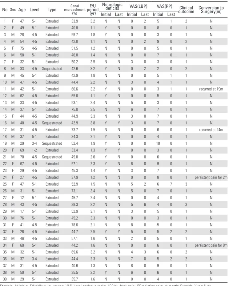
The occurrence region was most frequent at Lumbar 4-5 (L4-L5) with 19 cases, next was Lumbar 5 – Sacral 1 (L5- S1) with 17 cases, next Lumbar 3-4 (L3-L4) with 2 cases, and then Lumbar 1-2 (L1-L2) with 1 case. Extruded type was in 35 cases, sequestrated type was in 4 cases; the highest degree of spinal canal encroachment measured from the axial view of the MRI image was on 49.7% (range of 33.4%-78.6%)(Table 2).
2. Pain and Clinical Results
Of the 39 cases, 4 cases had to be converted to surgery due to the failure of conservative treatment or recurrence during the follow-up period; the remaining 35 cases showed VAS at the time of initial diagnosis of on average 2.3 (0-8) and at the time of final follow-up 0.5 (0-6), VAS for radiating pain was on average 5.5 (3-10) and at the time of final follow-up 0.4 (0-7). (Fig. 2) The clinical results were, of the 35 cases that did not require surgery: 27 cases of “excellent”, 6 cases of “good”, 2 cases of “fair” at the time of the final follow-up; i.e., without surgery 85% (33 out of 39) of all the cases showed satisfactory results(Fig. 3).
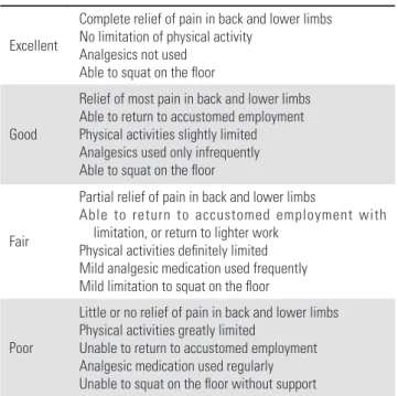
Fig. 2. VAS score at initial visit and the final follow-up Fig. 3. Clinical outcome at the final follow-up (four patients who Table.1. Kim & Kim’s criteria of clinical outcome
Excellent
Complete relief of pain in back and lower limbs No limitation of physical activity
Analgesics not used Able to squat on the floor
Good
Relief of most pain in back and lower limbs Able to return to accustomed employment Physical activities slightly limited Analgesics used only infrequently Able to squat on the floor
Fair
Partial relief of pain in back and lower limbs
Able to return to accustomed employment with limitation, or return to lighter work
Physical activities definitely limited Mild analgesic medication used frequently Mild limitation to squat on the floor
Poor
Little or no relief of pain in back and lower limbs Physical activities greatly limited
Unable to return to accustomed employment Analgesic medication used regularly Unable to squat on the floor without support
Byung-Joon Shin et al Volume 18 • Number 3 • September 2011
www.krspine.org 126
Table 2. Summary of cases
No Sex Age Level Type encroachmentCanal (%)
periodF/U (yr)
Neurologic
deficits VAS(LBP) VAS(RP) Clinical
outcome Conversion to Surgery(m) Initial Last Initial Last Initial Last
1 F 47 5-1 Extruded 33.9 3.2 N N 0 2 5 1 2 N
2 F 49 5-1 Extruded 40.8 1.1 Y N 0 0 8 0 1 N
3 M 28 4-5 Extruded 59.7 1.8 Y N 0 0 3 0 1 N
4 M 54 4-5 Extruded 42.0 1.1 N N 0 2 9 0 2 N
5 F 75 4-5 Extruded 51.5 1.2 N N 0 0 5 0 1 N
6 M 58 5-1 Extruded 46.8 1.4 N N 0 0 7 0 1 N
7 F 32 5-1 Extruded 50.2 3.5 N N 3 0 3 0 1 N
8 M 33 4-5 Sequestrated 42.6 3.2 Y N 0 2 2 0 2 N
9 M 45 5-1 Extruded 42.9 1.8 N N 0 0 5 1 1 N
10 M 47 4-5 Extruded 44.4 2.2 N N 3 0 4 1 1 N
11 M 42 5-1 Extruded 60.6 3.2 Y N 0 0 3 1 1 recurred at 19m
12 M 62 4-5 Extruded 65.0 1.1 Y N 0 0 5 0 1 N
13 M 33 4-5 Extruded 53.1 2.4 N N 5 0 3 0 1 N
14 M 37 5-1 Extruded 75.0 3.5 N N 6 0 7 0 1 N
15 F 44 4-5 Extruded 44.9 3.3 N N 3 0 7 0 1 N
16 M 40 4-5 Sequestrated 42.9 3.8 Y Y 3 0 7 0 1 N
17 M 31 4-5 Extruded 73.7 1.5 N N 0 0 6 0 1 recurred at 24m
18 M 37 5-1 Extruded 34.3 2.1 Y N 0 0 4 0 1 N
19 M 29 3-4 Sequestrated 52.4 1.9 Y N 0 0 10 0 1 N
20 F 69 1-2 Extruded 33.4 1.3 Y Y 0 0 3 0 1 N
21 M 70 4-5 Sequestrated 49.0 2.6 Y N 0 0 6 0 1 N
22 F 67 4-5 Extruded 57.1 2.3 Y N 6 0 9 0 1 N
23 F 29 4-5 Extruded 45.3 1.4 Y N 3 0 7 0 1 N
24 F 27 4-5 Extruded 37.9 1.2 N N 0 0 8 0 1 persistent pain for 2m
25 F 47 5-1 Extruded 52.9 1.5 N N 5 2 6 7 3 N
26 M 31 5-1 Extruded 73.1 3.4 N N 5 0 7 0 1 N
27 F 12 5-1 Extruded 45.7 2.4 N N 0 0 4 0 1 N
28 M 43 4-5 Extruded 38.3 2.2 N N 5 6 4 0 3 N
29 M 17 5-1 Extruded 52.9 3.1 N N 3 0 5 0 1 N
30 M 76 5-1 Extruded 45.2 3.3 N N 0 0 3 0 1 N
31 F 41 4-5 Extruded 78.6 2.1 N N 8 0 5 0 1 N
32 F 26 4-5 Extruded 44.7 2.5 Y Y 5 0 5 2 2 N
33 M 46 4-5 Extruded 57.1 1.6 N N 2 0 5 0 1 N
34 F 60 5-1 Extruded 44.2 1.6 N N 0 0 6 0 1 persistent pain for 8m
35 M 32 5-1 Extruded 69.6 3.2 N N 4 3 6 0 2 N
36 M 37 3-4 Extruded 44.4 2.3 N N 7 0 5 2 2 N
37 M 31 4-5 Extruded 40.6 1.3 N N 8 0 9 0 1 N
38 M 50 5-1 Extruded 35.5 2.2 Y N 6 0 6 0 1 N
39 M 29 5-1 Extruded 35.7 1.6 N N 0 0 4 0 1 N
F,female; M,Male; F/U:follow up; yr,year; VAS,visual analogue scale; LBP,low back pain; RP,radiating pain; m,month; Gr,grade; Y,yes; N,no
3. Analysis of Cases Requiring Surgery During Follow-Up Period
Surgeries for the 4 cases that required conversion to surgery were performed after 2, 8, 19, and 24 months after the initial diagnosis; the Case #24 was showing improvement but took a turn for the worse of radiating pain about 2 months after the initial diagnosis, thereby resorting to surgery. Case #34 was improving but developed back pain and radiating pain around 8 months after the initial diagnosis, thereby resorting to surgery, which showed severe adhesion of the herniated disc and nerve roots during intraoperative observations. In the Cases #11 and
#17, the symptoms disappeared with conservative treatment but they reoccurred: the Case #11 developed back pain and ipsilateral radiating pain after 19 months from the first visit, thereby resorted to surgery at another hospital; the symptoms in Case #17 completely disappeared after 2 months but they recurred and cauda equina syndrome complicated after 24 months, thereby requiring surgery.
The 3 cases that underwent surgery showed “excellent” clinical results at the final follow-up; the Case #17 that had cauda equina syndrome took over 1 year to regain urination and stool faculties.
4. Analysis of the Group with Neurological Deficits at the Initial Diagnosis
14 of the 39 cases showed neurological deficit such as muscle weakness: 1 case showed weakness in great toe extension down to Grade 3; the remaining 13 cases showed muscle weakness down to Grade 4. Although in the final follow-up every case
showed improvement in the neurological deficit, 3 cases showed mild paresthesias and mild muscle weaknesses remained. At the final follow-up, the back pain and radiating pain score was, respectively, 0.14 and 0.21, which were less than those (0.60 and 0.48) of the group without any neurological deficit; however, there was no statistical significance (p = 0.158, 0.516).
1 of the 14 cases required surgery due to recurrence during the follow-up period; there were 12 cases of “excellent”, and 1 case of “good”, which indicated a 93% (13 out of 14) satisfactory clinical result, which was higher than that of the group without neurological deficit, but there was no statistical significance (p = 0.391).
5. Analysis of the Group with Over 50% Spinal Canal Encroachment
16 of the 39 cases showed 50%-plus spinal canal encroachments, and the average degree of encroachment was 61.4%. There were 11 male cases and 5 female cases; the average age was 40.9 years old which was slightly younger than that (42.8 years old) of the total case population. The occurrence regions were: 8 cases at Lumbar 4-5 (L4-L5), 7 cases at Lumbar 5 – Sacral 1 (L5-S1), and 1 case at Lumbar 3-4 (L3- L4); extruded type was in 15 cases, sequestrated type was in 1 case. At the time of the initial diagnosis, back pain score was on average 2.9 (0-8); radiating pain on average was 5.5 (3-10); at the final follow-up the average back pain score was improved to 0.3 (0-6) and the average radiating pain was improved to 0.5 (0-7). 2 of the 16 cases required surgery during the follow- up period due to reoccurrence of symptoms. The clinical results
Byung-Joon Shin et al Volume 18 • Number 3 • September 2011
www.krspine.org 128
at the final follow-up were: “excellent” 12 cases, “good” 1 case, and “fair” 1 case, which indicated satisfactory results -- without surgery -- in 81% (13 out of 16) of the cases, which was a bit lower than that (87%, 20 out of 23) of the group with less than 50% encroachment, but there was no statistical significance (p = 0.674) (Fig.4).
6. Relationship Between Miscellaneous Factors at the Initial Diagnosis and Clinical Results
1) Age
The older age group (older than the average age of 42.6 years old), at the final follow-up, back pain and radiating pain scores were, respectively, 0.67 and 0.56, which were somewhat higher than those (0.24 and 0.24) of the not-old-age group, but they were not statistically significant. (p = 0.293, 0.421) The clinical results of the older age group was 83% (15 out of 18) satisfactory, which was similar to the 86% (18 out of 21) of the not-old-age group, thereby not showing any significant difference. (p = 0.837) The cases that required surgery during the follow-up period was 6% (1 out of 18), which was lower than that (14%, 3 out of 21) of the not-old-age group, but there was no statistical significance. (p = 0.370)
2) Gender
At the final follow-up, the back pain scores were on average 0.52 for males and 0.29 for females, and the radiating pain scores were on average 0.20 for males and 0.71 for females, which had no statistically significant difference (p = 0.562, 0.337).
The clinical results for males was 88% (22 out of 25) satisfactory (excellent and good), which was higher than that (79%, 11 out of 14) of the females, however, there was no statistical significance. (P = 0.647) Surgery was required in 8% (2 out of 25) of males and 14% (2/14) of females, but there was no statistically significant difference (p = 0.609).
3) Herniation Types
At the final follow-up, the back pain score for extruded type was on average 0.43 and sequestrated type was 0.50; the radiating pain score for extruded type was on average 0.43 and sequestrated type was 0.0, but there was no statistical difference (p = 0.911, 0.508). The clinical results for extruded type was 83% (29 out of 35) satisfactory, which was lower than the
100% (4 out of 4) for sequestrated type, but without statistical significance (p = 0.368). 11% (4 out of 35) of the extruded type required surgery, which was higher than that (0%, 0 out of 4) of sequestrated type, but without statistical significance (p = 0.475).
DISCUSSION
Hakelius,3) in his report on conservative treatment of patients with sciatica, reported that bed rest and the use of a corset resulted in a 93% improvement rate in 6 months, whereby a sound natural progress was suggested; however, since the report was published, controversies have continued to this day about the treatment results and natural progress of lumbar disc herniation. Although in this study only the cases with spinal canal encroachments of 1/3 or more based on MRI were used, during the 28-month follow-up period only 4 cases of the total 39 cases required surgery, and in most of the patients sound clinical results were observed. Without aggressive interventional procedures such as nerve block, 85% (33 out of 39) showed satisfactory results with conservative treatment alone, thereby demonstrating a good natural progress of disc herniation. This report was similar to the report by Saal et al.5,6) in which 90%
achieved satisfactory clinical results with aggressive rehabilitation efforts during a 31-month follow-up period. However, the results of this study is quite different from the results from the report by Weber et al.7) which stated that 17 cases (26%) of 66 conservative treatment cases required surgery within 1 year and the report by Atlas et al.8-10) which stated that 46% of group that underwent conservative treatment showed satisfactory results after 5-years. This could be attributable, as pointed out by Alaranta et al.11,12) to the inaccuracy in diagnostic methods e. g. , myelogram in the study by Weber et al.7) The diagnostic methods used in study by Atlas et al.8-10) as well included myelogram, CT scan, MRI, etc. and they were different and not standardized;
20% of the group that received conservative treatment were diagnosed only by physical examination, which in turn rendered the study somewhat limited. In contrast, this study, by using MRI as the only diagnosis method, should be considered as having sufficient clinical value.
As MRI is being used for measuring the location, type and size of herniated discs, a number of studies are being undertaken, and this study attempted to assess the differences in the clinical results by measuring the degrees of spinal canal encroachments
and types. Masui et al. stated that there was no association between the degree of spinal canal encroachment and clinical results, after following up on 21 cases for 7 years; there was no association found in this study either. However, Carlisle et al.14) reported in their retrospective study, which compared 44 cases that had undergone surgery and 44 cases that had undergone conservative treatment without surgery, that those patients who underwent surgery after the conservative treatment failed showed significantly large degree of spinal canal encroachment.
As a reason for the conflicting results, Carlisle et al.14) proposed a possibility of the degree of spinal canal encroachment’s affecting the decision to proceed with surgery. However, in the case of this study, although using only the cases with relatively large degrees of encroachments with more than 1/3 encroachment, it was possible to ascertain sound clinical results regardless of the degree of spinal canal encroachment.
In this study, even in the cases with more than 50% spinal canal encroachments, 81% (13 out of 16) achieved satisfactory clinical results, which weren’t significantly different from those (87% satisfactory results) of the cases with 33%-50%
spinal canal encroachments. These results are similar to the recent studies done using MRI; Cribb et al.15) reported that, by following up on 15 cases with more than 50% spinal canal encroachments for 24 months, in 14 of the 15 cases, improvement of clinical symptoms was shown without surgery, and reported that on average 80% reduction in size was shown, hence suggested that a careful consideration needs to be given regarding surgery due to potential surgical complications. In addition, Benson et al.16) by following up on 37 cases with more than 50% spinal canal encroachments for 23 months, reported that 83% satisfactory results were obtained, while only 4 cases required surgery. As such, regardless of the size of herniated discs, whether to undergo a surgery needs to be decided upon consideration of the duration and progress of the symptoms, and conservative treatment needs to be undertaken first before surgery.
The fact that sound natural progress was seen even in cases where herniation was accompanied by non-progressive neurological deficits regardless of treatment has been reported in numerous studies4,5,7,10,17,18) in this study as well, it was seen that 14 cases with neurological deficits at the initial diagnosis showed improvement of the neurological deficits at the final follow-up, and in 3 cases only mild residual muscle weakness
of Grade 4 was shown and resulted in a good progress. In addition, with the final clinical results showing a 93% (13 out of 14 cases) satisfactory result, it was suggested that, even in cases with neurological deficits at the initial diagnosis, as long as they are not of worsening pattern, conservative treatment should take precedence.
In this study, the factors at the initial diagnosis such as age, sex, and herniation type did not have an effect on the clinical results of conservative treatment. However, Takada et al.19) in their follow-up study of herniated discs using MRI, stated that the protruded type resulted in slower reduction in the size; since midsize-plus herniation only were used in this study, the protruded type disc herniation that were smaller than 1/3 spinal canal encroachment were excluded and only the extruded and sequestrated types were included. As such, in the future, additional studies are needed to investigate the differences in the results of conservative treatment among the protruded, extruded and sequestrated types. Many of the patients included in the study had been recommended for surgery by other hospitals, but using only conservative treatment they showed satisfactory clinical results. In addition, during the final interviews the patient satisfaction levels were high, and there were no cases of complications during the conservative treatment. Thus, even if the size of herniated disc is large in lumbar disc herniation cases and there are neurological deficits such as muscle weakness, as long as the symptoms are not too old and don’t show worsening pattern, conservative treatment should be considered first. The disadvantages of this study were that investigation from the initial diagnosis-to-improvement in symptoms was not done due to the follow-up period not being continued and only the final follow-up results were analyzed, which rendered this study somewhat limited; because of any additional MRIs not taken during the follow-up period, the extent of resorption of the disc was not ascertained. Recently, the reports that verified sequentially the extent of the nerve root compression and resorption of the herniated disc using MRI are being published20,21) at the current time where MRI has become mainstream for diagnosing lumbar disc herniation, if extensive, large-scale studies about the opinions of MRI, the natural progress and conservative treatment of lumbar disc herniation can be undertaken, they will become a great help in determining treatment guidelines.
Byung-Joon Shin et al Volume 18 • Number 3 • September 2011
www.krspine.org 130
CONCLUSION
In moderate to large size lumbar disc herniation cases, conservative treatment has resulted in most patients showing satisfactory results. Thus, when determining treatment methods, rather than surgery, conservative methods should be considered first, even if the herniated disc is large in size and there are neurological deficit. In addition, further studies, based on MRI findings, on the natural progress of lumbar disc herniation are deemed necessary.
REFERENCE
1. Deyo RA, Tsui-Wu YJ. Descriptive epidemiology of low- back pain and its related medical care in the United States.
Spine (Phila Pa 1976). 1987;12:264-8.
2. Taylor VM, Deyo RA, Cherkin DC, Kreuter W. Low back pain hospitalization. Recent United States trends and regional variations. Spine (Phila Pa 1976). 1994;19:1207- 12.
3. Hakelius A. Prognosis in sciatica. A clinical follow-up of surgical and non-surgical treatment. Acta Orthop Scand Suppl. 1970;129:1-76.
4. Costello RF, Beall DP. Nomenclature and standard reporting terminology of intervertebral disk herniation.
Magn Reson Imaging Clin N Am. 2007;15:167-74.
5. Saal JA, Saal JS. Nonoperative treatment of herniated lumbar intervertebral disc with radiculopathy. An outcome study. Spine (Phila Pa 1976). 1989;14:431-7.
6. Saal JA, Saal JS, Herzog RJ. The natural history of lumbar intervertebral disc extrusions treated nonoperatively. Spine (Phila Pa 1976). 1990;15:683-6.
7. Weber H. Lumbar disc herniation. A controlled, prospective study with ten years of observation. Spine (Phila Pa 1976).
1983;8:131-40.
8. Atlas SJ, Deyo RA, Keller RB, et al. The Maine Lumbar Spine Study, Part II. 1-year outcomes of surgical and nonsurgical management of sciatica. Spine (Phila Pa 1976).
1996;21:1777-86.
9. Atlas SJ, Keller RB, Chang Y, Deyo RA, Singer DE. Surgical and nonsurgical management of sciatica secondary to a lumbar disc herniation: five-year outcomes from the Maine Lumbar Spine Study. Spine (Phila Pa 1976). 2001;26:1179- 87.
10. Atlas SJ, Keller RB, Wu YA, Deyo RA, Singer DE. Long- term outcomes of surgical and nonsurgical management
of sciatica secondary to a lumbar disc herniation: 10 year results from the maine lumbar spine study. Spine (Phila Pa 1976). 2005;30:927-35.
11. Alaranta H, Hurme M, Einola S, et al. A prospective study of patients with sciatica. A comparison between conservatively treated patients and patients who have undergone operation, Part II: Results after one year follow- up. Spine (Phila Pa 1976). 1990;15:1345-9.
12. Hurme M, Alaranta H, Einola S, et al. A prospective study of patients with sciatica. A comparison between conservatively treated patients and patients who have undergone operation, Part I: Patient characteristics and differences between groups. Spine (Phila Pa 1976).
1990;15:1340-4.
13. Masui T, Yukawa Y, Nakamura S, et al. Natural history of patients with lumbar disc herniation observed by magnetic resonance imaging for minimum 7 years. J Spinal Disord Tech. 2005;18:121-6.
14. Carlisle E, Luna M, Tsou PM, Wang JC. Percent spinal canal compromise on MRI utilized for predicting the need for surgical treatment in single-level lumbar intervertebral disc herniation. Spine J. 2005;5:608-14.
15. Cribb GL, Jaffray DC, Cassar-Pullicino VN. Observations on the natural history of massive lumbar disc herniation. J Bone Joint Surg Br. 2007;89:782-4.
16. Benson RT, Tavares SP, Robertson SC, Sharp R, Marshall RW. Conservatively treated massive prolapsed discs: a 7-year follow-up. Ann R Coll Surg Engl. 2010;92:147-53.
17. Dubourg G, Rozenberg S, Fautrel B, et al. A pilot study on the recovery from paresis after lumbar disc herniation. Spine (Phila Pa 1976). 2002;27:1426-31.
18. Saal JA. Natural history and nonoperative treatment of lumbar disc herniation. Spine (Phila Pa 1976).
1996;21:1877-83.
19. Takada E, Takahashi M, Shimada K. Natural history of lumbar disc hernia with radicular leg pain: Spontaneous MRI changes of the herniated mass and correlation with clinical outcome. J Orthop Surg (Hong Kong). 2001;9:1-7.
20. Autio RA, Karppinen J, Niinimäki J, et al. Determinants of spontaneous resorption of intervertebral disc herniations.
Spine (Phila Pa 1976). 2006;31:1247-52.
21. Jensen TS, Albert HB, Soerensen JS, Manniche C, Leboeuf- Yde C. Natural course of disc morphology in patients with sciatica: an MRI study using a standardized qualitative classification system. Spine (Phila Pa 1976). 2006;31:1605- 12.
요추부 추간판 탈출증의 보존적 치료 결과
- 척추관의 1/3이상을 침범한 추간판 탈출증에 대한 전향적 연구 - 신병준 • 이재철 • 이호형 • 장해동
순천향대학교 의과대학 정형외과학교실
연구 계획: 전향적 임상연구
목적: 자기 공명 영상으로 진단된 중등도 크기 이상의 추간판 탈출증에 대한 보존적 치료의 임상적 결과 및 결과에 영향을 미치는 요인에 대하여 알아보 고자 하였다.
선행 문헌의 요약: 요추부 추간판 탈출증의 보존적 치료 결과에 대한 문헌은 많지 않으며, 자기공명영상을 이용한 최근의 연구들은 양호한 임상 경과를 시사하고 있다.
대상 및 방법: 자기공명영상에서 척추관의 1/3이상을 침범하는 요추부 추간판 탈출증이 환자 중 1년 이상 추시가 가능하였던 39예를 연구 대상으로 하였다. 평균 나이는 42.6세(12-76)였으며, 평균 추시 기간은 28개월이었다. 초진 및 최종 추시 시의 신경학적 이상유무 및 요통과 방사통의 통증점수 (VAS)를 비교하였고 Kim과 Kim의 평가법에 의해 임상결과를 평가하였다.
결과: 39예 중 4예에서는 추시 기간 중 수술로의 전환이 필요하였으나, 나머지 35예에서는 최종 추시 시 우수 27예, 양호 6예, 보통 2예로, 보존 적 치료만으로도 전체 환자 중 85%(33/39)에서 만족스러운 결과를 보였다. 초진 시 신경학적 결손이 있었던 14예에서도 1예만이 수술이 필요하여, 93%(13/14)에서 만족스런 결과를 보였다. 추간판의 척추관 침범정도 및 환자의 나이, 성별, 탈출형태, 초진 시 신경학적 결손유무 등의 인자와 최종 추 시 시 임상결과, 추시 기간중의 수술로의 전환, 요통이나 방사통 잔존 정도 간에는 통계적으로 유의한 상관관계를 찾을 수 없었다.
결론: 요추부 추간판 탈출증에서 척추관 침범정도가 큰 경우라도 보존적 치료시 만족스러운 임상 결과를 보였다. 따라서, 치료 방법 결정시 수술적 치료 보다는 보존적 치료가 우선적으로 고려되어야 할 것으로 사료되었다.
색인 단어: 요추부 추간판 탈출, 보존적 치료, 자기 공명 영상, 척추관 침범 약칭 제목: 요추부 추간판 탈출증의 보존적 치료 결과