pISSN : 1229-5418
Implantology 2018; 22(2): 80-86
https://doi.org/10.32542/implantology.20180007
Received: June 11, 2018 Revised: June 19, 2018 Accepted: June 21, 2018
Copyright © 2018. The Korean Academy of Oral &
Maxillofacial Implantology
This is an Open Access article distributed under the terms of the Creative Commons Attribution Non-Commercial License (http://creativecommons.
org/licenses/by-nc/4.0/) which permits unrestricted non-commercial use, distribution, and reproduction in any medium, provided the original work is properly cited.
OPEN ACCESS
Purpose: Radiological evaluation of bone quality as well as the shape of the alveolar bone of the edentulous part to be placed is very important f or successf ul implant placement. Recently, three- dimensional analysis using computed tomography and cone beam computed tomography has been actively used before implant surgery, but periapical and panoramic radiographs have been used for a long time to evaluate alveolar bone using low radiation doses. The purpose of this study was to compare the Hounsfield unit value of CT after dividing maxillary, mandibular, anterior, and posterior teeth into whether periapical and panoramic radiographs could be effectively used for bone quality evaluation.
Materials and Methods: The periapical x-ray, panoramic x-ray, and conventional CT were taken from a total of 100 patients who have undergone the implant treatment. For 3D analysis, the virtual implants with a diameter of 4 mm and a length of 10 mm were placed in the implant planning software and the bone quality of 1 mm circumference was measured by Hounsfield unit value.
Periapical x-ray and panoramic x-ray were also evaluated for bone quality. The degree of agreement among them was evaluated.
Results: In 3D image analysis, reformatted cross-sectional, panoramic image analysis, bony quality evaluation similar to CT analysis was possible in maxillary anterior/posterior teeth with periapical
Abstract
치근단 방사선사진, 파노라마 방사선 사진 및 전산화 단층 영상을 이용한 임플란트 시술 부위 골질의 평가
신동호1+, 박경미2+, 황재준3, 박원서4, 김기덕4*
1수련의, 연세대학교 치과대학 통합치의학과
2연구원, 연세대학교 치과대학 통합치의학과
3조교수, 부산대학교 치의학전문대학원 영상치의학과
4교수, 연세대학교 치과대학 통합치의학과
*Corresponding author: Kee-Deog Kim, [email protected]
+
These authors contributed equally to this work.
Assessment of Bone Quality in Pre-dental Implant Site Using Periapical, Panoramic and Computed Tomography
Dongho Shin1+, Kyeong-Mee Park2+, Jae Joon Hwang3, Wonse Park4, Kee-Deog Kim4*
1)Resident, Department of Advanced General Dentistry, Yonsei University College of Dentistry, Seoul, Korea
2)Researcher, Department of Advanced General Dentistry, Yonsei University College of Dentistry, Seoul, Korea
3)Assistant Professor, Department of Oral and Maxillofacial Radiology, Pusan National University School of Dentistry, Pusan, Korea
4)Professor, Department of Advanced General Dentistry, Yonsei University College of Dentistry, Seoul, Korea
and panoramic radiographs.
Conclusion: Periapical and panoramic radiographs could be used to evaluate the implant bone quality of the maxillary anterior and maxillary posterior.
Keywords: Bone quality, CT, CBCT, Periapical radiography, Panoramic radiography, Dental implant
I. 서론
성공적인 임플란트 치료를 위해서는 임플란트 수술 시 정확한 위치선정, 계획된 위치에 식립이 중요 한데, 계획된 위치에 식립하기 위해서는 해당부위에 임플란트의 초기 고정이 매우 중요하다1-3. 즉, 보철 적인 개념에 의해 결정된 위치의 골질을 수술 전에 면밀히 평가하고, 골질에 적합한 디자인을 가진 임플 란트를 이용하고, 그에 맞는 수술 방법을 적용하여 식립해야 한다. 따라서 성공적인 임플란트 식립을 위 해서는 수술 부위의 적절한 골질 평가가 수술 전 단계에서 필수적이다4-6.
임플란트를 위한 치과 방사선 사진은 전통적인 치근단 방사선 사진(Periapical radiography)부터 파노 라마 방사선 사진(panoramic radiography), 전산화 단층 촬영(Multislice computed Tomography, MSCT or Multidetector computed tomography, MDCT), 콘빔 전산화 촬영(Cone beam computed tomography, CBCT) 등이 다양히 사용될 수 있는데, 최근 CBCT의 사용이 점차 증가하고 있다7-10. 치과용 CBCT는 비교적 적은 방사선 조사를 통해 3차원적인 평가, 즉 길이 계측 등 Quantitative evaluation이 가능한 것 뿐만 아니라, 골질 평가와 같은 Qualitative evaluation이 가능하다고 알려져 있다11,12. 그러나 치과용 CBCT의 특성상 저선량으로 인해 noise가 발생할 수 있고, 낮은 대조도(low contrast)가 문제가 되며, 촬 영 조건에 따라 흑화도가 달라지며, Hounsfield unit (HU)값을 적용하기 어려운 단점도 존재한다13-15. 또한 모든 환자를 MDCT나 CBCT를 촬영할 수 있는 것이 아니므로, CT나 CBCT 외에 기존의 치근단 방사선 사진이나 파노라마 사진에서 비교적 정확하게 골질을 평가할 수 있다면 기본적인 방사선 사진 에서 초기고정을 예측할 수 있을 것이다.
본 연구의 목적은 기존에 사용되는 방사선 사진, 즉 치근단 사진과 파노라마 방사선 사진, 그리고 conventional CT (MDCT)가 임플란트 수술 시 골질 평가에 어떠한 차이를 갖는지 비교해보고자 하였다.
II. 연구방법
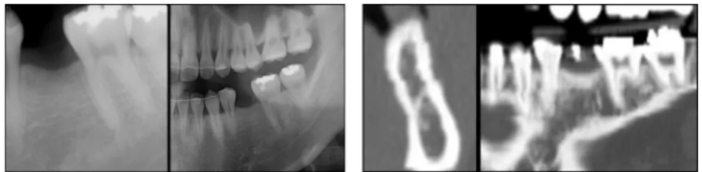
총 100명의 환자를 대상으로 하였다. 모든 환자는 연세대학교 치과대학병원 통합치의학과에 결손치 를 주소로 내원하여 임플란트를 식립한 환자로 2010년 1월 1부터 2015년 5월까지 내원한 환자에서 무 작위로 선택하였다. 모든 환자는 치근단 사진과 파노라마 방사선 사진, 그리고 전산화 단층촬영(conven- tional CT; MDCT)을 시행한 환자로 하였으며, 모든 환자의 방사선학적 평가는 두 명의 치과의사가 암 맹(blinded) 상태로 평가하였다(Fig. 1).
두 명의 치과의사가 Misch Bone density classification16에 따라 분석하였고, 두 관찰자 간의 오류가 있 을 때 다른 구강악안면방사선과 전문의가 추가로 평가하여 classification의 오류를 줄였다. 상악에서 상 악동(maxillary sinus)이나 관골(zygomatic arch)이 중첩된 경우에는 중첩되지 않은 부분만을 평가하였 다. 전산화 단층촬영 영상은 DICOM파일 형식으로 저장되어 임플란트 시뮬레이션 소프트웨어(Onde- mand®, CyberMed, Seoul, Korea)에서 reformatted cross-sectional 이미지와 reformatted panoramic section이미지를 분석하였다.
Reformatted 이미지들은 기술한 대로 2-3명의 평가자가 평가하였다. 3차원적인 분석을 위해 Onde- mand 소프트웨어상에서 가상으로 4 mm 직경, 10 mm 길이를 가진 임플란트를 무치악 부위에 위치시 켰다. 이후 임플란트 주위 1 mm의 골을 region of interest (ROI)로 설정하고 골질을 Hounsfield unit (HU)값을 측정하였다(Fig. 2). 측정된 HU값에 따라 D1에서 D4까지 골질을 분류하였다(D1, >1,250 HU; D2, 850-1,250 HU; D3, 350-850 HU; D4, 150-350 HU). ROI value는 reformatted cross section- al, panoramic 영상에서 평가하였으며 designating polygonal area를 포함하였다.
치근단 사진과 파노라마 사진에서도 골질을 평가하였다. 골질의 평가는 Misch의 골질 분류 시 cortical bone과 trabecular bone의 형태에 따라 분류하였다. 즉 cortical bone이 두껍거나 충분한 경우 D1, corti- cal bone의 형태가 거의 관찰되지 않고 trabecular bone도 충분하지 않고 loose한 pattern을 보이는 경우 D4로 분류하였다.
Fig. 1. Periapical, panoramic, reformatted cross sectional and reformatted panoramic view of missing tooth area.
Dongho Shin et al. : Assessment of bone quality in pre-dental implant site using periapical, panoramic and computed tomography. Implantology 2018
Fig. 2. 3D simulation of 4.0 mm X 10 mm virtual dental implant placement and Hounsfield unit of surrounding 1mm thickness bone.
Dongho Shin et al. : Assessment of bone quality in pre-dental implant site using periapical, panoramic and computed tomography. Implantology
2018
계측치와의 일치도를 3차원 평가와 치근단, 파노라마 사진, reformatted cross sectional ROI와 치근단, 파노라마 사진, reformatted panoramic ROI와 치근단, 파노라마 사진을 비교하였으며, 상악 전치, 상악 구치, 하악 전치, 하악 구치로 나누어 평가하였다.17
III. 결과
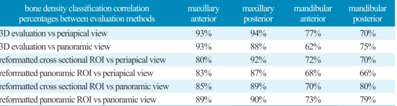
3차원 평가와 치근단, 파노라마 방사선 사진에서의 일치도를 평가하였을 때 상악 전치에서는 모두 93%, 상악 구치에서 94%, 88%로 높은 일치도를 보였다. 그러나 하악 전치는 77%, 62%, 하악 구치에서 는 70%, 75%로 낮은 일치도를 보였다(Table 1).
Reformatted cross sectional ROI와 치근단, 파노라마 방사선 사진에서의 일치도를 평가하였을 때 상 악 전치 80%, 85% 상악 구치에서 92%, 89%로 3차원 평가보다는 전치부에서 낮고 구치부에서 높은 일 치도를 보였다. 그러나 하악 전치와 하악 구치는 3차원 평가와 마찬가지로 72%, 70%, 70%, 80%로 비교 적 낮은 일치도를 보였다(Table 1).
Reformatted panoramic ROI와 치근단, 파노라마 방사선 사진에서의 일치도를 평가하였을 때 상악 전 치 85%, 89% 상악 구치에서 87%, 90%로 3차원 평가보다는 전치부에서 낮고 구치부에서 높은 일치도를 보였다. 하악 전치와 하악 구치 모두 68%, 73%, 66%, 79%로 계측되어 낮은 일치도를 보였다(Table 1).
IV. 토의
성공적인 임플란트 식립을 위해서는 다양한 방사선 검사를 통하여 환자의 골질을 정확하게 평가하는 것이 중요하지만, 환자를 위하여 방사선 피폭을 최소화하는 것이 의료인으로서의 기본 자세일 것이다
18-20. 치과용 CBCT는 적은 방사선 양으로 3차원적인 영상을 평가할 수 있지만, 표준화된 HU값을 적용
Table 1. Correlations between conventional CT, periapical view and panoramic view
Dongho Shin et al. : Assessment of bone quality in pre-dental implant site using periapical, panoramic and computed tomography. Implantology 2018
bone density classification correlation
percentages between evaluation methods maxillary
anterior maxillary
posterior mandibular
anterior mandibular posterior
3D evaluation vs periapical view 93% 94% 77% 70%
3D evaluation vs panoramic view 93% 88% 62% 75%
reformatted cross sectional ROI vs periapical view 80% 92% 72% 70%
reformatted panoramic ROI vs periapical view 83% 87% 68% 66%
reformatted cross sectional ROI vs panoramic view 85% 89% 70% 80%
reformatted panoramic ROI vs panoramic view 89% 90% 73% 79%
할 수 없기 때문에 골질평가의 절대적인 양을 평가하기에는 한계를 가질 수 있다5,7,13. 본 연구에서는 2차 원적인 치근단 사진과 파노라마 방사선 사진에서의 골질평가가 HU값을 가지는 Conventional CT의 골 질평가와의 비교를 통해 기본적인 방사선 사진들이 얼마나 골질을 예측할 수 있는지 검증하고자 하였 다. 2차원적인 방사선의 한계는 조사되는 해부학적 부위가 겹치기 때문에, 겹치는 부위의 3차원적인 해 부학적 형태가 2차원적으로 어떻게 나타나는지가 중요한 요소가 될 것이다.
본 연구에서도 가장 높은 일치도를 보인 것은 상악 전치와 상악 구치이며, 특히 3차원 평가와 상악 전 치, 상악 구치에서 88-94%의 가장 높은 일치도를 보였다. 이는 상악 전치와 상악 구치에서 겹친 해부학 적인 구조가 가장 적어서 2차원적인 영상에서 평가하는 골질이 3차원 평가에서 얻은 골질 평가와 유사 하다는 것으로, 상악 치아에서는 치근단 사진과 파노라마 방사선 사진이 골질 평가에 유용하게 사용될 수 있다는 것을 의미한다. 이에 비해서 하악 전치와 하악 구치는 낮은 일치도를 보였으며, 특히 하악 전 치에서 파노라마 방사선 사진이 더 낮은 일치도를 보였다. 이는 하악 치아에서 협측, 설측 피질골이 비 교적 두껍게 있기 때문에, 치근단 방사선 사진이나 파노라마 방사선 사진에서 3차원적인 골질을 예측하 는 데에 한계가 있다는 것을 보여준다. 특히 하악 전치에서는 파노라마 방사선 영상의 상층 오류로 인해 전치부가 잘 관찰되지 않는데, 이로 인한 오류가 발생할 수 있다고 추측된다.
이러한 내용은 reformatted cross sectional 이미지 분석과 reformatted panoramic 이미지 분석에도 비 슷하게 나타나는데, 3차원 분석에 비해서는 상악 전치와 구치가 조금 더 낮게 나타나게 된다. 이는 3차 원 분석은 virtual 3D volume분석을 통한 임플란트의 주변골 모두를 평가하게 되지만 reformatted 이미 지들인 경우 sectional 이미지로 영상을 만드는 단면이 어떻게 위치되는지에 따라 골질평가가 다를 수 있다는 것을 시사한다. Reformatted 영상 분석에서 하악 전치와 구치는 모두 60-70%로 낮은 일치도를 보이지만, 하악 구치의 reformatted cross sectional/reformatted panoramic 이미지와 파노라마 방서선 사 진은 80%, 79%로 다소 높다. 이는 하악 전치에서는 파노라마 방사선 사진 분석이 의미가 없지만, 하악 구치에서는 reformatted cross sectional 영상과 비슷하게 골질을 예측할 수 있다는 것이 되므로, 파노라 마 사진으로 하악 구치의 골질 평가는 이루어질 수 있다는 것으로 해석할 수도 있다. 그러나 3차원적인 분석에서는 70%, 75%의 일치도를 보였기 때문에 전적으로 이 분석을 신뢰하기는 어렵고 정확한 것은 CT분석을 통한 골질 평가가 있어야 한다고 판단된다.
본 연구는 모두 conventional CT자료를 이용하였다. CBCT를 이용한 연구가 아닌 conventional CT를 이용한 것은 HU을 이용한 정량적인 분석이 가능하다는 것이므로 사용하였으나, 이로 인해 비교적 오 래전의 데이터들로 분석이 이루어진 것이 이 연구의 한계라고 할 수 있다. 그러므로 비교적 해당된 숫자 가 적어 나이와 성별로 나눈 분석이 부족하며, conventional CT촬영 시 1mm thickness로 촬영한 촬영조 건 역시 isometric한 CBCT 분석에 비해 부족하다고 볼 수 있다. 차후 CBCT촬영 시 calibration이 가능 한 reference marker를 이용한 분석, 나이와 성별, 전신 질환 등과 관련된 분석이 추가적으로 이루어진다 면 CBCT와 2차원 사진의 장단점을 고려한 연구가 진행될 수 있을 것으로 기대된다.
V. 결론
CT에서 계측되는 HU를 기반으로 한 골질평가와 치근단, 파노라마 방사선 사진을 분석한 결과, 해부 학적인 특성에 따라 상악 전치와 상악 구치에서 높은 일치도를 보였으며, 하악 전치와 하악 구치에서는 낮은 일치도를 보였다. 이 결과를 근거로 상악 전치와 상악 구치의 임플란트 식립 시에는 CT평가 뿐만 아니라 치근단 사진이나 파노라마 방사선 사진을 이용한 골질 평가가 가능할 수 있으나, 하악치아에서 는 CT 혹은 CBCT를 이용한 골질평가가 더 유용하리라 판단된다.
Acknowledgement
본 연구는 연세대학교 치과대학 2015년도 교수 연구비에 의하여 이루어졌음 (과제번호 : 6-2015- 0141).
References
1.
Al-Ekrish AA. Radiology of Implant Dentistry. Radiol Clin North Am. 2018; 56(1): 141-156.
2.
Buser D, Sennerby L, De Bruyn H. Modern implant dentistry based on osseointegration: 50 years of progress, current trends and open questions. Periodontol 2000. 2017; 73(1): 7-21.
3.
Greenberg AM. Digital technologies for dental implant treatment planning and guided surgery. Oral Maxillofac Surg Clin North Am. 2015; 27(2): 319-340.
4.
Jeong KI, Kim SG, Oh JS, et al. Consideration of various bone quality evaluation methods. Implant Dent. 2013; 22(1): 55-59.
5.
Lindh C, Oliveira GH, Leles CR, et al. Bone quality assessment in routine dental implant treatment among Brazilian and Swedish specialists. Clin Oral Implants Res. 2014; 25(9): 1004-1009.
6.
Monsour PA, Dudhia R. Implant radiography and radiology. Aust Dent J. 2008; 53 Suppl 1: S11-25.
7.
Parsa A, Ibrahim N, Hassan B, et al. Bone quality evaluation at dental implant site using multislice CT, micro-CT, and cone beam CT. Clin Oral Implants Res. 2015; 26(1): e1-7.
8.
Ritter L, Elger MC, Rothamel D, et al. Accuracy of peri-implant bone evaluation using cone beam CT, digital intra-oral radiographs and histology. Dentomaxillofac Radiol. 2014; 43(6): 20130088.
9.
Shelley AM, Glenny AM, Goodwin M, et al. Conventional radiography and cross-sectional imag- ing when planning dental implants in the anterior edentulous mandible to support an overdenture: a systematic review. Dentomaxillofac Radiol. 2014; 43(2): 20130321.
10. Toia M, Stocchero M, Cecchinato F, et al. Clinical Considerations of Adapted Drilling Protocol by Bone Quality Perception. Int J Oral Maxillofac Implants. 2017; 32(6): 1288-1295.
11.
Baciut M, Hedesiu M, Bran S, et al. Pre- and postoperative assessment of sinus grafting procedures using cone-beam computed tomography compared with panoramic radiographs. Clin Oral Implants Res. 2013; 24(5): 512-516.
12.
Ibrahim N, Parsa A, Hassan B, et al. Diagnostic imaging of trabecular bone microstructure for oral implants: a literature review. Dentomaxillofac Radiol. 2013; 42(3): 20120075.
13.
Pauwels R, Jacobs R, Singer SR, et al. CBCT-based bone quality assessment: are Hounsfield units applicable? Dentomaxillofac Radiol. 2015; 44(1): 20140238.
14.
Valiyaparambil JV, Yamany I, Ortiz D, et al. Bone quality evaluation: comparison of cone beam computed tomography and subjective surgical assessment. Int J Oral Maxillofac Implants. 2012;
27(5): 1271-1277.
15.
Wakimoto M, Matsumura T, Ueno T, et al. Bone quality and quantity of the anterior maxillary tra- becular bone in dental implant sites. Clin Oral Implants Res. 2012; 23(11): 1314-1319.
16.
Norton MR, Gamble C. Bone classification: an objective scale of bone density using the computer- ized tomography scan. Clin Oral Implants Res. 2001; 12(1): 79-84.
17.
Cicchetti DV, Nelson LD. Re-examining threats to the reliability and validity of putative brain-be- havior relationships: new guidelines for assessing the effect of patients lost to follow-up. J Clin Exp Neuropsychol. 1994; 16(3): 339-343.
18.
Bornstein MM, Scarfe WC, Vaughn VM, et al. Cone beam computed tomography in implant den- tistry: a systematic review focusing on guidelines, indications, and radiation dose risks. Int J Oral Maxillofac Implants. 2014; 29 Suppl: 55-77.
19.
El Sahili N, Nasseh I, Berberi A, et al. Impact of Cone Beam Computed Tomography Dose in Pre-Surgical Implant Analysis. Open Dent J. 2018; 12: 94-103.
20.