ABSTRACT
Tough ectopic male breast cancer is extremely rare, non-axillary ectopic male breast cancer is even rare. To date, the natural course and prognosis of this disease are not fully understood.
Consequently, the appropriate treatment for this disease has not been established. We report on a patient with ectopic male breast cancer in the suprapubic area that relapsed with hematogenous metastasis 3 years after complete surgical resection and adjuvant treatment despite an early diagnosis. This unusual case highlights the need for new prognostic factors such as genomic profiling to predict whether ectopic male breast cancer is aggressive and to guide on the duration between follow-ups and the appropriate method for conducting them.
Keywords: Breast neoplasms, male; Neoplasm metastasis; Prognosis
INTRODUCTION
Ectopic male breast cancer is extremely rare with an unknown prognosis and etiology that may be confused with that of other soft tissue cancers [1]. The most common site of ectopic breast cancer in both men and women is the axilla, though ectopic breast cancer can occur anywhere along the milk line, which is an embryologic landmark from bilateral axillae to the inguinal region and medial thigh [1]. To date, non-axillary ectopic breast cancer in male patients has only been reported in 2 English articles, where the sites were the abdominal wall and perineum [2].
The prognosis of ectopic male breast cancer is unknown owing to its rarity. Therefore, there is no clear consensus on the prognosis and follow-up of male patients with ectopic breast cancer after complete surgical resection [3,4]. We report the case of a 65-year-old male patient with ectopic breast cancer in the suprapubic area that relapsed with hematogenous metastasis despite an early diagnosis. Our aim was to suggest the necessity of new prognostic factors such as genomic profiling for appropriate follow-up for metastasis or recurrence after complete surgical resection of ectopic male breast cancer.
Case Report
Received: Jun 2, 2020 Revised: Nov 16, 2020 Accepted: Mar 3, 2021 Correspondence to Eun Jung Choi
Department of Radiology, Research Institute of Clinical Medicine of Jeonbuk National University-Biomedical Research Institute of Jeonbuk National University Hospital, Jeonbuk National University Medical School, 20 Geonji- ro, Deokjin-gu, Jeonju 54907, Korea.
E-mail: [email protected]
© 2021 Korean Breast Cancer Society This is an Open Access article distributed under the terms of the Creative Commons Attribution Non-Commercial License (https://
creativecommons.org/licenses/by-nc/4.0/) which permits unrestricted non-commercial use, distribution, and reproduction in any medium, provided the original work is properly cited.
ORCID iDs Jung Hee Byon
https://orcid.org/0000-0002-5139-552X Ae Ri An
https://orcid.org/0000-0002-6047-1627 Jin Yong Shin
https://orcid.org/0000-0002-9304-8868 Eun Jung Choi
https://orcid.org/0000-0002-2339-4327 Conflict of Interest
The authors declare that they have no competing interests.
Jung Hee Byon
1, Ae Ri An
2, Jin Yong Shin
3, Eun Jung Choi
11
Department of Radiology, Research Institute of Clinical Medicine of Jeonbuk National University- Biomedical Research Institute of Jeonbuk National University Hospital, Jeonbuk National University Medical School, Jeonju, Korea
2
Department of Pathology, Research Institute of Clinical Medicine of Jeonbuk National University- Biomedical Research Institute of Jeonbuk National University Hospital, Jeonbuk National University Medical School, Jeonju, Korea
3
Department of Plastic and Reconstructive Surgery, Research Institute of Clinical Medicine of Jeonbuk National University-Biomedical Research Institute of Jeonbuk National University Hospital, Jeonbuk National University Medical School, Jeonju, Korea
Ectopic Male Breast Cancer in
Suprapubic Area That Relapsed with
Hematogenous Metastasis
Author Contributions
Conceptualization: Byon JH, Choi EJ;
Investigation: An AR; Resources: Shin JY;
Writing - original draft: Byon JH; Writing - review & editing: Byon JH, Choi EJ.
CASE REPORT
This study was approved by the Institutional Review Board (IRB) of Jeonbuk National University Hospital, Jeonju, Korea. The IRB waived the requirement for obtaining informed consent.
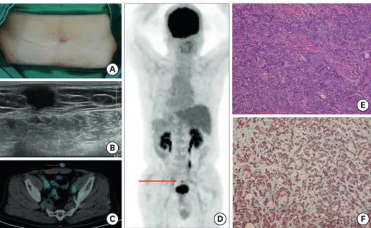
A 65-year-old man was referred to our hospital, presenting with a painless and slowly growing suprapubic mass in the last 2 years. He had no history of other cancers. Physical examination revealed a solitary, hard, non-tender, and immobile mass. The mass showed an accessory nipple and slight redness (Figure 1A). The patient's breasts and bilateral axillae were symmetrical without abnormality.
Ultrasound (US) and US-guided biopsy were performed at our hospital. US revealed an irregularly shaped hypoechoic mass, 1.4 × 1.0 cm in size, with a microlobulated margin at the subcutaneous layer of the suprapubic area (Figure 1B). The mass was abutting the skin. The biopsy result showed a moderately differentiated metastatic adenocarcinoma. Preoperative magnetic resonance imaging of the breasts revealed no abnormal findings. Preoperative positron emission tomography-computed tomography (PET-CT) was performed. However, there was only a fluorodeoxyglucose-avid mass in the suprapubic area with a standardized uptake value (SUV)
maxof 5.04 and SUV
avgof 1.43 (Figure 1C). There were no other signs of malignancy in other parts of the body (Figure 1D).
The patient underwent wide local excision of the tumor with advancement flaps at our hospital. On gross examination, a whitish and firm mass measuring 1.6 × 1.2 cm was found under the skin with an unclear margin. Histopathology results showed tumor cells arranged in clusters with lumen formation in the fibrotic stroma and mild pleomorphism, consistent
D
E
F A
B
C
Figure 1. Clinical, imaging, and pathologic findings of ectopic male breast cancer. (A) Accessory nipple with mild erythema of the mass. (B) Ultrasonography showing a hypoechoic mass with a microlobulated margin in the subcutaneous layer of the suprapubic area. (C) A mass with an abnormal radioactive concentration (arrow) is found on PET-CT. (D) There is no other lesion except for the suprapubic mass (arrow) on PET-CT. (E) Hematoxylin and eosin staining of a surgical specimen showing tumor cells arranged in clusters and associated with limited stroma. The cytoplasm is abundant and eosinophilic. Nuclei are relatively uniform with prominent nucleoli (×200). (F) Immunostaining is positive for the expression of GATA3 (×200).
PET-CT = positron emission tomography-computed tomography.
with findings of grade 2 invasive ductal carcinoma (Figure 1E). Immunohistochemically, the tumor cells were positive for the expression of the estrogen receptor (ER), the progesterone receptor (PR), and GATA (Figure 1F). Using the Allred scoring system, the score of ER and PR was 5+3 and the score of human epidermal growth factor-2 (HER-2) was 2+. A fluorescent in situ hybridization study was negative for the expression of HER-2. Ki-67 was 5%. The Bloom
& Richardson Grade was 1 with differentiation grade 2, nuclear grade 1, and mitotic index 1. Collectively, the histopathological and immunohistochemical findings supported the diagnosis of invasive ductal carcinoma from ectopic breast tissue. He had no family history of gynecological cancer. Tamoxifen (20 mg/day) was administered from the time of diagnosis as adjuvant endocrine treatment. A serum tumor marker (carcinoembryonic antigen [CEA]) test was performed every 3 months. Imaging tests such as abdominal computed tomography (CT), chest CT, and a bone scan were performed every 3–5 months.
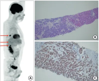
Three years later, multiple metastases to the liver and pulmonary lymph nodes were found on abdominal CT, chest CT, and PET-CT (Figure 2A). US-guided biopsy of the liver mass revealed a metastatic adenocarcinoma positive for the expression of ER (a 5+3 score of on the Allred scoring system), suggesting metastasis from ectopic breast cancer (Figure 2B and C).
Other immunohistochemistry test results were negative for the expression of PR and HER-2 as well as CK20 and TTF-1 and positive for the expression of CK7, PAS, and MTS. Markers for hepatocyte were negative presenting its origin was not liver. These clinical and pathological findings confirmed the diagnosis of metastatic breast cancer from ectopic breast tissue.
In addition to tamoxifen, 4 cycles of paclitaxel and carboplatin were initiated. Six cycles of anthracycline, 3 cycles of capecitabine, and 2 cycles of eribulin were initiated owing to the progression of liver metastases. A serum tumor marker (CEA) test was performed every month. For restaging, abdomen and chest CT examinations were performed every 3 months and a bone scan was performed every 5 months.
A
B
C
Figure 2. Imaging and pathologic findings of multiple metastases in the liver and pulmonary lymph node. (A)
There are 3 metastatic lesions (arrows) with high fluorodeoxyglucose uptake on positron emission tomography-
computed tomography. (B) Hematoxylin and eosin staining showing tumor cells arranged in clusters in the liver
parenchyma (×100). (C) Immunostaining is positive for the expression of the estrogen receptor (×100). These
findings are suggestive of metastatic carcinoma.
DISCUSSION
To the best of our knowledge, this is the first case report of accessory breast cancer in the suprapubic area in a male patient whose condition relapsed with distant metastasis despite early diagnosis. Ectopic breast tissue is found in up to 6% of the population, most commonly in the axilla along the milk line; however, aberrant breast tissue could also exist outside the milk line, such as in the face, posterior thigh, and vulva [1]. The incidence of male breast cancer is less than 1% and that of ectopic breast cancer is approximately 0.3%–0.6% of all breast cancers [2,5].
Knowledge about the ectopic breast tissue and pathologic processes that can occur in either sex can help with early diagnosis and appropriate treatment. In the English literature, only 10 cases of ectopic male breast cancer have been reported till date [2-11]. The most common site of ectopic male breast cancer is the axilla [3,4,6-11]. Until now, only 2 cases of non-axillary ectopic male breast cancer (in the lower abdominal wall and perineum) have been reported [2,5]. This is the third case of non-axillary ectopic male breast cancer in the English literature.
The prognosis of ectopic male breast cancer is unknown owing to its rarity. To date, only conventional prognostic factors such as anatomic-based tumor-node-metastasis (TNM) staging, molecular subtype, histologic grade, and Ki-67 are used to predict the prognosis of ectopic male breast cancer as with female breast cancer. Among reported ectopic male breast cancers [2-11], 30% (3/10) cases were already at the T4 stage or had bone metastasis when the diagnosis was established [3,10,11]. One patient had pathologic stage pT1N1M0 cancer and had relapsed with multiple distant metastases [6]. In all previous cases, a conventional prognostic factor could predict a poor prognosis. In this case, however, the TNM stage was T1 (1.6 cm) N0M0, and the molecular subtype was luminal A-like cancer (high ER/PR and low Ki-67, histologic grade 1), resulting in the diagnosis of pathologic prognostic stage IA.
Despite this case having favorable prognostic factors, hematogenous metastasis relapsed unexpectedly. Though ectopic male breast cancer has a favorable prognosis with respect to conventional prognostic factors, investigation of other prognostic factors is necessary.
Recently, the genomic profiles from multigene panel tests, such as Oncotype DX and MammaPrint, were incorporated into prognostic staging as new prognostic factors.
Oncotype DX has been shown to predict the likelihood of distant recurrence in tamoxifen- treated patients with node-negative, ER-positive breast cancer [12]. MammaPrint has shown to predict the likelihood of distant metastasis within 5 years in patients with lymph node- negative disease [13]. A previous study reported that the presence of clones with 2 or more amplifications of pluripotent genes in patients with luminal-like breast cancer T1N×M0 determines an unfavorable prognosis of distant metastasis [14]. In addition, there are many ongoing studies on profiling of genes, such as ESR1, FGFR1, TP53, and NYC, to predict poor prognosis in luminal cancers [15]. The prognosis of ectopic male breast cancer may be predicted more accurately by combining conventional prognostic factors and genetic factors, regardless of cancers being luminal-like or early-stage cancers. Next-generation sequencing studies are needed for ectopic male breast cancer in the future. When these genetic factors are identified, guidelines with respect to the follow-up method and durations between follow-ups could be established.
In conclusion, though non-axillary ectopic male breast cancer is extremely rare, the
possibility of metastasis and a poor prognosis should be considered even if conventional
prognostic factors are favorable. This highlights the need for further prognostic factors, such
as genomic profiling, for appropriate follow-up of ectopic male breast cancer.
REFERENCES
1. DeFilippis EM, Arleo EK. The ABCs of accessory breast tissue: basic information every radiologist should know. AJR Am J Roentgenol 2014;202:1157-62.
PUBMED | CROSSREF
2. Eom HJ, Ko BS, Song IH, Gong G, Kim HH. Ectopic male breast cancer in the perineum: a case report. J Breast Cancer 2017;20:404-7.
PUBMED | CROSSREF
3. Cheng Y, Li N, Eapen A, Parajuli R, Mehta R. Somatic BRCA2 mutation-positive concurrent accessory male breast cancer (BC) and non-small cell lung cancer (NSCLC): excellent efficacy of palbociclib, fulvestrant and leuprolide in platinum-exposed and endocrine-refractory BC associated with cyclin D1 and FGFR1 amplification and of carboplatin, paclitaxel and radiation in NSCLC. Case Rep Oncol 2019;12:494-9.
PUBMED | CROSSREF
4. Wang CX, Guo SL, Han LN. Successful treatment of accessory breast cancer with endocrine therapy. J Zhejiang Univ Sci B 2017;18:70-5.
PUBMED | CROSSREF
5. Zhong GB, Ye XQ, Liu JL, Xiao SZ, Huang QH, Wei W. Male accessory breast cancer on the abdominal wall: a case report and literature review. Onco Targets Ther 2018;11:6625-31.
PUBMED | CROSSREF
6. Bi L, Li J, Shi Z, Zhu Z, Lu Z. Male accessory breast cancer successfully treated with endocrine therapy: a case report. Oncol Lett 2015;10:2495-8.
PUBMED | CROSSREF
7. Bi M, Li D, Su Y, Sun P, Gao Y. Male axillary accessory breast cancer: a case report. Medicine (Baltimore) 2020;99:e19506.
PUBMED | CROSSREF
8. Lin Y, Wang Y. Case report of a male primary breast carcinoma of axillary accessory mammary gland. Clin Breast Cancer 2012;12:142-4.
PUBMED | CROSSREF
9. Takeyama H, Takahashi H, Tabei I, Fukuchi O, Nogi H, Kinoshita S, et al. Malignant neoplasm in the axilla of a male: suspected primary carcinoma of an accessory mammary gland. Breast Cancer 2010;17:151-4.
PUBMED | CROSSREF
10. Yamamura J, Masuda N, Kodama Y, Yasojima H, Mizutani M, Kuriyama K, et al. Male breast cancer originating in an accessory mammary gland in the axilla: a case report. Case Rep Med 2012;2012:286210.
PUBMED | CROSSREF
11. Yoshida Y, Sakakibara A, Watanabe T, Noto K, Sakita K, Sakai Y, et al. Extraordinarily large protruding accessory breast cancer in a man. J Am Acad Dermatol 2012;67:e230-1.
PUBMED | CROSSREF
12. Paik S, Shak S, Tang G, Kim C, Baker J, Cronin M, et al. A multigene assay to predict recurrence of tamoxifen-treated, node-negative breast cancer. N Engl J Med 2004;351:2817-26.
PUBMED | CROSSREF
13. van de Vijver MJ, He YD, van't Veer LJ, Dai H, Hart AA, Voskuil DW, et al. A gene-expression signature as a predictor of survival in breast cancer. N Engl J Med 2002;347:1999-2009.
PUBMED | CROSSREF
14. Tsyganov MM, Ibragimova MK, Pevzner AM, Doroshenko AV, Slonimskaya EM, Litviakov NV.
Amplification of stem genes: new potential metastatic makers in patients with an early form of breast cancer. J Korean Med Sci 2019;34:e312.
PUBMED | CROSSREF
15. Jeselsohn R, Buchwalter G, De Angelis C, Brown M, Schiff R. ESR1 mutations—a mechanism for acquired endocrine resistance in breast cancer. Nat Rev Clin Oncol 2015;12:573-83.
PUBMED | CROSSREF