사상체질의학회지
J. of Sasang Canst. M뼈.
Vol. 13. No 1. 2001
Studies on the Cytokine Production Regulation in Human
Astrocytes by Yuldahansotang
Choi Ji-sook* . Kim Kyoung-yo* . Kim Hyoung-min- . Ju ‘Jong-chon*
| 초 혹 l
A 間腦星狀細뼈에서 熱多寒少漫에 의한 細뼈活性物質 生빼 調節에 關한 iff 究
최지숙,* . 김경요* . 김형민 ** . 주종전
원광대학교한의과대학 사상체질과
.. 원광대학교 한의과대학
사상의학적 견지에서 太陰A의 중풍 , 치매와 같은 신경계질환에 다용되고 있는 熱多寒少陽은 최근에 그 엄
상적 효과를 뒷받청할 다각적인 연구들이 이루어지고 있음에도 불구하고 그 정확한 약리학적 기전에 대해서 는 밝혀지지 않고 있다.
본 연구에서는 인간성상세포를 이용하여 熱多寒少楊이 substance P (SP) 와 lipopolysaccharide αPS) 에 의해 유
도되는 다양한 세포활성물질의 분비량의 조절올 검토함으로써 熱多寒少陽의 약리기전을 연역학적 측면에서
보다 세밀하게 살펴보고자 하였다 .
熱多寒少楊 수침액은 인간 뇌 성상세포로 부터 LPS 와 SP 의 동시자극에 의해 생성되는 세포활성물질중
interleukin (IL)-I, IL-4, IL-6 및 tumor necrosis faccor-a (TNF- 미의 분비를 농도의존적으로 억제했다 . 그러나
interferon- r (lFN- r) 및 IL-2 의 분비 조절에는 영향을 미치지 않았다
그리고 항 IL-I P 항체에 의해 SP 유도성 TNF-a 분비의 증가가 억제되기 때문에 IL-I 은 TNF-a 증가를 매개 하는 역할을 하는 것으로 사료된다.
이상의 결과는 熱多寒少陽에 의한 급성기 중풍환자 치료 효과가 뇌 성상세포로부터 분비되는 세포활성물
질의 조절과 밀접한 관련성이 있다는 것올 암시하고 있다.
Keywords: Yulda-Hanso-Tang, Ascrocytes, Tumor necrosis faccor-a, Substance P, Lipopolysaccharide, Interleukin-I
I. INTRODUCTION constitutions inco four cy야s, according co the strengths
and weaknesses in funccions of the internal organs. They
are Taeyangin, Tae 띠run Soyangin 뼈d Soumin. Sasang
The Sasang Constitutional medicine class 퍼g
야ople
’s
. Dept. of Sasang ConstitUtional m뼈마Ie, College of Oriental M 때icine, Wonkwang Univ.
.. College of Oriental M벼icine, Wonkwang Univ
교신저자 최지숙 주소) 광주광역시 북구 일곡용49 천지인한의원 천화)062-575-4333 E-mail)ANAlOOO@ch 빼n.net
- 사상책짙의학회지 찌13권 쩌 1 호때1 -
Constitutional philosophy forms the basis of treatment
by correcting the imbalance of the internal organs
caused by the constitutional properties in each body
type. For example, Taeumin is thought to have a higher
rate of stroke, hypertension 뻐d hyperlipidemia than the
other types because he or she has a large liver and
small lungs. Thus, a pe πon of this body type has a
weak dispersing qi and excessive gathering qi I.2)
From the viewp 이 nt of the Sasang Constitutional
Medicine, Y띠dahansotang αH-Tang) is a prescription
which onen has been 따ed clinically for neurological
diseases, such as Taeumin's stroke and dementia
Recently, many researches support the clinical effect of
3-’). I _I'
YH-TangJ'''. Among them, Choj"' has reported that the
serum level of cytokine was regulated by YH-Tang in
an acute cerebral infarction (0) of Taeumin.
Astrocytes, one type of the neurological cells, have an
important role in maintaining central ne πous system
(CNS) homeostasis. To carty out homeostatic function,
astrocytes synthesize various immune-mediated cytokines
and interact with those substancel-IO). It is known that
cycokines are involved in various neuropathological
diseases, such as Alzheimer ’s disease, multiple sclerosis,
and acquired immunodeficiency syndrome (AIDS) 11-13)
Also the change of the s야cific cytokine level was
reported in an acute 0 patientI4). Astrocytes were
induced to secrete cytokines after interacting with
lipopolysaccharide αPS) or virus.
Substance P (SP) is a neurotransmitter and nerve-
originating chemical that mediates neurogenic infl 입n-
’. 16)
mation u. ,u'. In addition, SP stimulaces che production
of inflammatoty cycokines, such as cumor necrosis
factor-Q (TNF-Q), interleukin (IL)-117. 18) and IL-617), and
affects che number of SP receptors when che CNS 잉
injured IW"'. SP 얀 widely distribuced in che CNS and is
believed co scimulate cytokines to influence che
pachological process in the CNS I
In chis study, the reg 띠 acing effect of YH-Tang on
cycokine secretion induced by LPS and SP in astr εyces
was invescigated. An experiment on che regulating effect
of YH Tang on cycokine secretion in astrocytes was
conduCted co determine how the reg 띠 acing mechanism
providechebasisforcheclinicalcherapeuciceffeCton
YH-Tanginacuce0 patients
II. 써ATER 뻐A 해D METHODS
1. Materials 1) Reagents
Fetal bovine serum (FBS), SP, LPS,
penicillin/screpcomycin, 때d cween-20 were obcained
from Sigma Chemical Co. (Chicago, IL). Cycokines, such
as IFN-r, 1L-1, 1L-2, ι,-4, and 1L-6 were obcained
from R & D systems (Minneapolis, MN). Human
recombinanc TNF- a (rTNF- a), polyclonal anti-mouse
止-I a and anci-human TNF- a anci 야dy were obcained
from Genzyme (Cambridge, MA). Dulbecco ’s m 여ified
Eagle ’s me φum (DMEM) was obtained from Life
Technologies (Grand Island, NY). Enzyme-linked
immunosorbent assay (EUSA) places were obcained from
Nunc (Baperville, IL).
2) Cell line
Human 싫trocytes, CCF-STIG 1 cells were used
3) Preparation of YH- Tang
The plant sample was obtained from che Kwangju
Oriencal Medicine Hospital of Wonkwang University.
A prescripcion of YH-Tang weighs 48g, consiscing of
16 g of Puerariae Radix (Ge Gen), 8g of Scucellariae
R찌Dc (Huang Qin), 8g of Ligustici Tenuissimae Radix
(Gao Ben), 4g of Raphani Semen (Lai Fu Zi), 4g of
Angelicae Dahuricae Radix (Bai Zhi), 4g of Cimicifugae
Rhizoma (Sheng Ma), and 4g of Platycodi Radix Oil'
Geng). A water excract of YH-Tang was prepared by
de α>cting che prescripcion of dried herbs in discilled hot
wacer. The excract was mtered through a 0.45 띠nfih«
and freeze-dried.
The YH -Tang was preserved ac 4"C and used in this
study. The yield of che extracc was abouc 10% (w/w).
- 최지숙 외 3: A뻐g
훌狀뼈뼈에서 多흉少홉에 의한JlllII 홉↑
*빼it 호 해II애 뻐한 6]11,: -
2. Methods
1) Astrocytes were cultured at 4 X 10⁵ per well
and were grown for 3 days with DMEM containing
10% FBS in C02 inαbator.
2) SP preparation:
Special care was taken with SP to avoid possible iPS
contamination. Fiπt, peptide SP was dissolved in 0.01 %
acetic acid. Acetic acid was made of 1/1ááoo glacial
acetic acid and f1ltered through 2.2 띠n f1lter. SP stock
solution was kept in a refrigerator at -20'C after being
diluted in iPS non-contaminated dist 피ed water
immediately before use.
3) Measurement of cytokines :
Assays of cycokines were performed under modified
EUSA according to the procedure outlined by Scuderi et
al20) ηlat is, anti-cytokine monoclonal antibody was ‘
treated with a coating buffer solution [0.02% s여ium
azide contained phosphate buffered saline (PBS) pH = 7.
2) on flat-bottomed 9ιwell plate (Coming, Rochester,
NY) at a final concentration of 6.25 ng in each well,
and coated at 4'C for 12 h. After coating, to reduce
non-spec 퍼c binding, blocking buffered solution, PBS
containing 2% BSA, was added and coated at 37
'C for
2 h. Each well was added to 100 μ of recombinant
cytokine standard solution and su 야matant of each
specimen, and after washing 4 띠les with wash buffer
(PBS containing 0.05% Tween 20), were subsequently
incubated at 37
'C for 2 h.
The wells were then washed 4 times again with PBS
containing 0.05% rween 20. The wells were treated
with anti-cytokine antibody which had been diluted by
PBS containing 0.05% rween 20, and again incubated
at 37
'C for 2 h. After washing repeatedly 7 times with
a wash buffer, each well was treated with 100 ng/mL of
phosphatase-bound anti IgG antibody (Sigma Co.) and
incubated at 37.C for 2 h, then washed 7 t띠les agam.
After the last washing step, 100 때 of p-nirro phenyl
phosphate was added to each well, which was 버ssolved
in buffer solution of the mixture of 0.05M N 상K03
and 0.05mM MgCI2. Ten min after the color changed,
the absorbance of each cycokine was measured at 405
run wave-length in the EUSA reader. Appropriate
specificity controls were included.
3. Statistical analysis
D염a are given as means :!:: S.E. Statistical 뻐혀ys 잉
was performed with Student ’s t-test. Results with
P<0.05 were considered statistically significant.
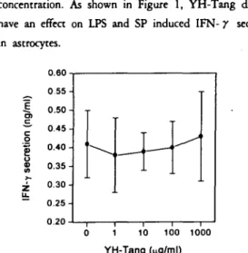
삐 . RESULTS
1. The effect of YH-Tang on LPS and SP
induced in’erferon- r (lFN- r) sec-
retion in as ’rocytes
To evaluate the effect of YH-Tang on IFN- r
secretion in 잃trαγtes, the amount of IFN- r secretion
was measured after 18 h of incubation of astrocyres
which was added by iPS and SP at ’ various
concentration. As shown in Figure I, YH-Tang did not
have an effea on iPS and SP induced IFN- r secretion
m 싫trocytes.
0.60 0.55
%%M%써
mω
%“때
nununu
”
u
”
ununu -EaI)=。
;eu@”←
zι
-
사캠 h 버니
l
o 10 100 1000
YH-Tang (
μg/ml)
Fig. 1. Effect 이 YH- Tang on LPS and SP induced IFN-r
secretion in astr< α;ytes. The cells (4 x 10⁵ cel 때ml)
were incubated for 24 h in medium ∞ntaining LPS
(1 μwml) plus SP (2 μ잉ml) with various con-
centrations of YH- Tang and the supernatants were
∞lIected and frozen at -8(re until assayed for IFN-
r. Each datum value indicates the mean :t S.E.
of six separated experiments.
*: statistically significant differences from the control values at P <
0.05
- 사상채짙의학회지 제13권 제 1 효 2X)\ -
2. The effect of YH- Tang on LPS and SP
induced IL -1 secretion in astrocytes
The analysis of the effeer of YH-Tang on U엄 and
SP induced IL-l secretion in astrocytes showed that
YH-Tang decreased IL-l concentration dependently, as
shown in Figur ξ 2. The εffeer of YH-Tang was
significant at a concentration of 100-1000 μpjrnLe
0.55
0.50
“”찌
%
nununu
(-FISt)
ζ。
;@」
Q@m
‘-
닐 0.30
0.25
o 1 10 100 1000
YH-Tang (
따잉ml)
Fig. 2. Effect 이 YH- Tang on LPS and SP induced IL-1
않cretion in astrl α:ytes. The ∞lis (4 x 10⁵ ∞II 의ml)
were incubated for 24 h in medium ∞ntaining LPS
(1 뼈ml) plus SP (2 빼ml) with various ∞ncen-
trations of YH- Tang and the supematants were
∞lIected and frozen at -BOt until assayed for
IL -1. Each datum value indicates the mean :.t S.E.
of' six separated experiments
*; sratisrically significanc differences from rhe concrol values at P <
0.05.
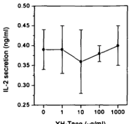
3. The effect of YH-Tang on LPS and SP
induced IL-2 secretion in astrocytes
A5 shown in Figure 3. 1L-2 secretion was not
increased significantly and the regulatory effeer of
YH- Tang on 1L-2 secretion was not significant.
0.50
“”애
%%
nunununu
=EBI)c。
;eu잉”
N」-
t-Y
0.25
o 10 100 1000
YH-Tang (vg/ml)
Fig. 3. Effect 이 YH- Tang on LPS and SP induced IL-2
secretion In astr ∞ytes. The ∞lis (4 x 10⁵ ∞II 잉ml)
were incubated for 24 h in medium ∞ntaining LPS
(1 μ잉ml) plus SP (2 μ밍'ml) with various ∞n ∞n-
!rations 이 YH- Tang and the supematants were
∞lIected and froze.n at -BOt until assayed for
IL-2. Each datum value indicates the mean :.t S.E.
이 six separated experiments.
*; sta 뼈에Iy 맹1ificanr diffcrcnccs from the conad 때‘JfS at P < 0.05.
4. The effect of YH-Tang on LPS and SP
induced IL-4 secra ’ion in astrocytes
The effect of YH-Tang on LPS and SP i뼈따ed 1L-4
secretion 띠 astrocytes was evaluated. As shown in Figure 4,
YH-Tang decreased 1L-4 concentration dependently.
0.46
””때애뼈”””“
”
u!unu
”
u
”
u
”
u
=E‘
@C)=。
;p8$*」
-
0.30
o 10 100 1000
YH-Tang (
μg/ml)
Fig. 4. Effect 이 YH- Tang on LPS and SP indu α퍼 IL-4
secretion in astr, ∞ytes. The ∞|잉 (4 x 10⁵ ∞II 잉ml)
were incubated for 24 h in medium ∞ntaining LPS
(1 μml) plus SP (2 μ밍ml) with various ∞ncen-
!rations of YH- Tang and the supematants were
∞lIected and frozen at -BOt until assayed for
IL-4. Ea 어1 datum value indicates the mean :.t S.E.
이 SIX se 며rated experiments.
*; stat ‘srically sisnificanc differences from rhe concrol v꾀ucs at
P < 0.05.
- 최지숙 외 3: A뻐뼈훌狀뼈뼈애서 !t 少훌에 의한IIII 효양. 호 여WI 에 뻐한 iiI'9.: -
0.55 0.60
0.50
0.45
0.40
0‘35
-E~aZ)t。
;밑
Q@” gι
z←
5. The effect of YH-Tang on LPS and SP
induced IL-6 secretion in astrocytes
The effect of YH-Tang on LPS and SP induced IL-6
secretion in ascrocytes was evaluated. As shown in
Figure 5, YH-Tang decreased 1L-6
dependently.
concentration
0.55
0.50 0.30
1000 100
o 10 0.45
YH-Tang (ng/ml)
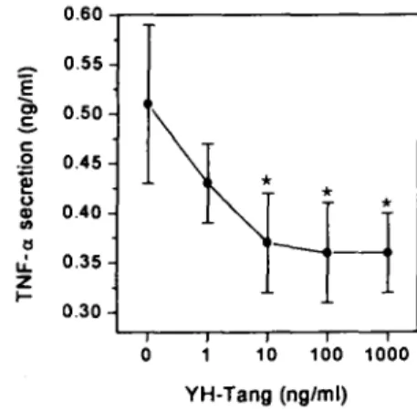
F밍. 6. Effect 이 YH- Tang on LPS and SP induced TNF- a
똥crehon in astr α:;ytes. The ∞lis (4 x 10⁵ cell 의mO
were inωbated for 24 h in medium ∞ntaining LPS (1 μ밍ml) plus SP (2 μwml) 에th various ∞ncen-
trations of YH- Tang and the supematants were
∞lIected and frozen at -BOt until assayed for
TNF- a. Each datum value indicates the mean ::t
S.E. of six separated experiments.
1000 100 1 10
o
0.40
0.35
0.30
0.25
(-ESZ)t。
;@』
Q%
얘」
-
*: statistically significant differences from the control values at P <
0.05
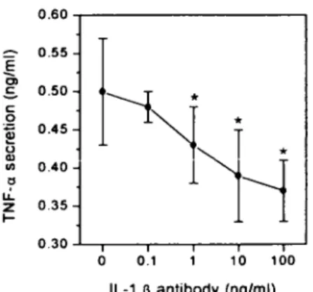
7. The effect of anti IL-1 β antibody on
LPS and SP induced TNF- a secretion
in astrocytes
To ev외uate whether YH- Tang ’s inhibirory effect on
TNF- a secretion was mediated via IL-I, the effect of
anti IL-I /3 antilx 삐y was as양ssed. After LPS( IμglmL)
and SPwere introduced ro an 양trocyte incubating
solution, anti IL-l /3 antibody was added and the
amount of TNF- a secretion was me 싫따ed after 24 h.
As shown in Figure 7, TNF- a concentration decreased
dependently in the anti IL-I /3 anti φdy treated group.
The inhibitory effect of the anti IL-I /3 antibody was
significant at a concentration of 1-100 뼈/mL
Fig. 5. Effect 이 YH- Tang on LPS and SP indu ∞d IL-경
secretion in astr ∞ytes. The ∞lis (4 x 10⁵ ∞II 잉ml)
were incubated for 24 h in medium ∞ntaining LPS
(1 μ밍ml) plus SP (2 μ잉ml) with various ∞ncen-
trations of YH- Tang and the supematants were
∞lIected and frozen at -BOt until assayed for IL용. Each datum value indicates the mean ::t S.E.
of six separated experiments.
YH-Tang (μg/ml)
*: statistically significant differences from the control values at P <
0.05.
6. The effect of YH-Tang on LPS and SP
induced TNF- a secretion in astro-
cytes
The effect of YH-Tang on LPS and SP induced
TNF- a secretion in astrocytes was evaluated. TNF- a
is an important inflammatory cyrokine. TNF- a sec-
retion by ascrocytes is induced by LPS and SP. As
shown in Figure 6, YH-Tang decreased TNF- a concen-
tration dependently
- 사상쩨짙의학회지 째13권 채’효 ::m1 -
0.60
%%
“””
%%
n“
nunununu (-E~@C)
ζ。
;@』
U@maι
ZLF
0.30
o 0.1 10 100
IL-1 II antibody (ng/ml)
Fig. 7. EHect of anti IL-1 β anti α잉y on LPS and SP
induced TNF-a 똥cretion in astr αγtes. The ∞lis
(4 x 10⁵ cell 의ml) were incubated for 24 h in
medium ∞ntainlng LPS (1 μ잉ml) plus SP (2 μg/ml)
with various ∞ncentrations 이anti IL-1 β antitx: 찌.
The supematants were ∞lIected and frozen at -8
O.C until assayed for TNF-a. Each datum value
indicates the mean :t S.E. 이 four separated
experiments.
*: statistically significant differences from the concrol values at P <
0.05.
N. DISCUSSION
In this study, the regulatory effect of YH-Tang on
LPS and SP induced cytokine secretion in astrocytes was
investigated. It has been reported that asrrocytes in
CNS pathophysiologically are likely to involve in
cytokines, both as stimulators and mediatots of asrrocyre
10)
function ,u', with which CNS homeostasis is maintianed.
LPS controls many biological properties of gtam-
negative bacteria. A serological spec 퍼ciry exists in the
part of varied polysaccaride and this part belongs to
O-antigen of gram-negative bacteria. O-antigen displays
a specific receptor of bacteriophage. LPS controls
endotoxic acciviry. If ba αeria loses its ability to
synthesize LPS, the pathogenicity of bacteria is often
lose. The properties of endotoxin include pyrogenicity,
leth 외ity, complement activity, B-cell mitogen activity in
the mouse, adjuvant acitivity, anti-rumor activity, etc2l).
SP has been reported to induce inflammatory
cytokine production in human neuroglial cells and
peripher 쇠 lymphoid cells as well 22)
It was noted thar YH-Tang inhibited the secretion of
IL-I, ι4, IL-6, and TNF- a significantly among the
various cyrokines produced by simultaneous stimulation
of LPS and SP in asrrocyres. The effect of YH-Tang
w’as concentration-dependem. It was derermined that
TNF- a and IL-I secretion in asrrocyres needed LPS
stimulation, and LPS stimulation was augmented by SP.
Because anti-IL-I p antibody inhibited the secretion of
SP-induced TNF- a, IL-l was considered as a mediator
of TNF- a incre 앓. These results are the evidence thar
SP, a neurotransmitter produced in the ne πe of the
CNS, is an important molecule involved in i바la-
mmarion. IL-l is an endogenous pyrogen, the activating
factor of lymph αyre products. It is made in many cells,
especially macrophage cells, where it is made most. IL-l
activates T-cell 뻐d B-cell, 뼈d causes the inflammatory
response. In the brain, IL-l causes heat and increases
the secretion of corticosteroid 23)
According 【o T ouzani et aI’s recenrrepor(24), IL-I has
an important role in the development of brain damage
following cerebral ischemia. The expression of IL-l in
the brain is dramatically increased during the early and
chronic stages of infarction. The most direct evidence
that IL-I conrributes significantly to ischemic injury is
that (I) cenrral administration of IL-I p exacerbates
brain damage, and (2) injection or over- expr δsion of
IL-l receptor antagonist, and blockade of IL-I P
converting enzyme activity dramatically reduce infarction
and improve behavioral deficit. The mechanisms
underlying IL-I actions in srroke have not been clearly
리 ucidated, and it seems likely that its effects are
mediated through stimulation and inhibition of a wide
range of pathophysiological processes.
2))
According to Yamasaki et aI’s report"', the conrribution
of cytokines in an inflarrunatory cascade on cerebral
reperfusion injury are characterized as consisting of
typical phases; leukocytes invasion, microglial activation
and remodeling. Within 1-2 days, IL-I and TNF- a
induce the expression of adhesion molecules that cause
leukocytes to adhere to endothelial cells.