Magnetoencephalography in Pediatric Lesional Epilepsy Surgery
This study was performed to assess the usefulness of magnetoencephalography (MEG) as a presurgical evaluation modality in Korean pediatric patients with lesional localization- related epilepsy. The medical records and MEG findings of 13 pediatric patients (6 boys and 7 girls) with localization-related epilepsy, who underwent epilepsy surgery at Seoul National University Children’s Hospital, were retrospectively reviewed. The hemispheric concordance rate was 100% (13/13 patients). The lobar or regional concordance rate was 77% (10/13 patients). In most cases, the MEG spike sources were clustered in the proximity of the lesion, either at one side of the margin (nine patients) or around the lesion (one patient); clustered spike sources were distant from the lesion in one patient. Among the patients with clustered spike sources near the lesion, further extensions (three patients) and distal scatters (three patients) were also observed. MEG spike sources were well lateralized and localized even in two patients without focal epileptiform discharges in the interictal scalp electroencephalography. Ten patients (77%) achieved Engel class I post- surgical seizure outcome. It is suggested that MEG is a safe and useful presurgical evaluation modality in pediatric patients with lesion localization-related epilepsy.
Key Words: Magnetoencephalography; Epilepsy Surgery; Localization; Spike Source;
Magnetic Source Imaging Hunmin Kim1, Byung Chan Lim2,3,
Woorim Jeong4, June Sic Kim4,5, Jong-Hee Chae2,3, Ki Joong Kim2,3, Chun Kee Chung4,5, Yong Seung Hwang2,3, and Hee Hwang1
1Department of Pediatrics, Seoul National University Bundang Hospital, Seongnam; 2Department of Pediatrics, 3Pediatric Clinical Neuroscience Center, Seoul National University Children’s Hospital, Seoul National University College of Medicine, Seoul;
4MEG Center, Seoul National University Hospital, Seoul; 5Department of Neurosurgery, Seoul National University Hospital, Seoul National University College of Medicine, Seoul, Korea
Received: 17 January 2012 Accepted: 13 March 2012 Address for Correspondence:
Hee Hwang, MD
Division of Pediatric Neurology, Department of Pediatrics, Seoul National University Bundang Hospital, 166 Gumi-ro, Bundang-gu, Seongnam 463-707, Korea
Tel: +82.31-787-7284, Fax: +82.31-787-4054 E-mail: [email protected]
http://dx.doi.org/10.3346/jkms.2012.27.6.668 • J Korean Med Sci 2012; 27: 668-673
INTRODUCTION
Magnetoencephalography (MEG) records the extracranial mag- netic field generated by the electrical activity of the brain (1).
Because MEG records continuous magnetic activity through multiple sensors that cover the entire brain, it has excellent tem- poral and spatial resolution compared with other modalities used to evaluate epilepsy. Important advantages of MEG over electroencephalography (EEG) are that the magnetic field de- tected from the scalp is not distorted or attenuated (2) and that accurate localization of the epileptic spike source is possible.
Overlaying this localized spike source in the patient’s structural imaging (magnetic source imaging, MSI) provides valuable in- formation about the localization of the epileptogenic foci (3).
The usefulness of MEG as a presurgical evaluation tool has been demonstrated in previous studies. The accuracy of MEG spike source localization was satisfactory in a study of 455 patients (4).
MEG spike sources localized well in temporal and extratempo- ral lesional epilepsy, and sometimes provided additive localiza- tion information to brain MRI or interictal scalp EEG (5). MEG also added diagnostic yield to scalp video-EEG (6, 7).
Although the usefulness of MEG has been well demonstrated and its application has yielded numerous promising results, the
superiority of MEG over EEG is not widely accepted. Moreover, there is no solid evidence that MEG can be substituted for inva- sive intracranial EEG in epilepsy surgery (8). Although the num- ber of reports on the use of MEG in pediatric epilepsy surgery in Asia is increasing, there is no report on the use of MEG in Korean pediatric patients with epilepsy. We examined the usefulness of MEG in evaluating Korean pediatric patients with lesional local- ization-related epilepsy. We also sought to analyze the pattern of MEG spike source distribution in relation to the lesions found by brain magnetic resonance imaging (MRI) and the surgical outcomes.
MATERIALS AND METHODS
Of 41 pediatric patients who underwent MEG spontaneous mag- netic activity analysis at the MEG Center of Seoul National Uni- versity Hospital between May 2005 and December 2010, 13 pa- tients (6 boys and 7 girls) received epilepsy surgery for the diag- nosis of lesional localization-related epilepsy and were included in the analysis. The electronic medical records were reviewed retrospectively. Data about sex, age, onset age, age at MEG ex- amination, seizure semiology, pre- and post-operative seizure frequency, brain MRI, brain fluorodeoxyglucose positron emis-
sion tomography (FDG PET) and the pathology report were re- viewed.
A 128-channel Grass Telefactor® digital EEG was used for the interictal scalp and long-term video-EEG monitoring. Scalp electrodes were placed according to the international 10-20 sys- tem. If necessary, electromyography (EMG) electrodes were also placed during the long-term video-EEG monitoring. Inter- ictal EEGs, video-EEGs, and intracranial EEGs were reviewed independently by three pediatric epileptologists. The seizure semiology was described based either on the parents’ or the caregiver’s reports or on the video-EEG monitoring findings.
The seizure semiology description and classification were based on the International League Against Epilepsy classification of epileptic seizures (9, 10). The sampling rate was 200 Hz for all recordings.
Spontaneous brain magnetic activity was recorded using a helmet-shaped, 306-channel, whole-head neuromagnetometer (VectorViewTM; Elekta Neuromag Oy, Helsinki, Finland). The MEG sensors comprised 2 planar gradiometers and 1 magne- tometer. The recording time was 50-90 min and most of the re- cordings comprised of spontaneous or sedated sleep records.
The sampling rate for data acquisition was 600 Hz. A band-pass filter (0.1-200 Hz) and a notch filter (60 Hz) were applied. Chlo- ral hydrate (50 mg/kg; maximum, 1,500 mg) was used when con- scious sedation was required for infants and preschool-aged patients.
The MEG recordings were analyzed and the spike sources were localized using the equivalent current dipole method and current density imaging. The data was processed using US FDA- approved Neuromag software (Elekta Neuromag Oy, Helsinki, Finland). MEG spike sources with goodness of fit values > 85%
were considered significant. When more than half of the sources were localized to the hemisphere with MRI lesions, the results were considered concordant. Similarly, when more than half of the sources were localized to a certain lobe or the anatomical region of MRI abnormality, the results were considered concor-
dant. MEG spike sources were classified as clustered around the lesion with or without extension, clustered distant from the le- sion, or scattered.
The postsurgical seizure frequency noted in the outpatient visit was reviewed and was classified according to Engel’s clas- sification (11).
Ethics statement
This study protocol was reviewed and approved by the institu- tional review board (IRB) of the Seoul National University Hos- pital (IRB No.H-1111-052-386). Waiver of informed consent was approved by the IRB after reviewing the study design, which was a retrospective review of medical record and MEG data.
RESULTS
The mean age at the onset of epilepsy was 6.4 yr (range, 10 months-12.1 yr) and mean age at the examination was 8.4 yr (range, 2.8-13.0 yr). The preoperative seizure frequency ranged from multiple daily seizures to monthly seizures. The most com- mon seizure semiology was dialeptic or hypomotor seizure, fol- lowed by versive, focal tonic, focal clonic, hypermotor, and gen- eralized tonic-clonic seizures. The most common location of MRI lesion was the temporal lobe (n = 8; six medial and two lateral), followed by frontal lobe (n = 3) and occipital lobe (n = 2) (Table 1).
The hemispheric concordance rate was 100% (13/13 patients) and the lobar or regional concordance rate was 77% (10/13 pa- tients). The number of MEG spikes analyzed was 2-52 (mean, 18.8). More than eight spikes were analyzed in 10 patients, but only a few spikes were recorded in two patients (patients 3 and 13). With regard to distribution and location, the MEG spike sources were clustered around or in proximity to the MRI lesion in 10 patients (77%). Among these patients, the MEG spike sources were extended from the cluster in three patients and distant scatters coexisted in another three patients. The MEG
Table 1. Clinical characteristics of 13 patients who underwent MEG as a presurgical evaluation
Patient No. Age at onset (yr) Age at exam. (yr) Sex Seizure semiology Preop. seizure frequency Lesion location in brain MRI
1 12.1 12.5 F Dialeptic 2/d Left inferior F
2 8.8 12.3 F Dialeptic 1-2/d Left medial T
3 2.9 3.9 F Hypomotor, versive 2/m Right O
4 3.8 4.1 M Face tonic-clonic 1/m Right inferior F
5 10.8 13.0 F Dialeptic, versive, GTCS 8/m Right lateral T
6 10.9 12.1 F Dialeptic, OAA, face clonic 1/d Right medial T
7 7.6 7.9 M Dialeptic 2-3/w Right F
8 2.6 2.8 F Dialpetic 1/m Right medial T
9 10.2 10.8 M Dialeptic, OAA 1/m Left lateral T
10 0.9 5.9 M Hypomotor, face tonic 1-2/d Left medial T
11 4.1 11.5 F Dialeptic, tonic 3-4/w Left medial T
12 5.5 8.3 M Dialeptic, versive 1/m Left medial T
13 2.5 4.4 M Right arm tonic, hypermotor 3/d Left O
GTCS, generalized tonic clonic seizure; OAA, oroalimentary automatism; F, female; M, male; d, day; w, week; m, month; F, frontal; T, temporal; O, occipital.
spike sources were scattered in two patients and were clustered distant from the lesion in one patient (Table 2).
MEG spike source clusters were observed in 11 patients, and these clusters were located in the proximity of the lesion in 10 patients. In most patients, the MEG spike sources were located on the margin of the lesion: superior (patients 4, 5, and 6), later- al (patients 1, 7, and 8), and superior and lateral (patients 10, 11, and 12). In patient 2, the spike sources were located around the lesion (Fig. 1). Among these patients, distant scatters were also identified in the contralateral hemisphere in two patients and ipsilateral hemisphere in one patient (Fig. 2). The MEG spike source cluster was distant from the lesion in patient 3. In the
two patients without clusters, the MEG spike sources were scat- tered in both hemispheres.
EEG abnormalities of epileptiform discharges or background abnormalities were present in 11 patients. Focal epileptiform discharges were present in 10 patients (77%), and the MEG spike source locations were concordant with these EEG discharges.
In two patients with a normal EEGs (patients 4 and 8) and in one patient with no focal epileptiform activity (patient 1), MEG detected well-lateralized and localized MEG spike sources. MEG spike sources were concordant in eight of the 10 patients whose PET findings were abnormal. MEG findings were concordant with the ictal onset zone identified by long-term video-EEG Table 2. Summary of lesion locations, interictal EEG findings, FDG PET, MEG spike source distribution, pathology, and postsurgical Engel classification of 13 patients
Patient
No. Lesion location EEG abnormality
PET Magnetoencephalography spike sources
Pathology Engel classification
Spikes Slowings L:R Distribution Location
1 Left inferior F N F7T3 N 13:0 C + E Left P (9), T (4) DNT I A
2 Left medial T T5 T5 N 20:0 C Left T medial (16), lateral (4) DNT I A
3 Right O O2, O1 N Right O 0:2 C* Right insula (1), P (1) FCD IIB I A
4 Right inferior F N N Right F 0:8 C Right F (6), P (2) GGL I A
5 Right lateral T F7T3
F8T4
N Right T 4:14 C + S Right T (8), P (3), F (3)
Left insula (2), T (1), F (1)
DNT I A
6 Right medial T F8T4 F8T4 Right T 0:52 C + E Right T (26), F (17), insula (9) FCD IIA I A
7 Right F Fp2F4 Fp2F4 Right F 0:25 C Right F (23), T (1), P (1) FCD IIB I A
8 Right medial T N N Right T 0:13 C + E Right T (10), P (2), F (1) FCD IB I A
9 Left lateral T F7 N N 9:4 S Left P (4), T (5)
Right P (4) DNT I A
10 Left medial T T3 N Left PT 14:4 C + S Left T (13), P (1)
Right R (3), T (1)
FCD IA I B
11 Left medial T T5O1 N Left T 10:0 C Left posterior T (9), F (1) GGL II A
12 Left medial T F7 N Left T 21:3 C + S Left T (18), cingulate (2), F (1)
Right F (3)
FCD IB III A
13 Left O T5 N Left O 2:1 S Left P (2)
Right P (1) FCD IB III A
Number in parenthesis shows the number of magnetic spike sources located to the specific brain region. *MEG spike source cluster was located distant from the lesion. L, left;
R, right; F, frontal; T, temporal; O, occipital; P, parietal; N, no abnormality; C, cluster; E, extension; S, scatter; DNT, dysembryoplastic neuroepithelial tumor; FCD, focal cortical dysplasia; GGL, ganglioglioma.
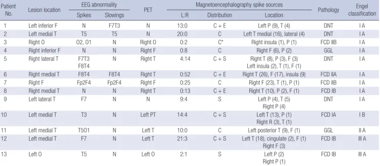
Fig. 1. Distribution and location of clustered MEG spike sources. (A) T1-weighted axial brain magnetic resonance imaging (MRI) shows MEG spike sources clustered around the margin of the lesion. Because the spike sources are overlaid in a single axial image, spike sources that are inside the lesion are located superior or inferior to the lesion (patient 2). (B) T2-weighted axial brain MRI shows MEG spike sources clustered on the margin of the lesion (overlaid image of patient 7). (C) T1-weighted axial brain MRI shows MEG spike sources clustered at the margin lateral to the lesion (overlaid image of patient 12). Epileptogenic lesion (arrow) is visible in brain MRI. R, right; L, left; A, anterior; P, posterior.
A B C
A
P
R L
A
P
R L
A
P
R L
monitoring (patients 7, 11, and 13) and invasive intracranial EEG monitoring (patients 4 and 13).
Lesionectomy was performed in all patients. Anterior tem- poral lobectomy was performed in two patients (patients 5 and 10). The surgeons determined the resection margin using the preoperative brain MRI findings and intraoperative ultrasono- grams. An invasive intracranial EEG-based resection margin was applied in 2 patients (patients 4 and 13). The pathology of the lesions included focal cortical dysplasia (n = 7), dysembryo- plastic neuroepithelial tumor (n = 4), and ganglioglioma (n = 2).
The mean follow-up duration after the surgery was 39.2 months (range, 26-64 months). The postoperative seizure outcomes in- cluded Engel class I (n = 10), class II (n = 1), and class III (n = 2).
Nine patients (69%) were completely seizure free (Table 2).
DISCUSSION
Identification and accurate localization of the epileptogenic area are critical for successful epilepsy surgery. In this study, the hemi- spheric concordance rate of the MEG spike sources was 100%.
Localization was also satisfactory at the level of the anatomical region because the MEG spike sources were distributed exactly at or in the vicinity of the lesion in most patients. The hemi- spheric lateralization rate is similar to or higher than that report-
ed for patients with intractable focal epilepsy in previous stud- ies: 68% (when compared to brain MRI and intracranial ictal onset zone) (12), 82% (lobar concordance to anatomical lesion- al in brain MRI) (7), and 89% (when compared to the final sur- gical decision) (4). The lobar or regional localization rate is sim- ilar to the 72% rate reported in patients with intractable focal epilepsy (7). Although the number of patients in this study is small, our data suggest that MEG is useful in identifying the lat- eralization and localization of the structural lesions in pediatric epilepsy patients. We observed good outcomes of 69% of pa- tients free from seizures and 77% of patients with an Engel class I outcome after a sufficient follow-up period. Favorable post- surgical seizure outcomes of MEG have also been shown in pre- vious studies (7, 13, 14). Although we did not compare the MEG spike source foci with the intracranial EEG ictal onset in this study, complete seizure freedom after surgery can be considered as indirect evidence that the epileptogenic area was removed or the epileptogenic condition was resolved. These findings allow us to infer that MEG is effective in identifying the lateralization and localization of epileptogenic areas in patients with lesional epilepsy.
In a study comparing the diagnostic sensitivity of presurgical evaluation modalities, MSI had the highest sensitivity (58%-64%), as well as the highest predictive value for identifying the ictal
A B
C D
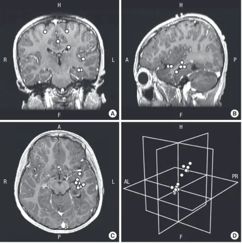
Fig. 2. MEG spike sources clustered around the le- sion and scattered in patient 12. (A-C) Axial, sagittal, and coronal contrast-enhanced T1-weighted brain magnetic resonance imaging (MRI) show clusters and distant scatters (overlaid on a single brain MRI image). (D) MEG spike sources presented in an imag- inary three-dimensional plane show the distribution of MEG spike sources of clusters and scatters. R, right; L, left; H, head; F, foot; AL, anterior left; PR, posterior right; A, anterior; P, posterior.
H
F
R L
A
P
R L
H
F
A P
H
F
AL PR
onset zone (15). Sutherling et al. reported additional benefits of MSI for preoperative planning (16). In our study, three patients had an interictal FDG PET result showing no decrease in glucose uptake. In two of these patients (patients 1 and 2), MEG identi- fied 13 and 20 MEG spike sources that recognized the lesional hemisphere and localized the spike source clusters. In two pa- tients with normal interictal scalp EEGs (patients 4 and 8), MEG also identified the lesional hemisphere and the anatomical lobe that was concordant with the lesion. These patients were com- pletely seizure free for 26-45 months after surgery. It is well known that MEG and EEG are not mutually exclusive in discriminating abnormal electrical activity (17). A recent study showed that a multimodality approach can improve surgical outcome even in pediatric patients with intractable nonlesional epilepsy (18).
Although we did not collect quantitative data, we demonstrated that MEG provides additive information in patients with nor- mal FDG PET or interictal scalp EEG results. Our findings sug- gest that MEG should be performed in patients undergoing pre- surgical evaluation, especially in those with discordant or insuf- ficient information on lateralization or localization.
The distribution of MEG spike sources in relation to the MRI lesions varied and included clusters, clusters with extension, or scatters. The clusters were located around the lesion or at the margin of the lesion. In most patients, the clusters were located on one side of the lesion and sometimes extended further to the same side. Similar findings were reported in previous studies that showed surrounding MEG spike sources in patients with focal cortical dysplasia (13, 19, 20). Although we found no dif- ferences in the MEG spike source distribution according to dif- ferent pathologies, we observed the typical distribution of clus- ters located at the margin of the lesion either around the lesion or at one side of the margin. This finding suggests that the irrita- tive zone in lesional focal epilepsy may reside in the margin of the lesion, and hence indicates one possible pathophysiologi- cal mechanism underlying the epileptogenesis of lesional epi- lepsy. These findings should be clarified in future research with more patients and objective methodology.
This is the first study performed to assess the value of MEG in pediatric patients from the tertiary epilepsy surgery center soon after utilizing magnetoencephalography. Although we could not demonstrate the additional benefit of MEG in lesional epilepsy surgery, this was shown in previous studies. Removal of the dys- plastic cortex that has not been characterized by brain imaging but by MEG was required for complete seizure freedom (13). In cases of discordant presurgical evaluation, concordant lateral- ization was related to good seizure control (12). Multiple MEG spike source clusters required identification of the multiple or extensive epileptogenic zone prior to accurate localization and delineation of the resection margin (21). Concordant lateraliza- tion and precise localization of interictal MEG spike sources that were noted in this study may provide additional information in
the presurgical evaluation and planning in lesional epilepsy sur- gery. MEG spike sources demonstrate and represent interictal irritative zone, this may partly explain the poor outcome of three patients in this study. Ictal onset zone should be verified by the intracranial EEG and interictal spike sources of MEG may pro- vide compensatory information. MEG and EEG are not mutu- ally exclusive (17), so both modalities should be used together to achieve the best information regarding the epileptogenic zone.
Young patients were included in this study. The youngest pa- tient reported to have undergone MEG analysis was 5 months (12), and another study included a patient who had received surgery at 4 yr of age (22). The age of patients included in the pe- diatric MEG studies usually range from 4 to 18 yr, and the mean age of patients were usually 10 to 12 yr (13, 20, 22). The young- est patient in this study was 2.8 yr old when she underwent MEG analysis, and other patients were 3.9, 4.1, and 4.4 yr old at the time of MEG analysis. Conscious sedation with oral chloral hy- drate, instead of general anesthesia or parenteral sedatives, was sufficient for these younger children and we found no limita- tions in processing and analyzing the MEG data. We emphasize that MEG can be performed safely and effectively even in young- er pediatric patients.
There are limitations to this study. We could not demonstrate directly that the MEG spike sources represent the ictal onset zone, and we found no difference between the MEG spike source dis- tribution and pathology of the epileptogenic lesion. This should be verified in future studies.
In conclusion, this is the first Korean report of MEG in the field of pediatric epilepsy surgery. MEG is safe and effective for analyzing pediatric patients with lesional localization-related epilepsy. The hemispheric and regional concordance is satis- factory. The favorable surgical outcomes in this study provide indirect evidence that the irritative zone defined by MEG can provide valuable information about the epileptogenic area.
REFERENCES
1. Cohen D. Magnetoencephalography: evidence of magnetic fields pro- duced by alpha-rhythm currents. Science 1968; 161: 784-6.
2. Ricci GB, Romani GL, Salustri C, Pizzella V, Torrioli G, Buonomo S, Peres- son M, Modena I. Study of focal epilepsy by multichannel neuromagnet- ic measurements. Electroencephalogr Clin Neurophysiol 1987; 66: 358-68.
3. Gallen CC, Hirschkoff EC, Buchanan DS. Magnetoencephalography and magnetic source imaging. Capabilities and limitations. Neuroimag- ing Clin N Am 1995; 5: 227-49.
4. Stefan H, Hummel C, Scheler G, Genow A, Druschky K, Tilz C, Kalten- häuser M, Hopfengärtner R, Buchfelder M, Romstöck J. Magnetic brain source imaging of focal epileptic activity: a synopsis of 455 cases. Brain 2003; 126: 2396-405.
5. Knowlton RC, Laxer KD, Aminoff MJ, Roberts TP, Wong ST, Rowley HA.
Magnetoencephalography in partial epilepsy: clinical yield and localiza- tion accuracy. Ann Neurol 1997; 42: 622-31.
6. Pataraia E, Simos PG, Castillo EM, Billingsley RL, Sarkari S, Wheless JW, Maggio V, Maggio W, Baumgartner JE, Swank PR, et al. Does mag- netoencephalography add to scalp video-EEG as a diagnostic tool in epi- lepsy surgery? Neurology 2004; 62: 943-8.
7. Paulini A, Fischer M, Rampp S, Scheler G, Hopfengärtner R, Kalten- häuser M, Dörfler A, Buchfelder M, Stefan H. Lobar localization infor- mation in epilepsy patients: MEG - a useful tool in routine presurgical diagnosis. Epilepsy Res 2007; 76: 124-30.
8. Bast T. Magnetoencephalography. In: Wyllie E, Cascino G, Gidal B, edi- tors. Wyllie’s treatment of epilepsy: principles and practice. Philadelphia:
Lippincott Williams & Wilkins, 2011, p 869-75.
9. Commission on Classification and Terminology of the International League Against Epilepsy. Proposal for revised clinical and electroenceph- alographic classification of epileptic seizures. Epilepsia 1981; 22: 489-501.
10. Blume WT, Lüders HO, Mizrahi E, Tassinari C, van Emde Boas W, En- gel J Jr. Glossary of descriptive terminology for ictal semiology: report of the ILAE task force on classification and terminology. Epilepsia 2001; 42:
1212-8.
11. Engel J Jr, VanNess P, Rasmussen T, Ojemann L. Outcome with respect to epileptic seizures. In: Engel J Jr, editor. Surgical treatment of the epilep- sies. 2nd ed. New York: Raven Press, 1993, p 609-21.
12. Ochi A, Otsubo H, Iida K, Oishi M, Elliott I, Weiss SK, Kutomi T, Nakaya- ma T, Sharma R, Chuang SH, et al. Identifying the primary epileptogenic hemisphere from electroencephalographic (EEG) and magnetoencepha- lographic dipole lateralizations in children with intractable epilepsy. J Child Neurol 2005; 20: 885-92.
13. Otsubo H, Ochi A, Elliott I, Chuang SH, Rutka JT, Jay V, Aung M, Sobel DF, Snead OC. MEG predicts epileptic zone in lesional extrahippocam- pal epilepsy: 12 pediatric surgery cases. Epilepsia 2001; 42: 1523-30.
14. Minassian BA, Otsubo H, Weiss S, Elliott I, Rutka JT, Snead OC 3rd. Mag- netoencephalographic localization in pediatric epilepsy surgery: com- parison with invasive intracranial electroencephalography. Ann Neurol
1999; 46: 627-33.
15. Knowlton RC, Elgavish RA, Limdi N, Bartolucci A, Ojha B, Blount J, Burneo JG, Ver Hoef L, Paige L, Faught E, et al. Functional imaging: I.
Relative predictive value of intracranial electroencephalography. Ann Neurol 2008; 64: 25-34.
16. Sutherling WW, Mamelak AN, Thyerlei D, Maleeva T, Minazad Y, Phil- pott L, Lopez N. Influence of magnetic source imaging for planning in- tracranial EEG in epilepsy. Neurology 2008; 71: 990-6.
17. Funke M, Constantino T, Van Orman C, Rodin E. Magnetoencephalog- raphy and magnetic source imaging in epilepsy. Clin EEG Neurosci 2009;
40: 271-80.
18. Seo JH, Holland K, Rose D, Rozhkov L, Fujiwara H, Byars A, Arthur T, DeGrauw T, Leach JL, Gelfand MJ, et al. Multimodality imaging in the surgical treatment of children with nonlesional epilepsy. Neurology 2011;
76: 41-8.
19. Bast T, Oezkan O, Rona S, Stippich C, Seitz A, Rupp A, Fauser S, Zent- ner J, Rating D, Scherg M. EEG and MEG source analysis of single and averaged interictal spikes reveals intrinsic epileptogenicity in focal corti- cal dysplasia. Epilepsia 2004; 45: 621-31.
20. Iida K, Otsubo H, Matsumoto Y, Ochi A, Oishi M, Holowka S, Pang E, Elliott I, Weiss SK, Chuang SH, et al. Characterizing magnetic spike sourc- es by using magnetoencephalography-guided neuronavigation in epi- lepsy surgery in pediatric patients. J Neurosurg 2005; 102: 187-96.
21. Oishi M, Kameyama S, Masuda H, Tohyama J, Kanazawa O, Sasagawa M, Otsubo H. Single and multiple clusters of magnetoencephalographic dipoles in neocortical epilepsy: significance in characterizing epilepto- genic zone. Epilepsia 2006; 47: 355-64.
22. RamachandranNair R, Otsubo H, Shroff MM, Ochi A, Weiss SK, Rutka JT, Snead OC 3rd. MEG predicts outcome following surgery for intracta- ble epilepsy in children with normal or nonfocal MRI findings. Epilepsia 2007; 48: 149-57.