INTRODUCTION
Non-small cell lung cancer (NSCLC) represents both a bio- logically and histopathologically heterogenous group of tu- mors. Although complete surgical resection is the only cura- tive therapy for NSCLC patients, the survival rate, even of patients with early stage NSCLC, is not satisfactory. This has led to a search for prognostic parameters of lung cancer from clinical data or histological examination. Recently, the poten- tial prognostic roles of cancer cell proliferation and molecu- lar biological alterations in oncogenes and tumor suppressor genes have been investigated in NSCLC, but none has been found to yield unambiguous results (1).
Epidermal growth factor receptor (EGFR) is a 170 kDa transmembrane glycoprotein with an intracellular domain that exhibits tyrosine kinase activity. EGFR is involved in cellular proliferation and differentiation (2, 3), and overexpres- sion of mRNA and/or protein encoded by the EGFR gene has been observed in many types of human malignancies, includ- ing breast, gastric, colorectal, and bladder cancer, as well as in 30% to 70% of NSCLCs (4-8). Findings have suggested that assays of EGFR mRNA and/or protein may be useful for predicting the prognosis of tumors. For example, enhanced
expression of EGFR has been related to poorer prognosis in breast cancer, and advanced gastric carcinomas have shown a higher level of EGFR expression than early gastric carcino- mas (4, 6, 9, 10). Although some studies have reported an association between enhanced EGFR expression and poor prognosis in patients with lung cancer, the prognostic role of EGFR remains yet controversial (11-14).
In the present study, we assayed immunohistochemically EGFR expression in human NSCLC tissues, and we analyzed the relationships between EGFR expression and several pro- gnostic variables, including angiogenesis and tumor cell pro- liferation. Furthermore, we investigated the prognostic role of EGFR overexpression in relation to disease-free survival and overall survival.
MATERIALS AND METHODS Study population
From February 1991 to March 1996, ninety-four patients with stage II or IIIA NSCLC underwent curative resection and received 2 courses of adjuvant MVP [mitomycin C (8
Jin-Hee Ahn, Sang-We Kim, Seung-Mo Hong*, Cheolwon Suh, Woo Kun Kim, In Chul Lee*, Jung-Shin Lee
Department of Medicine, and Pathology*, Asan Medical Center, University of Ulsan College of Medicine, Seoul, Korea
Address for correspondence Sang-We Kim, M.D.
Department of Medicine, Asan Medical Center, University of Ulsan College of Medicine, 388-1 Poongnap-dong, Songpa-gu, Seoul 138-736, Korea Tel : +82.2-3010-3210, Fax : +82.2-3010-6961 E-mail : [email protected]
*This study was presented as an abstract at the Ann Meeting of the Korean Association of Internal Medicine, 2003.
529
Epidermal Growth Factor Receptor (EGFR) Expression in Operable Non-small Cell Lung Carcinoma
This study was performed to assay the expression of epidermal growth factor recep- tor (EGFR) in non-small cell lung carcinoma (NSCLC), and to investigate the rela- tionship between EGFR status and various clinicopathologic features of NSCLC, including angiogenesis and proliferative activity. The expression of EGFR, microves- sel count (MVC) measured by CD31 monoclonal antibody, and proliferative activity using Ki-67 labeling index were immunohistochemically analyzed in formalin-fixed and paraffin-embedded tissue specimens from 65 patients with completely resected stage II-IIIA NSCLC. Pathologic and clinical records of all patients were retrospec- tively reviewed. EGFR was expressed in 18 (28%) of 65 NSCLC samples. More squamous tumors (35%) were EGFR-positive than other NSCLCs (23%) (p-value 0.308). There was a statistically significant correlation between EGFR expression and Ki-67 labeling index (p-value 0.042), but no correlation was observed between EGFR expression and tumor histology, stage, or MVC. There were no differences between EGFR positive and negative tumors in 5-yr disease-free survival (60% vs.
52%, p-value 0.5566) and 5-yr overall survival (53% vs. 45%, p-value 0.3382) rates.
In conclusion, our findings suggest that NSCLC proliferative activity may be depen- dent on EGFR expression, but that EGFR expression had no significant impact on survival in curatively resected NSCLC.
Key Words : Lung Neoplasms; Receptor, Epidermal Growth Factor; Angiogenesis; Angiogenesis Inducing Agents; Ki-67 Antigen; Prognosis
Received : 4 February 2004 Accepted : 23 March 2004
mg/m2), vinblastine (8 mg/m2), and cisplatin (60 mg/m2)]
chemotherapy, followed by sequential radiotherapy (50 Gy) 3 weeks later. This protocol has shown relatively low recur- rence and improved overall survival rates in patients with stage II and IIIA NSCLC (15). We obtained 65 of these resected lung tumor tissue samples from the pathology department for this study. We also reviewed the medical records and pa- thology data of these patients in order to determine their clini- copathologic variables, including age, sex, TNM stage, and histologic grade and types of tumors, as well as their disease free survival and overall survival.
Immunohistochemical method
Paraffin-embedded blocks were sectioned at about 4- m thickness, and deparaffinized and rehydrated through a graded alcohol series. The slides were pretreated in the microwave with citrate buffer (pH 6.0) at 90℃for 10 min to retrieve antigen, immersed in 0.3% hydrogen peroxide for 20 min to block endogenous peroxidase activity, washed, and incu- bated overnight at 4℃with normal bovine serum to reduce nonspecific antibody binding. Subsequently, all tissue sections were incubated with mouse monoclonal antibody to EGFR (1:50; Novocastra Laboratories, Newcastle, U.K.) and CD31, a sensitive marker for endothelial cells (16) (JC70, 1:50; DA- KO, Denmark), and with rabbit polyclonal antibody to Ki-67 protein, a marker for tumor cell proliferation (1:100; DAKO, Denmark). The slides were subsequently incubated with bio- tinylated goat anti-mouse antibody, and then with peroxidase- conjugated streptavidin (DAKO LSAB+ kit; Dako Corp., Carpinteria, CA, U.S.A.). Reaction products were subjected to a color reaction with 3,3-diaminobenzidine tetrahydrochlo- ride containing 3% H2O2in Tris buffer and were lightly coun- terstained with Mayer’s hematoxylin.
Evaluation of the immunohistochemical staining
All slides were coded and evaluated by an experienced pa- thologist who had no knowledge of the patients’ identity or clinical status. EGFR assessment was based on cytoplasmic staining intensity was, which was scored as 0 (negative, <5%
of cells stained), 1+ (weak, 5-20% of cells stained), 2+ (mod- erate, 20-50% of cells stained), and 3+ (strong, >50% of cells stained). Only tumors exhibiting 2+~3+ staining were con- sidered positive for EGFR expression (17).
For microvessel count (MVC) assessment, each slide was first scanned by light microscopy at ×100 magnification to determine three ‘‘hot spots’’ defined as areas with the maxi- mum number of microvessels. The slides were then examined at×200 magnification, and three ×200 fields in each ‘‘hot spot’’ were evaluated by counting the number of positive cells.
The highest number was recorded for statistical analysis. MVC values were expressed as mean±standard deviation (SD) (18).
Ki-67-positive cells were evaluated by nuclear staining,
with the number of positive cells in 1000 tumor cells count- ed at high magnification (×400). The Ki-67 labeling index was calculated as positive nuclei ×100/total number of count- ed nuclei (%). For statistical analysis, a threshold value of 15%
Ki-67 positive tumor cells were defined as separating tumors with a high proliferation rate from those with a low prolifera- tion rate (19).
Statistical analysis
Using the SPSS 11.0 program, the relationships of EGFR expression to clinicopathologic variables, MVC, and Ki-67- based proliferative activity were analyzed by chi-square or Fisher’s exact test. The significance of MVC for each clinico- pathologic factor, including EGFR expression, was evaluated using Student’s t test. Univariate analysis was performed to identify prognostic factors for survival by modeling Kaplan- Meier survival curves, and the difference in survival curves was compared by the log-rank test. Multivariate analysis was performed by Cox’ proportional hazards regression model.
Disease-free survival (DFS) was defined as the time from the date of operation to a documented recurrence or death from any other cause. Overall survival (OS) was defined as the time from the date of operation to death or last follow-up. In all statistical analyses, the difference was considered to be signif- icant when the p-value was less than 0.05.
RESULTS
Patient characteristics and EGFR expression in 65 NSCLC
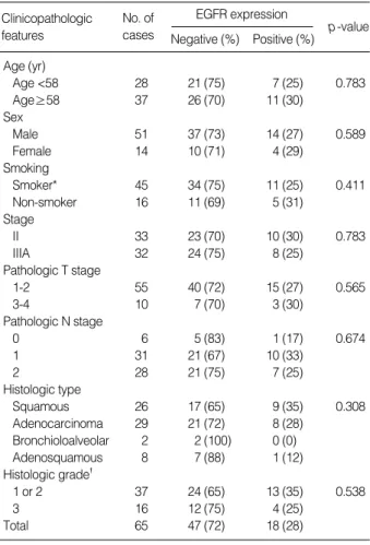
Sixty-five NSCLC patients (51 males and 14 females), with a median age of 58 yr (range, 34-73 yr) were studied. Of the 65 patients, 26 had squamous cell carcinoma, 29 had adeno- carcinoma, 2 had bronchioloalveolar carcinoma, and 8 had adenosquamous carcinoma. Thirty-three cases were stage II and 32 were stage IIIA. The median follow-up in 33 living patients was 85.3 months (range, 18-133 months). Table 1 shows patients’ characteristics and relationship with EGFR expression.
Typical immunostaining patterns for EGFR in NSCLC are shown in Fig. 1. Immunoreactivity was limited to cancer cells, and there was very little or no specific staining in the surrounding stroma. Staining for EGFR was strong in 11/65 (17%), moderate in 7/65 (11%), weak in 25/65 (38%), and negative in 22/65 (34%) tumor samples. Thus, 18/65 (28%) NSCLC tissues were positive for EGFR expression. Of the 26 squamous tumors, 9 (35%) were stained moderately or strong- ly, while of the 39 non-squamous tumors, 9 (23%) were pos- itive for EGFR. Overall, more squamous tumors were EGFR- positive than other NSCLCs, but the difference was not sta- tistically significant.
MVC, as an indicator of neovascularization in primary tu-
mors, was measured by immunohistochemical staining of endothelial vessels with an anti-CD31 monoclonal antibody.
The 65 tumors assayed had a median MVC of 95.0 per 200×
field (range, 38-209). MVC was significantly associated with histologic grade but not with any other clinicopathological or biological variable analyzed (data not shown).
We found no significant association between EGFR expres- sion and any clinicopathological parameters, including age, tumor size, pathologic stage, nodal status, histological type, and tumor angiogenesis (Table 1).
EGFR expression and proliferative activity
The proliferative activity of tumors was evaluated by Ki-67 expression. The mean Ki-67 labeling index was 13% (range 0-56%). We divided tumors into groups with low Ki-67 ex- pression (Ki-67 labeling index <15%) and high Ki-67 expres- sion (Ki-67 labeling index ≥15%). A high Ki-67 labeling index was found in 25/61 (40%) tumors. We observed a pos- itive correlation between EGFR expression and high rate of tumor proliferation (Table 2). EGFR expression was more fre- quently detected in the group with high Ki-67 expression than in the group with low Ki-67 expression (61.1% vs. 38.9
%, p-value 0.042).
Clinicopathologic features
No. of
cases EGFR expression p-value Negative (%) Positive (%) Age (yr)
Age <58 28 21 (75) 7 (25) 0.783
Age≥58 37 26 (70) 11 (30)
Sex
Male 51 37 (73) 14 (27) 0.589
Female 14 10 (71) 4 (29)
Smoking
Smoker* 45 34 (75) 11 (25) 0.411
Non-smoker 16 11 (69) 5 (31)
Stage
II 33 23 (70) 10 (30) 0.783
IIIA 32 24 (75) 8 (25)
Pathologic T stage
1-2 55 40 (72) 15 (27) 0.565
3-4 10 7 (70) 3 (30)
Pathologic N stage
0 6 5 (83) 1 (17) 0.674
1 31 21 (67) 10 (33)
2 28 21 (75) 7 (25)
Histologic type
Squamous 26 17 (65) 9 (35) 0.308
Adenocarcinoma 29 21 (72) 8 (28)
Bronchioloalveolar 2 2 (100) 0 (0)
Adenosquamous 8 7 (88) 1 (12)
Histologic grade�
1 or 2 37 24 (65) 13 (35) 0.538
3 16 12 (75) 4 (25)
Total 65 47 (72) 18 (28)
Table 1.Clinical characteristics and relationship with EGFR ex- pression
*Smokers include both ex-smoker and current smoker. �1, well differen- tiated; 2, moderately differentiated; 3, poorly differentiated.
EGFR expression
No. of
cases Ki-67 labeling index p-value
<15% ≥15%
Positive 18 7 (38.9%) 11 (61.1%) 0.042
Negative 47 29 (61.7%) 14 (38.3%)
Table 2.Correlation between EGFR expression and proliferative activity
Fig. 1.Microphotograph showing EGFR immunostaining in NSCLC tissue specimens. Positive immunoreactivity is evident in the neoplas- tic cells (A), whereas negative immunoreactivity is shown in (B) (×100).
A B
Survival
The prognostic impact of clinicopathological variables on patient survival was evaluated by univariate analysis. Advanced
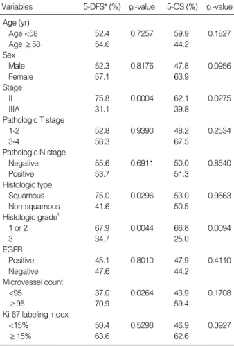
tumor stage, non-squamous histologic type, high grade tu- mors, and high level of MVC were significantly associated with lower DFS (Table 3). EGFR expression alone, however, could not be correlated with DFS (5-yr DFS, 45% vs. 48%, p-value 0.8010) or OS (5-yr OS, 48% vs. 44%, p-value 0.4110) (Fig. 2). By multivariate analysis, only tumor stage, but not histology, histologic grade, MVC level, or EGFR expression, had a significant prognostic impact on DFS and OS (Table 4).
DISCUSSION
EGFR expression is frequently detected in many types of tumors, including the majority of NSCLC. In our study, EGFR expression was immunohistochemically evaluated in 65 for- malin-fixed and paraffin-embedded human NSCLC tissues, and 18 (28%) were positive for EGFR. In previous studies by other investigators, EGFR was expressed in 30-70% of
Factors SE* p-value HR (95% CI)�
Stage 0.517 0.009 3.867 (1.405-10.643)
(II vs. IIIA)
Histology 0.496 0.060 0.394 (0.149-1.041)
(squamous vs.
non-squamous)
Histologic grade� 0.443 0.061 2.296 (0.964-5.470) (1 or 2 vs. 3)
EGFR expression 0.520 0.636 0.782 (0.282-2.168) (negative vs. positive)
Microvessel count 0.551 0.875 0.875 (0.297-2.578) (<95 vs. ≥95)
Ki-67 labeling index 0.436 0.333 0.656 (0.279~1.541) (<15% vs. ≥15%)
Table 4.Multivariate analysis of prognostic factors that influence the 5-yr overall survival
*Standard error. �Hazard ratio (95% confidence interval). �1, well differ- entiated; 2, moderately differentiated; 3, poorly differentiated.
Variables 5-DFS* (%) p-value 5-OS (%) p-value Age (yr)
Age <58 52.4 0.7257 59.9 0.1827
Age ≥58 54.6 44.2
Sex
Male 52.3 0.8176 47.8 0.0956
Female 57.1 63.9
Stage
II 75.8 0.0004 62.1 0.0275
IIIA 31.1 39.8
Pathologic T stage
1-2 52.8 0.9390 48.2 0.2534
3-4 58.3 67.5
Pathologic N stage
Negative 55.6 0.6911 50.0 0.8540
Positive 53.7 51.3
Histologic type
Squamous 75.0 0.0296 53.0 0.9563
Non-squamous 41.6 50.5
Histologic grade�
1 or 2 67.9 0.0044 66.8 0.0094
3 34.7 25.0
EGFR
Positive 45.1 0.8010 47.9 0.4110
Negative 47.6 44.2
Microvessel count
<95 37.0 0.0264 43.9 0.1708
≥95 70.9 59.4
Ki-67 labeling index
<15% 50.4 0.5298 46.9 0.3927
≥15% 63.6 62.6
Table 3.Univariate analysis of prognositc factors
*DFS, disease-free survival; OS, overall survival. �1, well differentiated;
2, moderately differentiated; 3, poorly differentiated.
Fig. 2.Disease-free survival (DFS) curve (A) and overall survival (OS) curve (B) according to EGFR expression.
Survival probability
1.0
0.8
0.6
0.4
0.2
0.0
0 20 40 60 80 100 120
Months
(a) EGFR (+)
p-value 0.8010
(a) median DFS: 56.7 months (95% CI=38.2-73.0) (b) median DFS: 50.8 months (95% CI=19.4-82.3)
(b) EGFR (-)
Survival probability
1.0
0.8
0.6
0.4
0.2
0.0
0 20 40 60 80 100 120 140
Months
(a) EGFR (+)
p-value 0.4110
(a) median OS: 60.3 months (95% CI=41.3-79.3) (b) median OS: 42.1 months (95% CI=22.3-62.0)
(b) EGFR (-)
A B
NSCLCs (4-8). In agreement with previous findings (11, 20- 22), we also found that EGFR expression was more frequent in squamous cell carcinomas than in other histological types of NSCLCs (35% vs. 23%), although the difference was not statistically significant (p-value 0.308). Theoretically, high EGFR expression in squamous cell carcinomas is expected because EGF promotes the proliferation and differentiation of epidermal-like tissues. Our series of NSCLC included a rel- atively small population of squamous cell carcinomas (26/65, 38%), which may have affected our low EGFR expression rate.
EGFR expression is generally low in normal bronchial epi- thelium, whereas it is enhanced in preneoplastic and neoplastic bronchial lesions (23). Attempts to establish a relationship between EGFR expression and other clinical and pathologi- cal prognostic parameters or prognosis in primary lung cancers, however, have led to conflicting results. One report showed no correlation between EGFR expression and pathological stage, nodal status, or tumor size, although EGFR concentra- tions were higher in neoplastic than in normal tissues (24).
A second report found that although EGFR expression was the highest in squamous cell carcinomas, it could not be corre- lated with other clinicopathological characteristics such as his- tologic grade or stage (25). In contrast, other investigators have reported significant correlations between EGFR expression and more aggressive tumor features, such as tumor stage (22) or nodal metastases (20).
In our study, no significant correlations were found between EGFR expression and several clinicopathological or biologi- cal parameters, including tumor size, nodal metastasis, his- tologic grade, and tumor angiogenesis. There was a correla- tion, however, between EGFR expression and tumor cell pro- liferative activity, as measured with the Ki-67 proliferation index. This index measures the proportion of cycling cells and is a potent biological marker for quantitative estimation of the growth of neoplasms (26). Binding of EGF or other lig- ands to EGFR is believed to trigger a series of complex sig- naling pathways leading to DNA synthesis and cell growth (27). Thus, our data also suggest that the proliferative activ- ity of NSCLC may be dependent on EGFR expression.
In this study, EGFR expression had no significant impact on DFS or OS in curatively resected stage II or IIIA NSCLC.
EGFR has been reported to act as a strong prognostic marker in head and neck cancers (28), genito-urinary carcinomas (29, 30) and esophageal cancers (31). In NSCLC, however, findings have been more ambiguous. Some previous studies have re- ported that survival rates of patients with high EGF or TGF- levels were significantly low only in EGFR-positive tumors (32, 33). In a recent study, 5-yr survival rate of EGFR positive tumors evaluated by immunohistochemistry (IHC) was sig- nificantly lower than EGFR negative ones (34). However, sev- eral studies using specimens from larger numbers of patients suggested that EGFR expression was not associated with poor outcome (17, 20, 35). These conflicting data on the relation- ship between EGFR expression and survival in NSCLC may
be due to heterogeneity of study populations or lack of a stan- dardized assay for determining EGFR status. More recently, the first report of EGFR gene copy number in NSCLC using fluorescence in situ hybridization (FISH) technology showed that there was no difference in survival between the two FISH groups (low and high copy numbers of the EGFR gene) (36).
However, high gene copy numbers combined with low level of EGFR protein expression by IHC had a trend toward poor prognosis. Because our study was retrospective and analyzed a relatively small number of tumors, we could not definitely determine the relationship between EGFR expression and long-term prognosis. When combined with previous results, our findings suggest that EGFR may be more important for lung tumor formation than for tumor progression. The pos- sibility that EGFR expression plays a prognostic role in pri- mary NSCLC should be further investigated.
Although EGFR expression may not be useful as a prognos- tic factor, it has potential clinical implications. The past few years have seen the rapid development of the EGFR inhibitors, and an increasing body of evidence suggests that selective inhibitors of EGFR are potential therapeutic agents for the treatment of NSCLC in adjuvant, metastatic and chemopre- ventive settings (37).
In conclusion, our data show no significant correlations between EGFR expression in NSCLC and other clinicopatho- logical parameters such as tumor size, nodal metastasis, angio- genesis and prognosis. EGFR may be a useful indicator of tumor cell proliferation in stage II or IIIA NSCLC. Our results suggest the need for additional prospective studies in patients with NSCLC.
REFERENCES
1. Strauss GM, Kwiatkowski DJ, Harpole DH, Lynch TJ, Skarin AT, Sugarbaker DJ. Molecular and pathologic markers in stage I non- small cell carcinoma of the lung. J Clin Oncol 1995; 13: 1265-79.
2. Downward J, Yarden Y, Mayes E, Scrace G, Totty N, Stockwell P, Ullrich A, Schlessinger J, Waterfield MD. Close similarity of epider- mal growth factor receptor and v-erb-B oncogene protein sequences.
Nature 1984; 307: 521-7.
3. Schreiber AB, Libermann TA, Lax I, Yarden Y, Schlessinger J. Bio- logical role of epidermal growth factor-receptor clustering: investi- gation with monoclonal anti-receptor antibodies. J Biol Chem 1983;
258: 846-53.
4. Sainsbury JRC, Nicholson S, Angus B, Farndon JR, Malcolm AJ, Harris AL. Epidermal growth factor receptor status of histological sub-types of breast cancer. Br J Cancer 1988; 58: 458-60.
5. Hendler FJ, Ozanne BW. Human squamous cell lung cancers express increased epidermal growth factor receptors. J Clin Invest 1984; 74:
647-51.
6. Yasui W, Sumiyoshi H, Hata J, Kameda T, Ochiai A, Ito H, Tahara E. Expression of epidermal growth factor receptor in human gastric and colonic carcinomas. Cancer Res 1988; 48: 137-41.
7. Smith K, Fennelly JA, Neal DE, Hall RR, Harris AL. Characteriza- tion and quantitation of the epidermal growth factor receptor in inva- sive and superficial bladder tumors. Cancer Res 1989; 49: 5810-5.
8. Veale D, Kerr N, Gibson GJ, Harris AL. Characterization of epider- mal growth factor receptor in primary human non-small cell lung cancer. Cancer Res 1989; 49: 1313-7.
9. Toi M, Nakamura T, Mukaida H, Wada T, Osaki A, Yamada H, Toge T, Niimoto M, Hattori T. Relationship between epidermal growth factor receptor status and various prognostic factors in human breast cancer. Cancer 1990; 65: 1980-4.
10. Nicholson S, Richard J, Sainsbury C, Halcrow P, Kelly P, Angus B, Wright C, Henry J, Farndon JR, Harris AL. Epidermal growth factor receptor (EGFr); results of a 6 year follow-up study in operable breast cancer with emphasis on the node negative subgroup. Br J Cancer 1991; 63: 146-50.
11. Volm M, Drings P, Wodrich W. Prognostic significance of the expres- sion of c-fos, c-jun and c-erbB-1 oncogene products in human squa- mous cell lung carcinomas. J Cancer Res Clin Oncol 1993; 119: 507- 10.
12. Veale D, Kerr N, Gibson GJ, Kelly PJ, Harris AL. The relationship of quantitative epidermal growth factor receptor expression in non- small cell lung cancer to long-term survival. Br J Cancer 1993; 68:
162-5.
13. Fujino S, Enokibori T, Tezuka N, Asada Y, Inoue S, Kato H, Mori A.
A comparison of epidermal growth factor receptor levels and other prognostic parameters in non-small cell lung cancer. Eur J Cancer 1996; 32A: 2070-4.
14. Fu XL, Zhu XZ, Shi DR, Xiu LZ, Wang LJ, Zhao S, Qian H, Lu HF, Xiang YB, Jiang GL. Study of prognostic predictors for non-small cell lung cancer. Lung cancer 1999; 23: 143-52.
15. Lee SW, Choi EK, Chung WK, Shin KH, Ahn SD, Kim JH, Kim SW, Suh C, Lee JS, Kim WS, Kim DS, Kim DK, Park SI, Sohn KH.
Postoperative adjuvant chemotherapy and radiotherapy for stage II and III non-small cell lung cancer (NSCLC). Lung cancer 2002; 37:
65-71.
16. Horak ER, Leek R, Klenk N, LeJeune S, Smith K, Stuart N, Greenall M, Stepniewska K, Harris AL. Angiogenesis, assessed by platelet/
endothelial cell adhesion molecule antibodies, as indicator of node metastases and survival in breast cancer. Lancet 1992; 340: 1120-4.
17. Rusch V, Klimstra D, Venkatraman E, Pisters PWT, Langenfeld J, Dmitrovsky E. Overexpression of the epidermal growth factor recep- tor and its ligand transforming growth factor is frequent in resec- table non-small cell lung cancer but does not predict tumor progres- sion. Clin Cancer Res 1997; 3: 515-22.
18. Dazzi C, Cariello A, Maioli P, Solaini L, Scarpi E, Rosti G, Lanzanova G, Marangolo M. Prognostic and predictive value of intratumoral microvessels density in operable non-small cell lung cancer. Lung cancer 1999; 24: 81-8.
19. Hommura F, Dosaka-Akita H, Mishina T, Nishi M, Kojima T, Hirou- mi H, Ogura S, Shimizu M, Katoh H, Kawakami Y. Prognostic sig- nificance of p27KIP1protein and Ki-67 growth fraction in non-small cell lung cancers. Clin Cancer Res 2000; 6: 4073-81.
20. Fontanini G, Vignati S, Bigini D, Mussi A, Lucchi H, Angeletti CA, Pingitore R, Pepe S, Basolo F, Bevilacqua G. Epidermal growth fac-
tor receptor (EGFr) expression in non-small cell lung carcinomas correlates with metastatic involvement of hilar and mediastinal lymph nodes in the squamous subtype. Eur J Cancer 1995; 31A: 178-83.
21. Hendler FJ, Ozanne BW. Human squamous cell lung cancers express increased epidermal growth factor receptors. J Clin Invest 1984; 74:
647-51.
22. Veale D, Ashcroft T, Marsh C, Gibson GJ, Harris AL. Epidermal growth factor receptors in non-small cell lung cancer. Br J Cancer 1987; 55: 513-6.
23. Rusch V, Klimstra D, Linkov I, Dmitrovsky E. Aberrant expression of p53 or the epidermal growth factor receptor is frequent in early bronchial neoplasia, and coexpression precedes squamous cell car- cinoma development. Cancer Res 1995; 55: 1365-72.
24. Dittadi R, Gion M, Pagan V, Brazzale A, Del Maschio O, Bargossi A, Busetto A, Bruscagnin G. Epidermal growth factor receptor in lung malignancies. Comparison between cancer and normal tissue.
Br J Cancer 1991; 64: 741-4.
25. Pfeiffer P, Nexo E, Bentzen SM, Clausen PP, Andersen K, Rose C.
Enzyme-linked immunosorbent assay of epidermal growth factor receptor in lung cancer: comparisons with immunohistochemistry, clinicopathological features and prognosis. Br J Cancer 1998; 78:
96-9.
26. Viberti L, Papotti M, Abbona GC, Celano A, Filosso PL, Bussolati G. Value of Ki-67 immunostaining in preoperative biopsies of car- cinomas of the lung. Hum Pathol 1997; 28: 189-92.
27. Carpenter G, Cohen S. Epidermal growth factor. Ann Rev Biochem 1979; 48: 193-216.
28. Maurizi M, Almadori G, Ferrandina G, Distefano M, Romanini ME, Cadoni G, Benedetti-Panici P, Paludetti G, Scambia G, Mancuso S.
Prognostic significance of epidermal growth factor receptor in laryn- geal squamous cell carcinoma. Br J Cancer 1996; 74: 1253-7.
29. Fischer-Colbrie J, Witt A, Heinzl H, Speiser P, Czerwenka K, Sevel- da P, Zeillinger R. EGFR and steroid receptors in ovarian carcino- ma: comparison with prognostic parameters and outcome of patients.
Anticancer Res 1997; 17: 613-9.
30. Mellon K, Wright C, Kelly P, Horne CH, Neal DE. Long-term out- come related to epidermal growth factor receptor status in bladder cancer. J Urol 1995; 153: 919-25.
31. Inada S, Koto T, Futami K, Arima S, Iwashita A. Evaluation of malig- nancy and the prognosis of esophageal cancer based on an immuno- histochemical study (p53, E-cadherin, epidermal growth factor recep- tor). Surg Today 1999; 29: 493-503.
32. Nicholson RI, Gee JMW, Harper ME. EGFR and cancer prognosis.
Eur J Cancer 2001; 37: S9-15.
33. Tateishi M, Ishida T, Mitsudomi T, Kaneko S, Sugimachi K. Immuno- histochemical evidence of autocrine growth factors in adenocarci- noma of the human lung. Cancer Res 1990; 50: 7077-80.
34. Selvaggi G, Novello S, Torri V, Leonardo E, De Giuli P, Borasio P, Mossetti C, Ardissone F, Lausi P, Scagliotti GV. Epidermal growth factor receptor overexpression correlates with a poor prognosis in completely resected non-small cell lung cancer. Ann Oncol 2004; 15:
28-32.
35. Pfieffer P, Clausen PP, Andersen K, Rose C. Lack of prognostic sig- nificance of epidermal growth factor receptor and the oncoprotein
p185HER-2 in patients with systemically untreated nonsmall cell lung cancer: an immunohistochemical study on cryosections. Br J Cancer 1996; 74: 86-91.
36. Hirsch FR, Varella-Garcia M, Bunn PA Jr, Di Maria MV, Veve R, Bremmes RM, Baron AE, Zeng C, Franklin WA. Epidermal growth factor receptor in non-small cell lung carcinioma: Correlation between
gene copy number and protein expression and impact on prognosis.
J Clin Oncol 2003; 21: 3798-807.
37. Richardson CM, Sharma RA, Cox G, O’Byrne KJ. Epidermal growth factor receptors and cyclooxygenase-2 in the pathogenesis of non-small cell lung cancer: potential targets for chemoprevention and systemic therapy. Lung cancer 2003; 39: 1-13.