Copyright © 2014 The Korean Society for Bone and Mineral Research
This is an Open Access article distributed under the terms of the Creative Commons Attribution Non-Commercial Li- cense (http://creativecommons.org/licenses/by-nc/3.0/) which permits unrestricted non-commercial use, distribu- tion, and reproduction in any medium, provided the original work is properly cited.
Regulation of NFATc1 in Osteoclast Differentiation
Jung Ha Kim, Nacksung Kim
Department of Pharmacology, Medical Research Center for Gene Regulation, Chonnam National University Medical School, Gwangju, Korea
Osteoclasts are unique cells that degrade the bone matrix. These large multinucleated cells differentiate from the monocyte/macrophage lineage upon stimulation by two es- sential cytokines, macrophage colony-stimulating factor (M-CSF) and receptor activator of nuclear factor-kappa B (NF-κB) ligand (RANKL). Activation of transcription factors such as microphthalmia transcription factor (MITF), c-Fos, NF-κB, and nuclear factor-activated T cells c1 (NFATc1) is required for sufficient osteoclast differentiation. In particular, NFATc1 plays the role of a master transcription regulator of osteoclast differentiation. To date, several mechanisms, including transcription, methylation, ubiquitination, acetylation, and non-coding RNAs, have been shown to regulate expression and activation of NFATc1.
In this review, we have summarized the various mechanisms that control NFATc1 regula- tion during osteoclast differentiation.
Key Words: Gene expression regulation, NFATc transcription factors, Osteoclasts, RANK ligand
INTRODUCTION
Bone is a highly dynamic tissue that undergoes continuous remodeling, which is regulated by various factors, including cytokines/chemokines, hormones, and mechanical stimuli.[1,2] Under normal conditions, bone homeostasis is controlled by the balance between bone formation and bone resorption, processes regulat- ed by osteoblasts and osteoclasts, respectively.[3] However, excessive bone re- sorption by osteoclasts compared to bone formation by osteoblasts, results in os- teopenic disorders such as osteoporosis, rheumatoid arthritis, Paget’s disease, and lytic bone metastases of malignancies.[2]
Osteoclast precursor cells of monocyte-macrophage lineage fuse to form tar- trate-resistant acid phosphatase (TRAP)-positive multinucleated cells. The multi- nucleated osteoclasts reorganize the actin cytoskeleton to attach to the bone sur- face and to resorb the bone.[4] Osteoclast differentiation is regulated by two es- sential cytokines, macrophage colony-stimulating factor (M-CSF) and receptor ac- tivator of nuclear factor-kappa B (NF-κB) ligand (RANKL). M-CSF is considered a crucial factor responsible for the survival and proliferation of osteoclast precursor cells. It also induces receptor activator of NF-κB (RANK) expression in osteoclast precursor cells to evoke efficient response to the RANKL-RANK signaling pathways.
[5-8] RANKL mediates biological effects in bone through its sole receptor, RANK.
Binding of RANKL to RANK receptor results in the recruitment of tumor necrosis Corresponding author
Nacksung Kim
Department of Pharmacology, Chonnam National University Medical School, 160 Baekseo-ro, Dong-gu, Gwangju 501-746, Korea
Tel: +82-62-220-4418 Fax: +82-62-223-4018 E-mail: [email protected] Received: September 17, 2014 Accepted: October 23, 2014
No potential conflict of interest relevant to this article was reported.
factor (TNF) receptor-associated factor 6 (TRAF6), which is involved in the activation of downstream signaling path- ways, such as NF-κB, c-Jun N-terminal kinase (JNK), p38, and extracellular signal-regulated kinase (ERK) pathways.
[2,9,10] In addition, RANKL activates various transcription factors such as NF-κB, microphthalmia transcription factor (MITF), c-Fos, and nuclear factor-activated T cells c1 (NFATc1), which are responsible for osteoclast differentiation.
In particular, NFATc1, a master regulator of osteoclast dif- ferentiation, regulates a number of osteoclast specific genes such as TRAP, cathepsin K, calcitonin receptor, and osteo- clast-associated receptor (OSCAR) through cooperation with MITF and c-Fos.[10-13] The essential role of NFATc1 in os- teoclast differentiation has been well established by sever- al studies performed on genetically modified mutant mice.
A transgenic mouse strain was generated by Winslow et al.[14] by crossing NFATc1-knockout mice with mice expre- ssing Tie2-promoter-driven NFATc1 in order to overcome a defect in cardiac valve formation in NFATc1- knockout mice.
These transgenic mice are viable and exhibit an osteope- trotic bone phenotype owing to a severe defect in the os- teoclastogenesis process.[14] Another report states that osteoclast-specific conditional NFATc1-deficient mice de- velop osteopetrosis owing to impaired osteoclastogenesis.
[15] NFATc1-deficient embryonic stem cells cannot differ- entiate into osteoclasts in response to RANKL. However, the ectopic expression of NFATc1 in osteoclast precursor cells induces osteoclast differentiation in these cells de- spite the absence of RANKL.[10] These results clearly indi- cate that NFATc1 is an indispensable factor for osteoclast differentiation in vitro and in vivo. Therefore, understand- ing the molecular mechanisms underlying the regulation of NFATc1 in osteoclasts may provide novel therapeutic stra- tegies for bone diseases associated with excessive osteo- clast differentiation and function.
NFATs
The NFAT gene family was originally identified around 25 years ago as a group of transcription factors that could bind to the interleukin 2 (IL-2) promoter in activated T cells.
Since the discovery of the first NFAT protein, NFATs have been discovered to be involved in immune cell activation, heart valve formation, cardiac hypertrophy, and osteoclast development.[16-19] However, the function and regula-
tion of NFAT proteins is best understood in T cells. The NFAT family consists of five members: NFAT1 (also known as NFATp or NFATc2), NFAT2 (also known as NFATc or NFATc1), NFAT3 (also known as NFATc4), NFAT4 (also known as NFATx or NFATc3), and NFAT5. All proteins from the NFAT family, ex- cluding NFAT5, are regulated by the calcium signaling path- way.[20] NFAT proteins are phosphorylated at the serine residues located in the regulatory domain, and exist in an inactive form in the cytosol in resting T cells. However, the signals induce the release of intracellular Ca2+, which acti- vates calcineurin, a ubiquitous serine-threonine phospha- tase, which dephosphorylates NFAT proteins. This leads to their translocation from the cytosol to the nucleus, where they regulate the target genes.[21,22] There is a high de- gree of structural similarity among the different members of the NFAT family, which allows for redundancy in some NFAT functions in T cells. Knockout mice deficient in indi- vidual NFAT proteins show only mild changes in immune function; however, elimination of more than one NFAT pro- tein in mice results in severe alterations in the immune sys- tem.[23-26] These results indicate some degree of redun- dancy among NFAT proteins in T cells.
Transcriptional regulation of NFATc1 in osteoclasts
NFATc1 expression is induced during osteoclast differen- tiation. Several transcription factors have been found to bind to NFATc1 promoter during osteoclastogenesis.
c-Fos, a member of the activator protein-1 (AP-1) family of transcription factors, is induced at an early stage during osteoclast differentiation. c-Fos-knockout mice develop osteopetrosis owing to defects in osteoclast lineage com- mitment.[27,28] In addition to osteoclast differentiation, the induction of NFATc1 mRNA by RANKL is also abrogated in c-Fos-deficient cells. These defects were corrected by overexpression of NFATc1 in c-Fos-deficient cells in vitro.
[10,29] Studies have also reported that c-Fos is recruited to the NFATc1 promoter at an early stage of osteoclast differ- entiation.[30,31] These results suggest that c-Fos is an in- dispensable factor involved in early induction of NFATc1 for osteoclast differentiation (Fig. 1).
RANKL also rapidly stimulates the activation of classical or alternative NF-κB pathway in osteoclast precursor cells.
[31] The NF-κB family, a group of dimeric transcription fac-
tors, consists of five members: cRel, RelA (p65), RelB, NF- κB1 (p50), and NF-κB2 (p52). In the classical pathway, in- hibitor of κB (IκB) kinase (IKK) complex phosphorylates and degrades IκB. Subsequently, the proteasomal degra- dation of IκB induces activation of the p50/RelA complex.
[32,33] In the alternative pathway, NF-κB-inducing kinase (NIK) and IKKα phosphorylate and process p100 to gener- ate p52 by proteasomes, resulting in the activation of the p52/RelB complex.[32-35] The important role played by NF-κB proteins in osteoclastogenesis has been verified by the severe osteopetrotic phenotype observed in p50 and
p52 double deficient mice.[36,37] Recently, it has been re- ported that dehydroxymethylepoxyquinomicin (DHMEQ), an NF-κB inhibitor, attenuates RANKL-induced osteoclasto- genesis through down-regulation of NFATc1.[38] Consis- tent with these results, ChIP experiments have demon- strated that the NF-κB components p50 and p65 are re- cruited to the NFATc1 promoter 1 hr after RANKL stimula- tion.[30] Although it is unclear whether the classical or al- ternative NF-κB pathway is dominant in osteoclast differ- entiation, it is certain that the NF-κB components p50 and p65 are important for the initial induction of NFATc1 dur- Fig. 1. Induction of nuclear factor-activated T cells c1 (NFATc1) in osteoclasts. Receptor activator of nuclear factor-kappa B (NF-κB) ligand (RANKL) induces the NFATc1 gene via the NF-κB and c-Fos signaling pathway. Cooperation between RANKL and the costimulatory signals for receptor ac- tivator of NF-κB (RANK) synergistically activates phospholipase Cγ (PLCγ) and calcium signaling, which are critical for NFATc1 induction and acti- vation. Activation of calcium signaling leads to the recruitment of NFATc1 to its own promoter for the robust induction of NFATc1. In contrast, RANKL downregulates anti-osteoclastogenic genes such as interferon regulatory factor 8 (IRF8), V-maf avian musculoaponeurotic fibrosarcoma oncogene homolog B (MafB), inhibitors of differentiation (Ids) and LIM homeobox 2 (Lhx2), which inhibit NFATc1 expression mediated by RANKL. RANKL, re- ceptor activator of nuclear factor-kappa B (NF-κB) ligand; OSCAR, osteoclast-associated receptor; PIR-A, paired immunoglobulin-like receptor-A;
RANK, receptor activator of nuclear factor-kappa B; TREM-2, triggering receptor expressed in myeloid cells-2; SIRPβ1, signal-regulatory protein β1; TRAF6, tumor necrosis factor receptor-associated factor 6; FcRγ, Fc receptor common γ subunit; DAP12, DNAX-activating protein 12; IRF8, in- terferon regulatory factor-8; MafB, V-maf avian musculoaponeurotic fibrosarcoma oncogene homolog B; Ids, inhibitors of differentiation; Lhx2, LIM homeobox 2; PLC-γ, phospholipase C-γ; NF-κB, nuclear factor-kappa B; GSK3, glycogen synthase kinase 3; NFATc1, nuclear factor-activated T cells c1.
ing RANKL-induced osteoclastogenesis (Fig. 1).
The NFAT protein family plays a redundant role in the immune system. NFATc1 and NFATc2, closely related due to the similarity in their DNA binding domains, are expressed in osteoclasts. However, NFATc1-deficient mice exhibit a severe osteopetrotic phenotype, while NFATc2-deficient mice show normal skeletal development.[15,30,39] To in- vestigate the unexpected non-redundant role played by NFATc1 in the skeletal system, Asagiri et al.[30] ectopically expressed NFATc1 and NFATc2 in NFATc1-deficient osteo- clast precursor cells. The defect in osteoclast formation by NFATc1-deficient osteoclast precursor cells is recovered by overexpression of NFATc1 and NFATc2. They also estab- lished that FK506, an inhibitor of NFAT activity, suppresses mRNA expression of NFATc1 but not of NFATc2.[30] ChIP experiments revealed that NFATc1 is consistently recruited to the NFATc1 promoter, but not the NFATc2 promoter, dur- ing the terminal differentiation of osteoclast.[30] Therefore, NFATc1 is suggested to be a unique NFAT protein, which is regulated at the transcriptional level through an autoregu- latory loop during osteoclast differentiation. Interestingly, NFAT-binding sites exist in NFATc2 as well as NFATc1 pro- moters. During osteoclast differentiation, transcriptional coactivators with histone acetylase activity, including the cyclic adenosine 3’,5’-monophosphate (cAMP) response el- ement-binding protein (CREB)-binding protein (CBP) and p300/CBP-associated factor (PCAF), are recruited to NFATc1 promoters but not NFATc2 promoters.[30] In addition, his- tone deacetylase 1 (HDAC1) gradually dissociates from NFATc1 promoters as the osteoclasts differentiate.[30] Therefore, the exclusive autoamplification of NFATc1 in osteoclasts is supported by epigenetic regulation of the NFATc1 promot- ers. In conclusion, the induction of high levels of NFATc1 transcriptional factor by RANKL induces the self-sustaining positive autoregulatory system to maintain sufficient NFATc1 expression and osteoclast differentiation (Fig. 1).
As NFATc1 is a master transcriptional factor for osteoclast differentiation, our research, as well as that of others, was focused on finding the negative regulators of NFATc1 in os- teoclasts. Our research has indicated that RANKL attenu- ates expression of the inhibitors of differentiation (Ids), V- maf musculoaponeurotic fibrosarcoma oncogene homo- log B (MafB), and LIM homeobox 2 (Lhx2), which act as negative regulators of osteoclastogenesis by downregulat- ing the expression of NFATc1 (Fig. 1).[40-42] We have also
found that protein inhibitor of activated STAT3 (PIAS3) in- hibits RANKL-mediated transcription of NFATc1 (Fig. 1).[43]
Interferon regulatory factor-8 (IRF-8) and B-cell CLL/lym- phoma 6 (Bcl-6) have also been identified as negative regu- lators of NFATc1.[44,45] RANKL inhibits the expression IRF-8 during osteoclastogenesis, and the overexpression of IRF-8 blocks RANKL-induced osteoclast differentiation.[44] Bcl-6 inhibits osteoclastogenesis by suppressing the expression of NFATc1 and other genes (Fig. 1).[45] In addition, the de- letion of Lhx2, IRF-8, and Bcl-6 in mice causes severe os- teoporosis.[42,44,45] These results demonstrate the im- portance of multi-pathway regulation of NFATc1 for main- tenance of normal bone homeostasis.
Epigenetic regulation of NFATc1 in osteoclasts
Epigenetics is defined as heritable change in the func- tion of genetic elements without changes in the DNA se- quence.[46] There are three classes of epigenetic markers, DNA methylation, histone modification, and noncoding RNAs. These play an important role in the determination of cell fate.[47]
Among the epigenetic modification methods, DNA meth- ylation is understood the best. Generally, hypermethylation of CpG-rich regions in gene promoters blocks gene expres- sion, while hypomethylation stimulates gene expression.
[47,48] Yasui et al.[49] recently used ChIP sequencing tech- nology to demonstrate that histone H3 lysine 4 (H3K4me3) is present in the NFATc1 gene in osteoclast precursor cells, but is markedly reduced in mature osteoclasts. During os- teoclastogenesis, expression and recruitment of jumonji domain-containing protein 3 (Jmjd3), a H3K27 demethyl- ase, around the transcription start site of NFATc1 is induced.
In addition, the down-regulation of Jmjd3 by using short hairpin RNA, inhibits demethylation of H3K27me3 at the transcription start site of NFATc1; thereby suppressing RA- NKL-induced osteoclast differentiation. These results raise the possibility that epigenetic regulation of NFATc1 by the mechanism underlining methylation or demethylation is essential for RANKL-induced osteoclast differentiation.
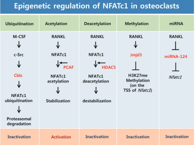
Post-translational modification such as ubiquitination and acetylation are important mechanisms of gene regula- tion.[50] Our previous studies have indicated that the sta- bility of NFATc1 proteins is controlled by ubiquitination and
acetylation during osteoclast differentiation (Fig. 2).[51]
NFATc1 proteins are downregulated by M-CSF during late stage osteoclastogenesis, and subsequently degraded through the ubiquitin-proteasome pathway in the cyto- plasm. In addition, NFATc1 interacts endogenously with c- Src, c-Cbl, and Cbl-b in the osteoclasts. Overexpression of c-Src induces down-regulation of NFATc1, and depletion of the Cbl proteins blocks NFATc1 degradation during late stage osteoclastogenesis. These results suggest that M-CSF induces NFATc1 ubiquitination and degradation via Cbl pro- teins in a Src kinase dependent method during late stage osteoclastogenesis.[51] Therefore, our previous research data suggest a negative regulatory mechanism through
ubiquitination of NFATc1, mediated by M-CSF signaling pathway, in osteoclast differentiation (Fig. 2).
RANKL increases the stability of the NFATc1 proteins thr- ough induction of NFATc1 acetylation. We demonstrated that RANKL could induce the accumulation of NFATc1, which is regulated by acetylation of the NFATc1 protein via a tran- scription-independent mechanism. NFATc1 acetylation is mediated by histone acetyltransferase (HAT) activity of PCAF through physical interaction with NFATc1.[52] Because acet- ylation of many proteins is a reversible post-translational modification, we have identified which HDAC can inhibit lysine acetylation by removal of the acetyl group from the NFATc1. Particularly, HDAC5 greatly inhibits NFATc1 acety-
Fig. 2. Epigenetic regulation of nuclear factor-activated T cells c1 (NFATc1) in osteoclasts. Macrophage colony-stimulating factor (M-CSF) induces phosphorylation of Cbl-b and c-Cbl in a Src kinase-dependent manner and subsequently induces NFATc1 ubiquitination and proteasomal degrada- tion. Receptor activator of nuclear factor-kappa B ligand (RANKL) induces acetylation of NFATc1 via p300/CBP-associated factor (PCAF), and NFATc1 acetylation induces its stability. This acetylation is reversed by histone deacetylase 5 (HDAC5). RANKL induces the Jmjd3 gene, which enhances demethylation of H3K27me3 at the transcription start site (TSS) of NFATc1 and enhances RANKL-induced NFATc1 expression. RANKL reduces miR-124 expression, which directly targets NFATc1. M-CSF, macrophage colony-stimulating factor; NFATc1, nuclear factor-activated T cells c1;
RANKL, receptor activator of nuclear factor-kappa B (NF-κB) ligand; PCAF, cyclic adenosine 3’,5’-monophosphate response element-binding pro- tein (CREB)-binding protein (CBP) and p300/CBP-associated factor; HDAC5, histone deacetylase 5; Jmjd3, jumonji domain-containing protein 3;
TSS, transcription start site.
lation, among the HDAC protein group. In addition, HDAC5 reduces the stability and transactivation of NFATc1, thereby attenuating RANKL-induced osteoclast differentiation (Fig.
2).[52] Therefore, our study proves that M-CSF and RANKL, two essential cytokines, induce ubiquitination and acetyla- tion of NFATc1 respectively, to control osteoclast differenti- ation, by modulating NFATc1 stability and activity.
miRNAs are short non-coding RNAs, approximately 22 nucleotides in length, that regulate diverse biological pro- cesses, including proliferation, apoptosis, and differentia- tion, through post-transcriptional regulation. Generally, miRNAs act as direct negative regulators of gene expres- sion that bind to the specific sequence at the 3’ untranslat- ed region (UTR) of the target mRNA.[53,54] The miRNA bio- genesis pathway has multiple steps: transcription, pri-miR- NA processing, transport to the cytoplasm, precursor miR- NA processing, strand selection, target transcription, and fate transcription.[55,56] Very little is known about the role of miRNAs during osteoclast differentiation. Recent reports have revealed the unprecedented regulation of NFATc1 by miRNA in osteoclasts. Lee et al.[57] have shown that miR- NA-124 regulates osteoclastogenesis by suppressing NFATc1 expression (Fig. 2). With emerging evidence suggesting the involvement of several miRNA in osteoclastogenesis, addi- tional studies will be required to find an miRNA that can directly regulate NFATc1 expression in osteoclasts.
Regulation of NFATc1 by Ca
2+signaling and costimulatory signaling in
osteoclasts
It has been substantially demonstrated that NFAT pro- teins are primarily regulated by calcium and calcineurin.
RANKL activates phospholipase C-γ (PLC-γ), which hydroly- ses phosphatidylinositol-4,5-bisphosphate to generate inositol-1,4,5-trisphosphate (InsP3) and diacylglycerol.
InsP3 induces the release of calcium from the endoplasmic reticulum Ca2+ stores. Calcium binds to calmodulin, which in turn activates the calmodulin-dependent phosphatase calcineurin. Dephosphorylation of serine residues in NFATc1 by activated calcineurin leads to nuclear translocation, and activation of NFATc1 proteins. Targeted deletion of PLC-γ2 in mice results in an osteopetrotic phenotype, and the os- teoclast precursor cells lacking PLC-γ2 do not sufficiently induce NFATc1 expression even in the presence of RANKL.
[58] Well known inhibitors of calcineurin, such as FK506 and cyclosporine A, strongly inhibit RANKL-induced osteo- clast differentiation by blocking NFATc1 translocation into the nucleus in vitro and in vivo. In addition, the Ca2+ chela- tor BAPTA-AM suppresses osteoclast differentiation medi- ated by RANKL through the inhibition of nuclear localiza- tion of NFATc1.[10] These results suggest that the activa- tion of calcium-calcineurin pathway plays a crucial role in the regulation of NFATc1 during osteoclastogenesis.
Several kinases such as glycogen synthase kinase 3 (GSK3), casein kinase 1 (CK1), p38, and JNK phosphorylate the vari- ous serine-rich motifs in NFAT proteins, thereby preventing translocation into the nucleus or promoting nuclear export of NFAT proteins.[59-63] A recent study has reported that the ectopic expression of the constitutively active GSK-3β (GSK3β-S9A) mutant in osteoclast precursor cells inhibits RANKL-mediated NFATc1 expression and Ca2+ oscillations.
Furthermore, Jang et al.[64] have also shown a significant defect in NFATc1 expression and nuclear localization in os- teoclast precursor cells isolated from transgenic mice ex- pressing GSK3β-S9A mutant. These findings indicate that GSK3β inactivation by RANKL is important for the expres- sion and activation of NFATc1 in osteoclasts (Fig. 1).
Since RANK receptor does not seem to directly initiate Ca2+ signaling, it has been studied for its role as an immu- noreceptor alongside other receptors such as OSCAR, trig- gering receptor expressed in myeloid cells-2 (TREM-2), paired immunoglobulin-like receptor-A (PIR-A), and signal-regu- latory protein β1 (SIRPβ1) in osteoclasts that are involved in calcium signaling in immune cells.[65] The association of immunoreceptors with immunoreceptor tyrosine-based activation motif (ITAM)-harboring adaptors has been ob- served in osteoclast precursor cells: OSCAR and PIR-A are associated with Fc receptor common γ subunit (FcRγ) and TREM-2 and SIRPβ1 are associated with DNAX-activating protein 12 (DAP12). The association of an immunoreceptor with the ITAM-harboring adapters, FcRγ and DAP12, stimu- lates calcium signaling. Although DAP12-deficient mice ex- hibit only mild osteopetrosis, FcRγ and DAP12 double knock- out mice (DAP12-/- FcRγ-/-) develop severe osteopetrosis due to defects in osteoclast differentiation.[65-67] Interestingly, the defect in osteoclast differentiation in DAP12-/- FcRγ-/- cells is fixed by the overexpression of DAP12, but not DAP12Y65F, which has a mutation in ITAM, suggesting that the ITAM- mediated signaling pathway is indispensable for osteo-
clastogenesis.[65] As expected, Ca2+ oscillation and the in- duction of NFATc1 were also suppressed in DAP12-/- FcRγ-/- cells, recovered by the overexpression of NFATc1, but not c- Fos or TRAF6.[65] These results suggest that the induction and activation of NFATc1 is regulated by calcium signaling pathway, mediated by ITAM-harboring adapters, FcRγ and DAP12, in the osteoclasts (Fig. 1).
The stimulation of ITAM-associated receptors alone, with- out RANKL, cannot induce osteoclast differentiation, alth- ough ITAM-associated receptors stimulate Ca2+-NFATc1 path- way through ITAM-harboring adaptors during osteoclasto- genesis. Therefore, ITAM-mediated signals may provide co- stimulatory signals to maximize the induction of NFATc1 for RANK.[9]
CONCLUSION
Since NFATc1 is indispensable for osteoclast differentia- tion, future studies based on currently available data about NFATc1 regulation could provide novel therapeutic strate- gies for bone diseases.
REFERENCES
1. Suda T, Takahashi N, Udagawa N, et al. Modulation of os- teoclast differentiation and function by the new members of the tumor necrosis factor receptor and ligand families.
Endocr Rev 1999;20:345-57.
2. Teitelbaum SL. Bone resorption by osteoclasts. Science 2000;289:1504-8.
3. Walsh MC, Kim N, Kadono Y, et al. Osteoimmunology: in- terplay between the immune system and bone metabo- lism. Annu Rev Immunol 2006;24:33-63.
4. Boyle WJ, Simonet WS, Lacey DL. Osteoclast differentiation and activation. Nature 2003;423:337-42.
5. Wiktor-Jedrzejczak W, Bartocci A, Ferrante AW Jr, et al. To- tal absence of colony-stimulating factor 1 in the macro- phage-deficient osteopetrotic (op/op) mouse. Proc Natl Acad Sci U S A 1990;87:4828-32.
6. Yoshida H, Hayashi S, Kunisada T, et al. The murine mutation osteopetrosis is in the coding region of the macrophage colony stimulating factor gene. Nature 1990;345:442-4.
7. Kong YY, Yoshida H, Sarosi I, et al. OPGL is a key regulator of osteoclastogenesis, lymphocyte development and lym- ph-node organogenesis. Nature 1999;397:315-23.
8. Dougall WC, Glaccum M, Charrier K, et al. RANK is essen- tial for osteoclast and lymph node development. Genes Dev 1999;13:2412-24.
9. Takayanagi H. Osteoimmunology: shared mechanisms and crosstalk between the immune and bone systems. Nat Rev Immunol 2007;7:292-304.
10. Takayanagi H, Kim S, Koga T, et al. Induction and activation of the transcription factor NFATc1 (NFAT2) integrate RANKL signaling in terminal differentiation of osteoclasts. Dev Cell 2002;3:889-901.
11. Matsumoto M, Kogawa M, Wada S, et al. Essential role of p38 mitogen-activated protein kinase in cathepsin K gene expression during osteoclastogenesis through association of NFATc1 and PU.1. J Biol Chem 2004;279:45969-79.
12. Kim K, Kim JH, Lee J, et al. Nuclear factor of activated T cells c1 induces osteoclast-associated receptor gene expression during tumor necrosis factor-related activation-induced cytokine-mediated osteoclastogenesis. J Biol Chem 2005;
280:35209-16.
13. Kim Y, Sato K, Asagiri M, et al. Contribution of nuclear fac- tor of activated T cells c1 to the transcriptional control of immunoreceptor osteoclast-associated receptor but not triggering receptor expressed by myeloid cells-2 during osteoclastogenesis. J Biol Chem 2005;280:32905-13.
14. Winslow MM, Pan M, Starbuck M, et al. Calcineurin/NFAT signaling in osteoblasts regulates bone mass. Dev Cell 2006;
10:771-82.
15. Aliprantis AO, Ueki Y, Sulyanto R, et al. NFATc1 in mice re- presses osteoprotegerin during osteoclastogenesis and dissociates systemic osteopenia from inflammation in che- rubism. J Clin Invest 2008;118:3775-89.
16. Hogan PG, Chen L, Nardone J, et al. Transcriptional regula- tion by calcium, calcineurin, and NFAT. Genes Dev 2003;
17:2205-32.
17. Graef IA, Chen F, Crabtree GR. NFAT signaling in vertebrate development. Curr Opin Genet Dev 2001;11:505-12.
18. Crabtree GR, Olson EN. NFAT signaling: choreographing the social lives of cells. Cell 2002;109 Suppl:S67-79.
19. Kiani A, Habermann I, Haase M, et al. Expression and regu- lation of NFAT (nuclear factors of activated T cells) in human CD34+ cells: down-regulation upon myeloid differentia- tion. J Leukoc Biol 2004;76:1057-65.
20. López-Rodríguez C, Aramburu J, Jin L, et al. Bridging the NFAT and NF-kappaB families: NFAT5 dimerization regu- lates cytokine gene transcription in response to osmotic
stress. Immunity 2001;15:47-58.
21. Horsley V, Pavlath GK. NFAT: ubiquitous regulator of cell differentiation and adaptation. J Cell Biol 2002;156:771-4.
22. Takayanagi H. The role of NFAT in osteoclast formation.
Ann N Y Acad Sci 2007;1116:227-37.
23. Hodge MR, Ranger AM, Charles de la Brousse F, et al. Hy- perproliferation and dysregulation of IL-4 expression in NF-ATp-deficient mice. Immunity 1996;4:397-405.
24. Xanthoudakis S, Viola JP, Shaw KT, et al. An enhanced im- mune response in mice lacking the transcription factor NFAT1. Science 1996;272:892-5.
25. Yoshida H, Nishina H, Takimoto H, et al. The transcription factor NF-ATc1 regulates lymphocyte proliferation and Th2 cytokine production. Immunity 1998;8:115-24.
26. Ranger AM, Hodge MR, Gravallese EM, et al. Delayed lym- phoid repopulation with defects in IL-4-driven responses produced by inactivation of NF-ATc. Immunity 1998;8:125- 34.
27. Johnson RS, Spiegelman BM, Papaioannou V. Pleiotropic effects of a null mutation in the c-fos proto-oncogene. Cell 1992;71:577-86.
28. Wang ZQ, Ovitt C, Grigoriadis AE, et al. Bone and haema- topoietic defects in mice lacking c-fos. Nature 1992;360:
741-5.
29. Matsuo K, Galson DL, Zhao C, et al. Nuclear factor of acti- vated T-cells (NFAT) rescues osteoclastogenesis in precur- sors lacking c-Fos. J Biol Chem 2004;279:26475-80.
30. Asagiri M, Sato K, Usami T, et al. Autoamplification of NFATc1 expression determines its essential role in bone homeo- stasis. J Exp Med 2005;202:1261-9.
31. Anderson DM, Maraskovsky E, Billingsley WL, et al. A ho- mologue of the TNF receptor and its ligand enhance T-cell growth and dendritic-cell function. Nature 1997;390:175-9.
32. Ghosh S, Karin M. Missing pieces in the NF-kappaB puzzle.
Cell 2002;109 Suppl:S81-96.
33. Hayden MS, Ghosh S. Signaling to NF-kappaB. Genes Dev 2004;18:2195-224.
34. Novack DV, Yin L, Hagen-Stapleton A, et al. The IkappaB function of NF-kappaB2 p100 controls stimulated osteo- clastogenesis. J Exp Med 2003;198:771-81.
35. Ruocco MG, Maeda S, Park JM, et al. IκB kinase (IKK)β, but not IKKα, is a critical mediator of osteoclast survival and is required for inflammation-induced bone loss. J Exp Med 2005;201:1677-87.
36. Franzoso G, Carlson L, Xing L, et al. Requirement for NF-
kappaB in osteoclast and B-cell development. Genes Dev 1997;11:3482-96.
37. Iotsova V, Caamaño J, Loy J, et al. Osteopetrosis in mice lack- ing NF-kappaB1 and NF-kappaB2. Nat Med 1997;3:1285-9.
38. Takatsuna H, Asagiri M, Kubota T, et al. Inhibition of RANKL- induced osteoclastogenesis by (-)-DHMEQ, a novel NF-ka- ppaB inhibitor, through downregulation of NFATc1. J Bone Miner Res 2005;20:653-62.
39. Koga T, Matsui Y, Asagiri M, et al. NFAT and Osterix cooper- atively regulate bone formation. Nat Med 2005;11:880-5.
40. Lee J, Kim K, Kim JH, et al. Id helix-loop-helix proteins neg- atively regulate TRANCE-mediated osteoclast differentia- tion. Blood 2006;107:2686-93.
41. Kim K, Kim JH, Lee J, et al. MafB negatively regulates RANKL- mediated osteoclast differentiation. Blood 2007;109:3253-9.
42. Kim JH, Youn BU, Kim K, et al. Lhx2 regulates bone remod- eling in mice by modulating RANKL signaling in osteoclasts.
Cell Death Differ 2014;21:1613-21.
43. Kim K, Lee J, Kim JH, et al. Protein inhibitor of activated STAT 3 modulates osteoclastogenesis by down-regulation of NFATc1 and osteoclast-associated receptor. J Immunol 2007;
178:5588-94.
44. Zhao B, Takami M, Yamada A, et al. Interferon regulatory factor-8 regulates bone metabolism by suppressing os- teoclastogenesis. Nat Med 2009;15:1066-71.
45. Miyauchi Y, Ninomiya K, Miyamoto H, et al. The Blimp1-Bcl6 axis is critical to regulate osteoclast differentiation and bone homeostasis. J Exp Med 2010;207:751-62.
46. Bird A. Perceptions of epigenetics. Nature 2007;447:396-8.
47. Yasui T, Hirose J, Aburatani H, et al. Epigenetic regulation of osteoclast differentiation. Ann N Y Acad Sci 2011;1240:
7-13.
48. Jaenisch R, Bird A. Epigenetic regulation of gene expres- sion: how the genome integrates intrinsic and environmen- tal signals. Nat Genet 2003;33 Suppl:245-54.
49. Yasui T, Hirose J, Tsutsumi S, et al. Epigenetic regulation of osteoclast differentiation: possible involvement of Jmjd3 in the histone demethylation of Nfatc1. J Bone Miner Res 2011;26:2665-71.
50. Bae SC, Lee YH. Phosphorylation, acetylation and ubiquiti- nation: the molecular basis of RUNX regulation. Gene 2006;
366:58-66.
51. Kim JH, Kim K, Jin HM, et al. Negative feedback control of osteoclast formation through ubiquitin-mediated down- regulation of NFATc1. J Biol Chem 2010;285:5224-31.
52. Kim JH, Kim K, Youn BU, et al. RANKL induces NFATc1 acet- ylation and stability via histone acetyltransferases during osteoclast differentiation. Biochem J 2011;436:253-62.
53. Ambros V. The functions of animal microRNAs. Nature 2004;
431:350-5.
54. Bartel DP. MicroRNAs: genomics, biogenesis, mechanism, and function. Cell 2004;116:281-97.
55. Chen K, Rajewsky N. The evolution of gene regulation by transcription factors and microRNAs. Nat Rev Genet 2007;
8:93-103.
56. Kapinas K, Delany AM. MicroRNA biogenesis and regula- tion of bone remodeling. Arthritis Res Ther 2011;13:220.
57. Lee Y, Kim HJ, Park CK, et al. MicroRNA-124 regulates os- teoclast differentiation. Bone 2013;56:383-9.
58. Mao D, Epple H, Uthgenannt B, et al. PLCgamma2 regu- lates osteoclastogenesis via its interaction with ITAM pro- teins and GAB2. J Clin Invest 2006;116:2869-79.
59. Beals CR, Sheridan CM, Turck CW, et al. Nuclear export of NF-ATc enhanced by glycogen synthase kinase-3. Science 1997;275:1930-4.
60. Chow CW, Rincón M, Cavanagh J, et al. Nuclear accumula- tion of NFAT4 opposed by the JNK signal transduction path- way. Science 1997;278:1638-41.
61. Okamura H, Garcia-Rodriguez C, Martinson H, et al. A con- served docking motif for CK1 binding controls the nuclear localization of NFAT1. Mol Cell Biol 2004;24:4184-95.
62. Gómez del Arco P, Martínez-Martínez S, Maldonado JL, et al. A role for the p38 MAP kinase pathway in the nuclear shuttling of NFATp. J Biol Chem 2000;275:13872-8.
63. Zhu J, Shibasaki F, Price R, et al. Intramolecular masking of nuclear import signal on NF-AT4 by casein kinase I and MEKK1. Cell 1998;93:851-61.
64. Jang HD, Shin JH, Park DR, et al. Inactivation of glycogen synthase kinase-3beta is required for osteoclast differenti- ation. J Biol Chem 2011;286:39043-50.
65. Koga T, Inui M, Inoue K, et al. Costimulatory signals medi- ated by the ITAM motif cooperate with RANKL for bone homeostasis. Nature 2004;428:758-63.
66. Kaifu T, Nakahara J, Inui M, et al. Osteopetrosis and thala- mic hypomyelinosis with synaptic degeneration in DAP12- deficient mice. J Clin Invest 2003;111:323-32.
67. Mócsai A, Humphrey MB, Van Ziffle JA, et al. The immuno- modulatory adapter proteins DAP12 and Fc receptor gam- ma-chain (FcRgamma) regulate development of function- al osteoclasts through the Syk tyrosine kinase. Proc Natl Acad Sci U S A 2004;101:6158-63.