Korean J Endocrine Surg 2013;13:157-164
Endocrine Surgery
Predictors of Lateral Lymph Node Metastasis in Papillary Thyroid Microcarcinoma: A Retrospective Review
Young Ju Jeong, Sung Hee Mun1, Jin Gu Bong, Sung Hwan Park
Departments of Surgery, 1Radiology, School of Medicine, Catholic University of Daegu, Daegu, Korea
Purpose: Cervical lymph node metastasis in patients with papillary thyroid micro- carcinoma (PTMC) is associated with an increased recurrence rate and distant metastases.
In this study, we analyzed the predictive factors of lateral metastasis in patients with PTMC.
Methods: We retrospectively reviewed the medical records of 1,030 patients with PTMC who underwent thyroidectomy. The clinicopathological characteristics and radiological findings upon ultrasonography (US) and computed tomography (CT) were then analyzed to evaluate the predictability of lateral metastasis of PTMC.
Results: The overall rate of lateral metastasis was 3.3% for the 1,030 patients with PTMC.
All patients underwent central lymph node dissection (CLND), and 119 of these patients (11.6%) underwent either prophylactic or therapeutic selective neck dissection (SND).
Among patients who underwent lateral node evaluation, 28.6% had lateral metastasis. We found that lateral metastasis was associated with larger tumor size, extrathyroidal extension, multiple tumors, bilateral tumors, CLN metastasis, and positive expression of thyroid peroxidase. Positive findings of CLN and lateral metastases on ultrasonography (US) and computed tomography (CT) were significantly associated with lateral metastasis.
Conclusion: This study demonstrated that, for patients with PTMC, larger tumor size, extrathyroidal extension, multiple tumors, bilateral tumors, CLN metastasis, positive expression of thyroid peroxidase, and positive CLN and lateral metastases identified on US and CT were significantly associated with lateral metastasis. Further studies with a large prospective study and longer follow-up are needed to clarify the predictive value of these factors.
Key Words: Papillary thyroid microcarcinoma, Cervical lymph node metastasis, Lateral metastasis, Predictive factors
Received August 30, 2013, Revised September 5, 2013, Accepted September 5, 2013 Correspondence: Young Ju Jeong Department of Surgery, College of Medicine, Catholic University of Daegu, 3056-6 Daemyoung-4-dong, Nam-gu, Daegu 705-718, Korea
Tel: +82-53-650-3006 Fax: +82-53-624-7185 E-mail: [email protected]
Copyright © 2013 Korean Association of Thyroid and Endocrine Surgeons; KATES. All Rights Reserved.
cc This is an Open Access article distributed under the terms of the Creative Commons Attribution Non-Commercial License (http://creativecommons.org/licenses/by-nc/3.0) which permits unrestricted non-commercial use, distribution, and reproduction in any medium, provided the original work is properly cited.
INTRODUCTION
Papillary thyroid carcinoma (PTC) is the most common histologic type of thyroid malignancy, and a papillary thyroid microcarcinoma (PTMC) is a PTC tumor of ≤1 cm diameter.(1) The guidelines for PTC treatment recommend near-total or total thyroidectomy, and the indications for lobectomy are restricted to small (<1 cm), low-risk, unifocal, intrathyroidal PTC in the absence of prior head and neck irradiation or radiologically or clinically involved
cervical nodal metastases.(2) In recent years, a consensus was reached on this issue: Fine-needle aspiration cytology (FNAC) is not recommend for suspicious PTMC smaller than 5 mm,(2) and there has been a trial of observation for PTMC, without immediate surgery.(3) However, the mana- gement of PTMC is continues to be debated, and the optimal extent of surgical removal of cervical lymph nodes for PTMC remains controversial. Patients with PTMC have a good prognosis and a low mortality rate, but appro- ximately 12.3% to 64.1% of such patients have cervical
lymph node metastasis,(3-5) which is associated with an increased recurrence rate and distant metastases. “Skip metastases,” defined as lateral metastases without central lymph node (CLN) involvement also rarely occur in pa- tients with PTMC.(6) Thus, to plan an optimal mana- gement for PTMC, the predictive factors of CLN metastases as well as lateral metastases should be considered.
Several studies have reported the frequency and pattern of lateral metastases from PTC,(7-9) but only a few studies have assessed the predictive factors of lateral metastasis only from PTMC and predictive value of ultrasonography (US) and computed tomography (CT) for the prediction of node metastases in PTMC. In this study, we analyzed the association of the clinicopathological characteristics and lateral metastasis in patients with PTMC.
METHODS
Our institutional review board approved this study, and this study required neither patient approval nor informed consent for the retrospective review of images and records.
We reviewed the medical records of 1,030 patients who underwent a surgery for PTMC at Daegu Catholic Univer- sity Medical Center from January 2006 to May 2011. The patients’ inclusion criterion was a histological diagnosis of PTC in a previously untreated tumor of ≤10 mm. Prior to surgery, the patients preoperatively underwent thyroid function tests, thyroid US and FNAC of primary tumors and suspicious cervical lymph nodes, and neck CT. US was performed using a 10∼12 MHz linear transducer (iU22;
Philips Medical Systems, Bothell, WA) and CT scans, using a multidetector CT (Lightspeed VCT; GE Medical systems, Milwaukee, WI).
Cervical lymph nodes were considered positive for metastasis if the US showed one of the following findings;
focal or diffuse hyperechogenicity, cystic change, micro- calcifications, cortical thickening, loss of central hilar echogenicity, or round shape (long/transverse diameter ratio <1.5).(10) On the CT scan, they were considered positive for metastasis if they showed one of the following findings; strong enhancement without hilar vessel en- hancement, heterogeneous enhancement, calcification,
cystic changes, or necrotic changes.(11) On combined US and CT (US/CT), nodal metastases were presumed to be present when the cervical nodes had been above positive findings on either US or CT.
The following clinicopathological characteristics were evaluated: age; sex; tumor size; tumor location; multi- plicity; bilaterality; tumor encapsulation; extrathyroidal extension; lymphatic invasion; vascular invasion; nodal status; associated thyroiditis; and presence of immu- nologic markers including galectin-3, thyroid peroxidase (TPO), HBME-1, and cyclin D-1.
Patients were divided into 2 groups based on the involvement of lateral cervical lymph nodes; Group 1 with initial lateral metastasis, and group 2 without initial lateral metastasis, regardless of whether the CLNs were involved.
Statistical analyses were performed using SPSS version 15.0 (SPSS Inc., Chicago, IL). The correlation between the 2 groups and their clinicopathological features were assessed using the chi-square test or Fisher’s exact test, or Student’s t-test. A P-value of less than 0.05 was considered statistically significant.
RESULTS
1) Clinicopathological characteristics of the patients 1,030 patients who underwent surgery for PTMC from January 2006 to May 2011 were enrolled. The study group consisted of 930 women and 100 men, with a mean age of 47.57±10.90 years (Table 1). The largest primary tumors ranged in size from 0.1 to 1.0 cm, with a mean of 0.52±
0.24 cm. Extrathyroidal extension by primary tumors was found in 189 (18.5%) patients and multiple and bilateral primary lesions, in 258 (25.0%) and 179 (17.4%) patients, respectively. CLN metastasis was found in 241 (23.4%) patients, and lateral metastasis, in 34 patients; the overall rate of lateral metastasis was 3.3%. Among the patients who underwent lateral node evaluation, 28.6% had lateral metastasis.
2) Surgical protocols and nodal status
Table 2 shows the extent of surgical therapy; all patients underwent thyroidectomy and among them, 875 (85.0%)
Variables Value (n=1,030) Age
Mean age (range) 47.57±10.90 years
(12∼78)
<45 years, n (%) 401 (38.9%)
≥45 years, n (%) 629 (61.1%)
Sex, n (%)
Male 100 (9.7%)
Female 930 (90.3%)
Tumor size
Mean size (range) 0.52±0.24 cm
(0.1∼1.0)
≤5 mm, n (%) 597 (58.0%)
>5 mm, n (%) 420 (40.8%)
Extrathyroidal extension, n (%) 189 (18.5%)
Multiplicity, n (%) 258 (25.0%)
Bilaterality, n (%) 179 (17.4%)
Central lymph node metastasis, n (%) 241 (23.4%) Lateral lymph node metastasis, n (%) 34 (3.3%) Number of metastatic lymph nodes
Mean (range) 2.60±3.27 (1∼31)
Central compartment 1.95±1.44 (1∼11) Lateral compartment 7.00±7.01 (1∼31) Mean number of removed lymph
nodes (range)
8.15±7.39
PTMC = papillary thyroid microcarcinoma.
Table 1. The demographics and the clinicopathological characteristics of 1,030 patients with PTMC
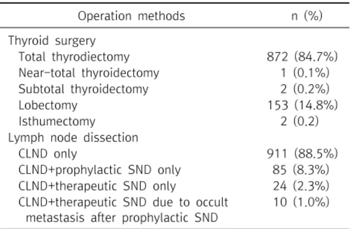
Operation methods n (%)
Thyroid surgery
Total thyrodiectomy 872 (84.7%)
Near-total thyroidectomy 1 (0.1%) Subtotal thyroidectomy 2 (0.2%)
Lobectomy 153 (14.8%)
Isthumectomy 2 (0.2)
Lymph node dissection
CLND only 911 (88.5%)
CLND+prophylactic SND only 85 (8.3%) CLND+therapeutic SND only 24 (2.3%) CLND+therapeutic SND due to occult
metastasis after prophylactic SND
10 (1.0%)
PTMC = papillary thyroid microcarcinoma; CLND = central lymph node dissection; SND = selective neck dissection.
Table 2. Extent of surgical therapy in 1,030 patients with PTMC
patients underwent total or near-total thyroidectomy and subtotal thyroidectomy, 153 (14.8%) patients underwent lobectomy, and 2 (0.2%) patients underwent isthmusec- tomy. Total or near-total thyroidectomy was performed when patients with PTMC had extrathyroidal extension or a pre- or intraoperative finding of lymph node metastasis.
Lobectomy was chosen for patients with intrathyroidal PTMC without clinical lymph node metastasis and for patients who requested a minimal necessary procedure. All patients underwent CLN dissection (CLND) for the affected side of thyroid cancer, and among them, 119 (11.6%) patients underwent either prophylactic or therapeutic selective neck dissection (SND).
Therapeutic CLND was performed for patients with biopsy-proven cervical lymphadenopathy; prophylatic CLND was performed even if a clinical involvement of the cervical lymph nodes was uncertain. Therapeutic SND (level II, III, IV, V) was performed for patients with metastatic lateral lymphadenopathy. Prophylactic SND (level III, IV) was performed if metastasis was suspected from the preoperative imaging studies, but US-guided
FNAC of sonographically suspicious lymph nodes could not prove lateral metastases. 188 patients revealed suspicious metastatic-positive findings of lateral nodes on preope- rative neck US or CT, and all of these patients underwent preoperative FNAC of suspicious lateral node. 24 patients underwent therapeutic SND for management of biopsy- proven lateral metastases; 95 patients underwent pro- phylactic SND and among them, 10 (10.5%) patients re- vealed lateral metastases by intraoperative frozen-section biopsy. Of the 164 patients with metastatic-negative lateral node on FNAC, 69 patients who revealed less suspicious findings of metastatic lateral node on combined US and CT did not underwent further evaluation for lateral metastases.
Table 3 shows the status of lymph node metastases according to the extent of lymph node dissection. Of the 1030 patients who underwent CLND, 241 (23.4%) had metastases to CLN. The overall incidence of skip metastasis was 1.3%. Among the 119 patients who underwent evaluation of the lateral nodes, lateral metastases were found in 34 (28.6%) patients, and the incidence of skip metastasis was 10.9% (13 of 119).
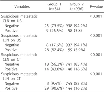
3) Diagnostic value of US and CT for detection of metastatic cervical lymph nodes
Suspicious metastatic-positive findings of CLN and LLN seen on US as well as CT were significantly associated with pathological LLN metastasis (P<0.001 for all) (Table 4).
Status of metastatic lymph node n (%) Central (-) / lateral (-) 776 (75.3%) Central (+) / lateral (-) 220 (21.4%) Central (-) / lateral (+) 13 (1.3%) Central (+) / lateral (+) 21 (2.0%) (+) = lymph node metastasis present; (-) = lymph node metastasis absent.
Table 3. Status of lymph node metastases according to lymph node dissection
Variables Group 1 (n=34)
Group 2
(n=996) P-value Suspicious metastatic
CLN on US
<0.001
Negative 25 (73.5%) 938 (94.2%) Positive 9 (26.5%) 58 (5.8) Suspicious metastatic
LLN on US
<0.001
Negative 6 (17.6%) 937 (94.1%) Positive 28 (82.4%) 59 (5.9%) Suspicious metastatic
CLN on CT
<0.001
Negative 18 (56.3%) 741 (83.4%) Positive 14 (43.8%) 148 (16.6%) Suspicious metastatic
LLN on CT
<0.001
Negative 3 (9.4%) 745 (83.8%) Positive 29 (90.6%) 144 (16.2%)
PTMC = papillary thyroid microcarcinoma; CLN = central lymph node; LLN = lateral lymph node; US = ultrasound; CT
= computed tomography.
Table 4. Association between radiologic findings and lateral cervical lymph node metastasis in pa- tients with PTMC
Imaging method Diagnostic values
Sensitivity (%) Specificity (%) PPV (%) NPV (%) Accuracy (%)
CLN levels US 26.5 94.2 13.4 97.4 91.9
CT 43.8 83.4 8.6 97.6 81.9
US/CT 21.9 85.4 5.6 97.1 92.8
LLN levels US 82.4 94.1 32.2 99.4 93.7
CT 90.6 83.8 16.8 99.6 84.0
US/CT 78.1 95.1 36.2 99.2 94.5
PTMC = papillary thyroid microcarcinoma; US = ultrasound; CT = computed tomography; US/CT = combined US and CT; PPV = positive predictive value; NPV = negative predictive value; CLN = central lymph node; LLN = lateral lymph node.
Table 5. Diagnostic accuracies of US and CT for the detection of metastatic cervical lymph nodes in patients with PTMC Table 5 shows the sensitivity; specificity; positive predic-
tive value (PPV); negative predictive value (NPV); and accuracy of US, CT, and US/CT for the detection of meta- static cervical nodes in patients with positive CLN and LLN metastases as observed on US and CT. US, CT, and US/CT had diagnostic accuracies of 91.9%, 81.9%, and 92.8% for CLN metastasis, and 93.7%, 84.0%, and 94.5% for LLN metastasis, respectively. For CLN metastasis, the false- positive rates of US, CT, and US/CT were 5.8%, 5.9%, and 4.5%, and, for LLN metastasis, the false-positive were 5.7%, 15.6%, and 4.8%, respectively.
4) Association of LLN metastasis with the clinicopa- thological features
Group 1 consisted of 34 patients who had lateral meta- stases, and group 2 consisted of 996 patients who did not have LLN metastasis. The mean tumor size of group 1 was significantly larger than that of group 2 (P<0.001), and the proportion of tumors larger than 5 mm in diameter was also higher in group 1 (P<0.001) (Table 6). Similarly, extra- thyroidal extension, multiplicity, and bilaterality of the thyroid tumors were also more common in group 1 (P < 0.001, P=0.027 and P=0.019, respectively). Furthermore,
LLN metastasis was associated with the mean number of metastatic lymph nodes, mean number of removed lymph nodes, and the presence of CLN metastasis (P<0.001 for all). However, in terms of the metastatic ratio (the ratio of the number of metastatic lymph nodes to the number of total lymph nodes acquired), the 2 groups did not show any statistically significant difference (P=0.928). Among the immunologic markers, only TPO showed a significantly higher positive expression in group 1 than in group 2 (P=
0.009).
Variables
With lateral metastasis
(n=34)
Without lateral metastasis
(n=996)
P-value
Age
Mean age (years) 47.82±13.45 47.56±10.82 0.890
<45 years, n (%) 14 (41.2%) 387 (38.9%) 0.785
≥45 years, n (%) 20 (58.8%) 609 (61.1%)
Sex, n (%) 0.317
Male 5 (14.7%) 95 (9.5%)
Female 29 (85.3%) 901 (90.5%)
Tumor size
Mean size (cm) 0.72±0.23 0.51±0.24 <0.001
≤5 mm, n (%) 8 (23.5%) 589 (59.9%) <0.001
>5 mm, n (%) 26 (76.5%) 394 (40.1%)
Main tumor location 0.106
Upper lobe, n (%) 14 (42.4%) 260 (26.6%)
Middle lobe, n (%) 13 (39.4%) 470 (48.0%)
Lower lobe, n (%) 3 (9.1%) 147 (15.0%)
Isthmus, n (%) 0 (0.0%) 50 (5.1%)
Multifocal, n (%) 3 (9.1%) 52 (5.3%)
Extrathyroidal extension, n (%) 15 (44.1%) 174 (17.6%) <0.001
Multiplicity, n (%) 14 (41.2%) 244 (24.5%) 0.027
Bilaterality, n (%) 11 (32.4%) 168 (16.9%) 0.019
Mean number of metastatic lymph nodes, n 7.00±7.01 1.95±1.44 <0.001
Mean number of Removed lymph nodes, n 34.47±13.86 7.25±5.07 <0.001
Presence of central lymph node metastasis, n (%) 21 (61.8%) 220 (22.1%) <0.001
Associated Hashimoto’s thyroiditis, n (%) 6 (17.6%) 189 (19.0%) 0.846
Positive expression of immunologic marker, n (%)
Galectin-3 29 (96.7%) 843 (96.2%) 0.902
TPO 2 (6.7%) 10 (1.1%) 0.009
HBME-1 26 (89.7%) 724 (90.3%) 0.912
Cyclin D-1 8 (26.7%) 274 (31.5%) 0.578
PTMC = papillary thyroid microcarcinoma; Group 1 = lateral cervical lymph node metastasis present on lymph node dissection;
group 2 = lateral cervical lymph node metastasis absent on lymph node dissection; TPO = thyroid peroxidase.
Table 6. Association between the clinicopathological factors and lateral cervical lymph node metastasis in patients with PTMC
DISCUSSION
PTMC has a good prognosis with adequate surgery and adjuvant treatment, and the recurrence rates of PTMC are reported to be 1.7∼8%.(3,12,13) Traditionally, cervical lymph node metastases have been thought to have no impact on prognosis of PTC and a variety of prognostic algorithms such as AGES,(14) AMES,(15) and MACIS(16) are used to define prognosis. However, several studies have demonstrated that the presence of cervical lymph node metastases is related to the increased locoregional re- currence rate and distant metastases.(4,5,12) It is now accepted that the presence of LLN metastases is an inde- pendent risk factor for locoregional recurrence, and therefore, it is usually treated aggressively with surgery and
with adjuvant radioactive iodine.
Cervical lymph node metastasis occurs in approxi- materly 12.3∼64.1% of patients with PTMC,(3-5) whereas LLN metastasis occurs in approximately 3.7∼30.8% of such patients.(7,17) In agreement with previous studies, the incidence CLN and LLN metastases in our patients with PTMC was 23.4% and 3.3% respectively.
Previous studies showed that PTMC tumors of >5 mm in diameter were more significantly associated with poor prognostic factors than smaller tumors.(18-20) Consistent with previous studies, our results showed that the larger the tumor size of the PTMC, especially if it is larger than 5 mm, the stronger association with LLN metastases. These findings suggest a need for the evaluation of LLN meta- stases in patients with PTMC for tumors larger than 5 mm,
even if there is no evidence of clinical node metastasis.
The appropriate management of cervical lymph nodes in PTMC patients is still being debated. The actual guidelines recommend systematic central lymphadenec- tomy regardless of clinical nodal metastasis because pre- vious studies suggest that this could reduce locoregional recurrence rates. Hisham et al.(21) showed that CLND during primary thyroidectomy is safe and does not lead to a higher morbidity rate when compared with thyroidec- tomy alone. Regardless of a preoperative diagnosis of CLN metastasis, we routinely remove the CLN in such cases.
Such dissection can be performed without extension of the wound, and re-operation would be difficult when CLN metastasis recurrence occurs. This study demonstrated that the presence of CLN metastasis is significantly correlated with LLN metastasis and that the sensitivity of US and CT for the detection of metastatic lymph node is low as the false negative rate for preoperative imaging is high. Our results suggest that routine CLND with the frozen biopsy may be considered to identify patients who may need or suitable for SND.
Recent study of a literature review reported that lateral neck dissection should be performed only as a therapeutic intervention for known disease, however adequate mana- gement for suspicious LLN metastasis and optimal extent of neck dissection remain controversial.(22) Lim et al.(9) performed prophylactic SND after ipsilateral CLN meta- stasis was confirmed by intraoperative frozen biopsy, and showed 55% of the incidence of occult LLN metastasis in patients with PTC. In this study, the incidence of occult LLN metastasis which is sonographically suspicious but not proved by preoperative FNAC was 10.5%. These results suggest that occult LLN metastases cannot be removed without prophylactic SND in these patients.
Previous studies indicated that for patients with PTC, the relationship between LLN involvement and CLN metastasis is predictable.(5,8,23-25) The first echelon lymph nodes are in the ipsilateral tracheoesophageal groove, with subsequent drainage to the jugular chain(26); this suggests that LLN metastasis may be accompanied by CLN me- tastasis from PTMC. We found that patients with LLN metastasis from PTMC were also likely to have CLN
metastasis (61.8%), but were unlikely to have lateral skip metastasis (8.7%); therefore, this finding advocates for routine CLND for patients with PTMC.
There are few studies of the predictive factors of LLN metastasis in patients with PTMC, although some studies have investigated the predicting factors that influence LLN metastasis in PTC(7,8,25) and several studies have shown the risk factors of cervical lymph node metastasis of PTMC.(4,5,19) Gülben et al.(4) reported that in PTMC lymph node metastasis is associated with multifocality and capsular invasion, and Chow et al.(19) reported that a tumor size larger than 5 mm and multifocal disease are related to CLN metastasis in patients with PTMC. Kwak et al.(17) reported an association between the presence of CLN metastases and US features of PTMC such as being adjacent to the capsule, presence of calcifications, upper pole location. Similarly, we found that tumor size larger than 5 mm, extrathyroidal extension, tumor multifocality, bilaterality, presence of CLN metastasis, and positive LLN metastasis on US and CT were significantly associated with LLN metastasis.
Although US and CT examination can be useful during the preoperative evaluation of cervical lymph node meta- stasis, there are only a few reports on the diagnostic accuracy of these imaging modalities for the detection of cervical lymph node metastasis from PTC. To our know- ledge, there are no published reports that address the diagnostic value of US and CT for the evaluation of lymph node metastasis in PTMC. Previous studies(27,28) reported that for US, the sensitivity, specificity, accuracy, PPV and NPV at the lateral neck level of patients with PTC were 64
∼65%, 82∼92%, 71∼82%, 83∼86%, and 59∼82%, respectively, while these diagnostic values for CT were 74
∼78%, 78∼95%, 78∼87%, 86∼89%, and 68∼86%. Kim et al.(27) reported a slight difference between the diagnostic accuracies of US and CT and that combined US and CT is superior to US alone in the detection of lymph node metastasis from PTC. Ahn et al.(28) reported that the sensitivity of CT was significantly higher than that of US for whole neck levels. We found that the sensitivity, specificity and accuracy for US and CT at the lateral neck level of patients with PTMC were comparable to that of the
previous studies.(27,28), but PPV and NPV showed varying value. In our study, the accuracy of combined US and CT was superior to US or CT alone, but there was no stati- stically significant difference. These results suggest that preoperative US is necessary for evaluation of LLN meta- stasis, and CT combined with US may be needed to evaluate LLN status preoperatively.
Our study has several limitations. First, the evaluation of LLN was done for only 119 (11.5%) of 1,030 patients, and the proportion of patients with LLN metastasis who underwent prophylactic or therapeutic SND was only 28.6%. The overall rate of LLN metastasis in this study was very low (3.3%), and this may decrease the significance of the statistical analysis. Second, patients without clinical evidence of LLN metastasis by preoperative US or CT did not undergo LLN evaluation, and the LLN pathology status was available for only 11.5% of all patients. Considering that subclinical node metastases can be found in patients with PTMC, LLN metastases can be underestimated, although the false-negative rate of US and CT is very low (0.6% and 0.3%, respectively) for detection of LLN meta- stasis. Third, we could not analyze the impact of LLN metastasis on long-term prognosis, because the follow-up period ranged from 6 months to 5 years, and this period is too short to deduce clear results of the follow-up for PTMC patients. Despite these limitations, this is one of a few studies that determined the predictive factors for LLN metastasis in patients with PTMC, and the presence of LLN metastasis is important factor for determination of the optimal surgical approach. To our knowledge, the present study is the first to show the diagnostic value of US and CT for the evaluation of lymph node metastasis in patients with PTMC.
CONCLUSION
For patients with PTMC, a tumor larger than 5 mm in diameter, extrathyroidal extension, multiple tumors, bila- teral tumors, the presence of central lymph node meta- stasis, positive expression of thyroid peroxidase, and positive CLN and LLN metastases findings on US and CT were associated with LLN metastasis. It seems that meti-
culous evaluation of LLN metastases may be considered for patients with these prognostic factors, although preope- rative US and US-guided FNA of sonographically suspi- cious lymph nodes failed to prove LLN metastases. Further studies with large prospective study and longer follow-up are needed to clarify the predictive value of these factors.
REFERENCES
1. Hedinger C, Williams ED, Sobin LH. Histological typing of thy- roid tumours. Series: WhoInternational Hitological Classifica- tion of Tumors. 2nd ed. Berlin: Springer; 1988.
2. American Thyroid Association (ATA) Guidelines Taskforce on Thyroid Nodules and Differentiated Thyroid Cancer, Cooper DS, Doherty GM, Haugen BR, Kloos RT, Lee SL, et al. Revised American Thyroid Association management guidelines for pa- tients with thyroid nodules and differentiated thyroid cancer.
Thyroid 2009;19:1167-214.
3. Ito Y, Miyauchi A. Therapeutic strategies for papillary micro- carcinoma of the thyroid. Current Cancer Therapy Reviews 2005;1:19-25.
4. Gülben K, Berberoğlu U, Celen O, Mersin HH. Incidental papil- lary microcarcinoma of the thyroid--factors affecting lymph node metastasis. Langenbecks Arch Surg 2008;393:25-9.
5. Wada N, Duh QY, Sugino K, Iwasaki H, Kameyama K, Mimura T, et al. Lymph node metastasis from 259 papillary thyroid micro- carcinomas: frequency, pattern of occurrence and recurrence, and optimal strategy for neck dissection. Ann Surg 2003;
237:399-407.
6. Machens A, Holzhausen HJ, Dralle H. Skip metastases in thyroid cancer leaping the central lymph node compartment. Arch Surg 2004;139:43-5.
7. Chung YS, Kim JY, Bae JS, Song BJ, Kim JS, Jeon HM, et al. Lateral lymph node metastasis in papillary thyroid carcinoma: results of therapeutic lymph node dissection. Thyroid 2009;19:241-6.
8. Roh JL, Kim JM, Park CI. Lateral cervical lymph node metastases from papillary thyroid carcinoma: pattern of nodal metastases and optimal strategy for neck dissection. Ann Surg Oncol 2008;15:1177-82.
9. Lim YS, Lee JC, Lee YS, Lee BJ, Wang SG, Son SM, et al. Lateral cervical lymph node metastases from papillary thyroid carcino- ma: predictive factors of nodal metastasis. Surgery 2011;150:
116-21.
10. Rosário PW, de Faria S, Bicalho L, Alves MF, Borges MA, Purisch S, et al. Ultrasonographic differentiation between metastatic and benign lymph nodes in patients with papillary thyroid carcinoma. J Ultrasound Med 2005;24:1385-9.
11. Som PM, Brandwein M, Lidov M, Lawson W, Biller HF. The var- ied presentations of papillary thyroid carcinoma cervical nodal disease: CT and MR findings. AJNR Am J Neuroradiol 1994;
15:1123-8.
12. Hay ID, Hutchinson ME, Gonzalez-Losada T, McIver B, Reinalda ME, Grant CS, et al. Papillary thyroid microcarcinoma: a study of
900 cases observed in a 60-year period. Surgery 2008;144:
980-7.
13. Yamashita H, Noguchi S, Murakami N, Toda M, Uchino S, Watanabe S, et al. Extracapsular invasion of lymph node metastasis. A good indicator of disease recurrence and poor prognosis in patients with thyroid microcarcinoma. Cancer 1999;86:842-9.
14. Hay ID, Grant CS, Taylor WF, McConahey WM. Ipsilateral lo- bectomy versus bilateral lobar resection in papillary thyroid carcinoma: a retrospective analysis of surgical outcome using a novel prognostic scoring system. Surgery 1987;102:1088-95.
15. Cady B, Rossi R. An expanded view of risk-group definition in differentiated thyroid carcinoma. Surgery 1988;104:947-53.
16. Hay ID, Bergstralh EJ, Goellner JR, Ebersold JR, Grant CS.
Predicting outcome in papillary thyroid carcinoma: develop- ment of a reliable prognostic scoring system in a cohort of 1779 patients surgically treated at one institution during 1940 through 1989. Surgery 1993;114:1050-7.
17. Kwak JY, Kim EK, Kim MJ, Son EJ, Chung WY, Park CS, et al.
Papillary microcarcinoma of the thyroid: predicting factors of lateral neck node metastasis. Ann Surg Oncol 2009;16:1348-55.
18. Lee J, Rhee Y, Lee S, Ahn CW, Cha BS, Kim KR, et al. Frequent, aggressive behaviors of thyroid microcarcinomas in Korean patients. Endocr J 2006;53:627-32.
19. Chow SM, Law SC, Chan JK, Au SK, Yau S, Lau WH. Papillary mi- crocarcinoma of the thyroid-Prognostic significance of lymph node metastasis and multifocality. Cancer 2003;98:31-40.
20. Machens A, Holzhausen HJ, Dralle H. The prognostic value of primary tumor size in papillary and follicular thyroid
carcinoma. Cancer 2005;103:2269-73.
21. Tisell LE, Nilsson B, Mölne J, Hansson G, Fjälling M, Jansson S, et al. Improved survival of patients with papillary thyroid cancer after surgical microdissection. World J Surg 1996;20:854-9.
22. Ferris R, Goldenberg D, Haymart MR, Shaha AR, Seth S, Sosa JA, et al. American Thyroid Association Consensus Review of the Anatomy, Terminology and Rationale for Lateral Neck Dissec- tion in Differentiated Thyroid Cancer. Thyroid 2012 [Epub ahead of print].
23. Gimm O, Rath FW, Dralle H. Pattern of lymph node metastases in papillary thyroid carcinoma. Br J Surg 1998;85:252-4.
24. Sivanandan R, Soo KC. Pattern of cervical lymph node meta- stases from papillary carcinoma of the thyroid. Br J Surg 2001;88:1241-4.
25. Kupferman ME, Patterson M, Mandel SJ, LiVolsi V, Weber RS.
Patterns of lateral neck metastasis in papillary thyroid carcinoma. Arch Otolaryngol Head Neck Surg 2004;130:
857-60.
26. Shaha AR. Management of the neck in thyroid cancer.
Otolaryngol Clin North Am 1998;31:823-31.
27. Kim E, Park JS, Son KR, Kim JH, Jeon SJ, Na DG. Preoperative di- agnosis of cervical metastatic lymph nodes in papillary thyroid carcinoma: comparison of ultrasound, computed tomography, and combined ultrasound with computed tomography. Thyroid 2008;18:411-8.
28. Ahn JE, Lee JH, Yi JS, Shong YK, Hong SJ, Lee DH, et al.
Diagnostic accuracy of CT and ultrasonography for evaluating metastatic cervical lymph nodes in patients with thyroid cancer. World J Surg 2008;32:1552-8.