http://dx.doi.org/10.3988/jcn.2014.10.2.101 J Clin Neurol 2014;10(2):101-107
Introduction
Hemispherectomy has been recognized as an effective treat- ment modality for refractory epilepsy in children with hemi- spheric epileptogenic lesions. In 1938, McKenzie1 introduced anatomical hemispherectomy for seizure control in infantile hemiplegia. Krynauw2 subsequently reported successful seizure
control in 10 of 12 cases in 1950. Anatomical hemispherecto- my has subsequently become a widely used surgical approach for intractable epilepsy secondary to hemispheric syndrome such as in Rasmussen encephalitis, Sturge-Weber syndrome, hemimegalencephaly, and extensive hemispheric infarct.3-5 Al- though this surgical technique has been associated with seizure- free rates of 70–80% in previous reports,3,4,6 early and delayed surgical complications including hydrocephalus and superficial cerebral hemosiderosis have also occurred following this pro- cedure, with associated high mortality rates.
To avoid these complications, functional hemispherectomy was introduced in 1983 by Rasmussen6 based on a combina-
Long-Term Outcomes of Hemispheric Disconnection in Pediatric Patients with Intractable Epilepsy
Yun-Jeong Lee,a* Eun-Hee Kim,a* Mi-Sun Yum,a Jung Kyo Lee,b Seokho Hong,b Tae-Sung Koa
aDivision of Pediatric Neurology, Department of Pediatrics, Asan Medical Center Children’s Hospital, Seoul, Korea
bDepartment of Neurosurgery, Asan Medical Center, University of Ulsan College of Medicine, Seoul, Korea
Received September 13, 2013 Revised October 18, 2013 Accepted October 22, 2013 Correspondence Tae-Sung Ko, MD Department of Pediatrics, Asan Medical Center, University of Ulsan College of Medicine, 88 Olympic-ro 43-gil, Songpa-gu, Seoul 138-736, Korea Tel +82-2-3010-3390 Fax +82-2-473-3725 E-mail [email protected]
*These two authors contributed equally in this work.
Background and PurposezzHemispherectomy reportedly produces remarkable results in terms of seizure outcome and quality of life for medically intractable hemispheric epilepsy in children. We reviewed the neuroradiologic findings, pathologic findings, epilepsy characteris- tics, and clinical long-term outcomes in pediatric patients following a hemispheric disconnection.
MethodszzWe retrospectively studied 12 children (8 males) who underwent a hemispherecto- my at Asan Medical Center between 1997 and 2005. Clinical, EEG, neuroradiological, and sur- gical data were collected. Long-term outcomes for seizure, motor functions, and cognitive functions were evaluated at a mean follow-up of 12.7 years (range, 7.6–16.2 years) after surgery.
ResultszzThe mean age at epilepsy onset was 3.0 years (range, 0–7.6 years). The following epilepsy syndromes were identified in our cohort: focal symptomatic epilepsy (n=8), West syn- drome (n=3), and Rasmussen’s syndrome (n=1). Postoperative histopathology of our study pa- tients revealed malformation of cortical development (n=7), encephalomalacia as a sequela of infarction or trauma (n=3), Sturge-Weber syndrome (n=1), and Rasmussen’s encephalitis (n=1).
The mean age at surgery was 6.5 years (range, 0.8–12.3 years). Anatomical or functional hemi- spherectomy was performed in 8 patients, and hemispherotomy was performed in 4 patients.
Eight of our 12 children (66.7%) were seizure-free, but 3 patients with perioperative complica- tions showed persistent seizure. Although all patients had preoperative hemiparesis and devel- opmental delay, none had additional motor or cognitive deficits after surgery, and most achieved independent walking and improvement in daily activities.
ConclusionszzThe long-term clinical outcomes of hemispherectomy in children with intracta- ble hemispheric epilepsy are good when careful patient selection and skilled surgical approach-
es are applied. J Clin Neurol 2014;10(2):101-107
Key Wordszz seizure, hemispherectomy, hemispherotomy, psychomotor outcomes.
Open Access
cc This is an Open Access article distributed under the terms of the Cre- ative Commons Attribution Non-Commercial License (http://creative- commons.org/licenses/by-nc/3.0) which permits unrestricted non-com- mercial use, distribution, and reproduction in any medium, provided the ori- ginal work is properly cited.
tion of the partial anatomic excision and disconnection of the remaining lobe. Furthermore, hemispherotomy incorporating partial cortical removal has reduced adverse events while still producing excellent results.7-10 These technological improve- ments have widened the spectrum of congenital substrates of intractable epilepsy in childhood and highlighted the issue of seizure and cognitive outcomes in pediatric candidates.
The present study examined long-term seizure rates and functional outcomes in a series of pediatric patients with hemi- spheric disconnections who were treated at a single tertiary center in Korea. Detailed clinical characteristics and surgical techniques were also reviewed to evaluate outcome predictors.
Methods
We retrospectively reviewed the medical records of patients aged 18 years or younger who underwent a hemispherectomy or hemispherotomy between 1997 and 2005 at the Asan Medi- cal Center. We collected demographic, clinical, imaging, and EEG data. All patients were confirmed as having intractable epilepsy secondary to hemispheric pathology by both a pediat- ric neurologist and a neurosurgeon. Presurgical evaluations in- cluded the family and personal histories, neurologic and neu- ropsychiatric examinations, 24-hour video EEG monitoring, and brain magnetic resonance imaging (MRI). Wada tests were performed in selected cases. We also collected data on the age at surgery, etiology of seizure, site of surgery, type of surgery, postoperative complications, postsurgical EEG, and brain MRI.
The final etiology of epilepsy was determined by preopera- tive brain MRI and postoperative histopathology. The postop- erative seizure status was determined at the last follow-up or by telephone interview in cases lost to follow-up. Engel’s classifi- cation was used to assess postsurgical seizure outcome. A neu- rological examination was performed by a pediatric neurolo- gist at the last follow-up in order to quantify the functional outcome. The Korean version of the Wechsler Intelligence Scale for Children was used to evaluate cognitive outcome.
Results
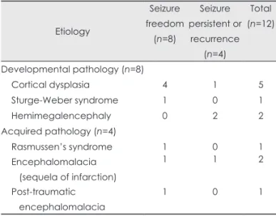
Twelve pediatric epilepsy cases (8 males) treated at our hospi- tal from 1997 to 2005 were reviewed. Their epilepsy onset age varied between 3 days and 7.6 years (mean, 3.0 years). The mean duration of seizure prior to surgery was 4.9 years (range, 0.6–11.2 years), and the mean age at surgery was 6.5 years (range, 0.8–12.3 years). All of the patients had symptomatic epilepsy with definite hemispheric etiologies: focal symptom- atic epilepsy was described in 8 patients, West syndrome in 3 patients, and Rasmussen’s syndrome in 1 patient following The International League Against Epilepsy classification (Table 1).
Eight patients had a developmental pathology: five with focal cortical dysplasia, two with hemimegalencephaly, and one with Sturge-Weber syndrome. Four patients had an acquired pathol- ogy: two with encephalomalacia as a sequela of infarction, one with posttraumatic encephalomalacia as the result of a traffic accident, and one with Rasmussen’s syndrome.
Three of our 12 patients had preoperative contralateral-side structural abnormalities on MRI that were associated with their diffuse pathologies. One patient showed cortical dysplasia as a sequela of congenital infection, one patient had posttraumatic encephalomalacia, and one had hemimegalencephaly showing contralateral structural lesions on brain MRI, but no contralat- eral EEG abnormalities. Preoperative interictal epileptiform abnormalities were identified in all 12 patients, but the affected hemisphere could be confirmed by ictal EEG in only 5 patients.
Contralateral interictal discharges were also reported in three patients.
Surgical procedures and postoperative complications
Our study cohort received six functional hemispherectomies, two anatomical hemispherectomies, three peri-insular hemi- spherotomies, and one vertical parasagittal hemispherotomy.
Five of these patients experienced early postoperative compli- cations (<7 days after surgery). One child presented with acute brain edema, and the associated increased intracranial pressure was successfully treated using barbiturate coma therapy and osmotic diuresis. Two of our patients experienced aseptic men- ingitis, which improved by 2 weeks after surgery. Delayed postoperative complications (with an onset at >7 days postsur- gery) including hydrocephalus or subdural hygroma requiring ventriculoperitoneal or subduroperitoneal shunts occurred in five patients. One of these cases developed a shunt-related brain abscess at 7 years postsurgery.
Table 1. Seizure outcomes according to etiology in the epilepsy cases of this study
Etiology
Seizure freedom (n=8)
Seizure persistent or
recurrence (n=4)
Total (n=12)
Developmental pathology (n=8)
Cortical dysplasia 4 1 5
Sturge-Weber syndrome 1 0 1
Hemimegalencephaly 0 2 2
Acquired pathology (n=4)
Rasmussen’s syndrome 1 0 1
Encephalomalacia (sequela of infarction)
1 1 2
Post-traumatic encephalomalacia
1 0 1
Seizure outcomes
During a long-term mean follow-up of 12.7 years (range, 7.6–
16.2 years), eight of the 12 patients in our study cohort (66.7%) remained seizure-free (Engel class I) and one patient showed a
>90% reduction in seizure frequency. Five patients (41.7%) were seizure-free after the surgery (Engel class Ia), with four of these cases achieving seizure freedom without the use of any antiepileptic drug (Table 2). The four remaining patients in our cohort (33.3%) continued to show persistent seizures or experi- ence seizure recurrences (Engel class IIa or IIIa) (Table 3). Eti- ological seizure freedom occurred in four of our five patients with cortical dysplasia but in neither of the two patients with hemimegalencephaly.
Motor and cognitive outcomes
All of our patients except for one infant with generalized hypo- tonia exhibited pre-existing hemiparesis at the preoperative evaluation. Additionally, one of our patients had preoperative facial palsy and another exhibited limited extraocular move- ment before surgery. Despite a transient deterioration of hemi- paresis during their early postoperative course, 11 patients were able to ambulate with or without an assistive device after sur- gery and no other neurological deterioration was reported at the last follow-up in all of these cases. One patient (patient 11) (Table 3) who underwent a vertical parasagittal hemispheroto- my suffered postoperative intraventricular hemorrhage and hy- drocephalus, and remained with spastic quadriplegia. However, the overall developmental severity category was unchanged following surgery in all of the patients in the current cohort (Table 2 and 3).
Discussion
Hemispheric disconnections have been reported to be efficient intervention approaches for epileptic seizures. These proce- dures are performed in children also to prevent additional cog- nitive injury to an otherwise healthy contralateral brain. How- ever, defining the effect of hemispheric disconnections in children remains a challenge since there have been no random- ized controlled trials of this method and the surgical candidates include a broad variety of epilepsy syndromes that arise at dif- ferent ages.
To improve our understanding of the impact of hemispheric disconnections in children, we carefully reviewed the long- term outcomes of 12 pediatric epilepsy cases that underwent hemispheric disconnection at our hospital. In this case series, 75% of the children who underwent hemispheric disconnection became seizure-free or showed a >90% reduction in seizure frequency, which is consistent with the success rates reported for previously reported series of this type.11-15 Although the eti-
ologies, epilepsy syndromes, spectra of brain imaging abnor- malities, and demographic data have differed among previous studies, hemispherectomy has been commonly reported as an effective treatment for intractable epilepsy. Recently, 136 of 186 hemispherectomized patients (73%) were reported to have achieved either seizure freedom or major reductions in seizure frequency.13 In another study of 92 pediatric epilepsy patients, 78 of the cases (85%) were reported to be seizure-free at their last follow-up.15
Some previous studies have found that seizure outcomes dif- fer significantly with the underlying etiology, and that they are significantly worse in epilepsy patients with a developmental pathology than in those with an acquired pathology.7,11,16,17 In contrast, another study found no correlation between seizure outcome and etiology.12 In our current pediatric series we found that seizure outcomes did not differ significantly between cases with developmental (5/8, 62.5%) and acquired (3/4, 75%) (Ta- ble 1) pathologies. However, neither of the two patients in our cohort with hemimegalencephaly achieved seizure freedom, whereas four of the five patients with cortical dysplasia achieved a seizure-free state. It has been found previously that the post- surgical seizure outcome and neurocognitive function were worse in patients with hemimegalencephaly than in those with other types of pathology.5 A previous study found that although the rate of seizure freedom was 82.5% in acquired-pathology patients at a 3.4-year median follow-up, only one of the five patients in that series with hemimegalencephaly became sei- zure-free.11 Similarly, the seizure-free rate in another 92 pa- tients was worse in those with hemimegalencephaly (60%) than in patients with cortical dysplasia (80%).15
While the appropriate indication of hemispherectomy is ex- pected to be a presentation with unilateral interictal and ictal EEG abnormalities, and structural brain lesions, some authors have argued that there is no relationship between the presence of contralateral EEG and brain MRI abnormalities and the sei- zure outcome.12,18,19 In our current series, children with interic- tal epileptiform discharges in a contralateral lesion (patients 1 and 4) (Table 2) also achieved seizure freedom, in accordance with previous findings. Two of three patients in our present co- hort with a preexisting contralateral brain MRI lesion–which was considered to be a nonepileptogenic focus (periventricular hyperintense signal or encephalomalacia)–became seizure-free after hemispheric disconnection. Because of the variable brain MRI lesions in such cases, some of which have uncertain clini- cal significance due to the dependence on the interpretation of the radiologist, various seizure outcomes for such lesions have been noted in previous studies.12,20 Thus, the decision to pro- ceed to hemisphere disconnection must be individualized based on various clinical features including the side of hemi- paresis, semiology, lateralization findings on EEG, and the risk
Table 2. Seizure-free patients in this study after hemispheric disconnection: preoperative clinical characteristics and psychomotor outcomes PatientEtiology/ epileptic syndrome
Epilepsy onset (years)
Age at surgery (years)
Surgery typeMotor outcome (pre→post)Development (pre→post)
Contralat. epileptiform discharge
Contralat. MRI abnormality
Postoperative complications
Seizure outcome (Engel class)
Antiepileptic drugs (pre→post) 1CD/SFE0.55.4Lt. PIHRt. hemiparesis (grade IV) →no change
GDD→NDYesNoNoIaPHT, VGB, VPA→none 2RS/SFE5.712.3Rt. FHLt. hemiparesis (grade III) →no change
FSIQ=60 →FSIQ=60NoNoFacial palsyIaVGB, CBZ, VPA→none 3EM after infarction/WS (Fig. 1A)
0.81.2Lt. AH (Fig. 1E)Rt. hemiparesis (grade IV) →no change
GDD→GDDNoNoHydrocephalus s/p VP shunt aseptic meningitis
IaVGB, TPM→none 4CD/SFE (Fig. 1B)0.79.2Rt. FH (Fig. 1F)Lt. hemiparesis (grade IV) →no change
ND→SQ=34YesNoHematoma at operative siteIaVPA, TPM, CBZ, CZP→none 5Post-traumatic EM/SFE4.810.5Rt. AHLt. hemiparesis (grade IV) →no change
FSIQ=47 →FSIQ<40NoLt. frontal EMHydrocephalus subdural hygroma s/p VP shunt
IaCBZ, VPA, PHB→CBZ 6SWS/SFE (Fig. 1C)0.34.1Rt. PIH (Fig. 1G)Lt. hemiparesis (grade IV) →no change
GDD →FSIQ<34NoNoNoIbPHB, VGB, LTG→PHB 7CD/SFE5.412.2Rt. FHLt. hemiparesis (grade IV) →no change
FSIQ=80 →NDNoNoBrain swellingIbCBZ, VPA, CZP→CBZ, CLB 8CD/SFE4.46.7Lt. PIHRt. hemiparesis (grade IV) →no change
GDD →FSIQ=50NoPV HSINoIbTPM, CLB, VGB→TPM AH: anatomical hemispherectomy, BA: background activity, CBZ: carbamazepine, CD: cortical dysplasia, CLB: clobazam, CZP: clonazepam, EM: encephalomalacia, FH: function- al hemispherectomy, FSIQ: full scale intelligence quotient, GDD: global developmental delay, Lt.: left-side, LTG: lamotrigine, ND: no data available, PHB: phenobarbital, PHT: phe- nytoin, PIH: peri-insular hemispherotomy, PV HSI: periventricular high signal intensity, RS: Rasmussen’s syndrome, Rt.: right-side, s/p: status post, SFE: simple focal epilepsy, SQ: social quotient, TPM: topiramate, VGB: vigabatrin, VP shunt: ventriculoperitoneal shunt, VPA: valproic acid, VPH: vertical parasagittal hemispherotomy, WS: West syndrome.
of a new prospective neurologic deficit.
Almost all of the children in our current series exhibited pre- existing hemiparesis, and the symptoms remained unchanged following surgery in all cases despite a transient worsening of the hemiparesis during the early postoperative period. Previous related findings are concordant with our current results, which suggests that hemiparesis is likely to remain unchanged in the majority of cases, with few patients likely to show either an improvement or a deterioration.2,4,11 Despite a young age at sur- gery and the achievement of walking with support after sur- gery, an infant in our present series who was hypotonic at the time of surgery was hemiparetic at the last follow-up. Consid- ering the motor complications in that case, this finding suggests that a decision to undergo a hemispheric disconnection may be much easier to make if the patient has pre-existing hemiparesis.
Cognitive development was substantially interrupted among all of the children in our cohort with severe epilepsy, particular- ly in cases with symptomatic epilepsy. All of the children ex- amined in this study had a hemispheric malformation or trau- matic lesions and had exhibited cognitive and developmental delay prior to surgery. The developmental and cognitive status of these patients did not deteriorate after surgery, and most of these individuals showed unchanged intelligence or develop- mental levels. It has also been previously reported that preoper- ative neurocognitive function is one of the most important de- terminants of postoperative cognitive function.11,12,21,22 These findings support that early surgical intervention can stabilize the cognitive function of the remaining hemisphere by arrest- ing further seizures, even though the already impaired cogni- tion cannot be regained. However, complete seizure control or elimination of traumatic seizures can improve the daily living activities and greatly improve the quality of life, which can be another important purpose of epilepsy surgery in children.
Despite its enormous efficacy against intractable seizures with hemispheric lesions, the postoperative complications of hemispheric disconnection procedures such as blood volume loss, intracranial hematoma, hydrocephalus, and superficial he- mosiderosis are critical aspects of this procedure that can se- verely affect the associated morbidity and mortality. Some of our patients experienced aseptic meningitis, brain swelling, and intracranial hematoma during the early postoperative period, or hydrocephalus requiring a shunt operation. All of the four chil- dren in our present cohort who had poor seizure outcomes had also experienced surgical complications, which strongly sug- gests an association between adverse events following surgery in epileptic patients and the seizure outcome.
To further decrease adverse events following such surgery, a new surgical procedure that is based on hemispherotomy has been developed that reduces the volume of brain removed and increases the ratio of disconnection to resection.23 Various Table 3. Patients in this study who were refractory to hemispheric disconnection: preoperative clinical characteristics and psychomotor outcomes PatientEtiology/ epileptic syndrome
Epilepsy onset (years)
Age at surgery (years)Surgery typeMotor outcome (pre→post)Development (pre→post)
Contralat. epileptiform discharge
Contralat. MRI abnormality
Postoperative complications
Seizure outcome (Engel class)
Antiepileptic drugs (pre→post) 9HME/SFE7.69.0Lt. FHRt. hemiparesis (grade IV) →no change
FSIQ=40 →FSIQ<45
NoNoHydrocephalus s/p VP shuntIIaVGB, CBZ→VPA 10EM after infarction/SFE3.67.7Lt. FHRt. hemiparesis (grade IV) →no change
SQ=34 →FSIQ<34YesNoAPOS, aseptic meningitisIIIaPHB, VGB, TPM→TPM, LEV, CLB, LTG 11
HME/WS (Fig. 1D)
2.32.6
Lt. VPH (Fig. 1H)
Rt. hemiparesis (grade IV) →spastic quadriplegia
GDD→NDNo
Mild brain atrophy
Hydrocephalus s/p VP shuntIIIaPHB, VGB, TPM→TPM, CLB, OXC 12CD/WS0.0 (3 days)0.7Lt. FHRt. hemiparesis (grade III) →no change
GDD→NDNoNoHydrocephalus s/p VP shuntIIIaVGB, TPM, LTG→LEV, CLB APOS: acute postoperative seizure (occurring <7 days after surgery), CBZ: carbamazepine, CD: cortical dysplasia, CLB: clobazam, EM: encephalomalacia, FH: functional hemi- spherectomy, FSIQ: full scale intelligence quotient, GDD: global developmental delay, HME: hemimegalencephaly, LEV: levetiracetam, Lt.: left-side, LTG: lamotrigine, ND: no data available, OXC: oxcarbazepine, PHB: phenobarbital, Rt.: right-side, s/p: status post, SFE: simple focal epilepsy, SQ: social quotient, TPM: topiramate, VGB: vigabatrin, VPA: valproic acid, VPH: vertical parasagittal hemispherotomy, WS: West syndrome.
modifications of hemispherotomy have been described,7,8 and the following four different surgical procedures were used for hemispherotomy in our current series by a single neurosurgeon (JKL): functional hemispherectomy (n=6), anatomical hemi- spherectomy (n=2), peri-insular hemispherotomy (n=3), and vertical parasagittal hemispherotomy (n=1). All of these surgi- cal procedures included callosotomy and disconnection of the frontal and occipital lobes. Anatomical hemispherectomy in- volves the complete removal of the hemisphere whilst sparing the thalamus and basal ganglia. Functional hemispherectomy involves a temporal lobectomy, resection of the frontoparietal cortex, callosotomy, and disconnection of the residual frontal and parieto-occipital lobe. Peri-insular hemispherotomy con- sists of insular resection or dissection through the supra- and infra-insular windows, and transventricular callosotomy through the window. A temporal lobectomy was performed during the infra-insular window stage. Vertical parasagittal hemispheroto- my is a modified surgical technique that reaches the lateral ventricle through a vertical parasagittal plane (Fig. 1). The type of surgery was carefully chosen for each patient through a multidisciplinary management consultation process based on the consideration of multiple factors, including etiology, ex- tent of brain abnormalities, and EEG findings.
Our retrospective review of a small pediatric cohort was subject to several noteworthy limitations. The small number of patients included in the review prevented an analysis of the correlation between clinical factors and outcomes. A lack of standardized functional outcome measures obscured the con-
sistent assessment of cognitive and motor function. However, the long-term seizure outcomes of hemispheric disconnection in our selected patients was excellent and provides valuable in- formation that should help in the future to prevent intractable seizures in children with a hemispheric pathology.
In conclusions, we have found hemispheric surgery to be a potent treatment option for children with medically intractable hemispheric epilepsy. This procedure can lead to seizure-free outcomes without significant neurocognitive deterioration. Op- timizing seizure outcomes requires the selection of appropriate candidates for hemispheric disconnection and application of the most suitable surgical technique for these patients.
Conflicts of Interest
The authors have no financial conflicts of interest.
Acknowledgements
The authors would like to thank Asan Epilepsy Team for their endeavor of the best decision making of these patients.
REFERENCES
1. McKenzie KG. The present status of a patient who had the right ce- rebral hemisphere removed. J Am Med Assoc 1938;111:168-183.
2. Krynauw RA. Infantile hemiplegia treated by removing one cerebral hemisphere. J Neurol Neurosurg Psychiatry 1950;13:243-267.
3. White HH. Cerebral hemispherectomy in the treatment of infantile hemiplegia; review of the literature and report of two cases. Confin Neurol 1961;21:1-50.
4. Wilson PJ. Cerebral hemispherectomy for infantile hemiplegia. A re- port of 50 cases. Brain 1970;93:147-180.
5. Bulteau C, Otsuki T, Delalande O. Epilepsy surgery for hemispheric Fig. 1. Presurgical MRI scans of pa- tients with different etiologies (A-D). Left encephalomalacia (patient 3) (A), right frontotemporal cortical dysplasia (pa- tient 4) (B), Sturge-Weber syndrome in- volving the right hemisphere (patient 6) (C), and left hemimegalencephaly (pa- tient 11) (D). Axial (upper) and coronal (lower) MRI scans demonstrating the main surgical procedure for hemispheric disconnection (E-H). Anatomical hemi- spherectomy produced by removal of the entire hemisphere including most of the deep structures in patient 3 (E).
Functional hemispherectomy based on a combination of partial anatomic exci- sion and disconnection of the remaining lobes in patient 4 (F). Modified peri-in- sular hemispherotomy and removal of the frontotemporoparietal operculum and underlying deep structures in pa- tient 6 (G). Vertical parasagittal hemi- spherotomy performed in patient 11 (H).
A B C D
E F G H
syndromes in infants: hemimegalencepahly and hemispheric cortical dysplasia. Brain Dev 2013;35:742-747.
6. Rasmussen T. Hemispherectomy for seizures revisited. Can J Neurol Sci 1983;10:71-78.
7. Delalande O, Bulteau C, Dellatolas G, Fohlen M, Jalin C, Buret V, et al. Vertical parasagittal hemispherotomy: surgical procedures and clinical long-term outcomes in a population of 83 children. Neuro- surgery 2007;60(2 Suppl 1):ONS19-ONS32; discussion ONS32.
8. Villemure JG, Mascott CR. Peri-insular hemispherotomy: surgical principles and anatomy. Neurosurgery 1995;37:975-981.
9. Schramm J, Kral T, Clusmann H. Transsylvian keyhole functional hemispherectomy. Neurosurgery 2001;49:891-900; discussion 900-901.
10. Shimizu H, Maehara T. Modification of peri-insular hemispherotomy and surgical results. Neurosurgery 2000;47:367-372; discussion 372- 11. Devlin AM, Cross JH, Harkness W, Chong WK, Harding B, Vargha-373.
Khadem F, et al. Clinical outcomes of hemispherectomy for epilepsy in childhood and adolescence. Brain 2003;126(Pt 3):556-566.
12. Boshuisen K, van Schooneveld MM, Leijten FS, de Kort GA, van Rijen PC, Gosselaar PH, et al. Contralateral MRI abnormalities af- fect seizure and cognitive outcome after hemispherectomy. Neurolo- gy 2010;75:1623-1630.
13. Moosa AN, Gupta A, Jehi L, Marashly A, Cosmo G, Lachhwani D, et al. Longitudinal seizure outcome and prognostic predictors after hemispherectomy in 170 children. Neurology 2013;80:253-260.
14. Kossoff EH, Vining EP, Pillas DJ, Pyzik PL, Avellino AM, Carson BS, et al. Hemispherectomy for intractable unihemispheric epilepsy etiology vs outcome. Neurology 2003;61:887-890.
15. Schramm J, Kuczaty S, Sassen R, Elger CE, von Lehe M. Pediatric
functional hemispherectomy: outcome in 92 patients. Acta Neurochir (Wien) 2012;154:2017-2028.
16. Villarejo-Ortega F, García-Fernández M, Fournier-Del Castillo C, Fabregate-Fuente M, Álvarez-Linera J, De Prada-Vicente I, et al.
Seizure and developmental outcomes after hemispherectomy in chil- dren and adolescents with intractable epilepsy. Childs Nerv Syst 2013;29:475-488.
17. Jonas R, Nguyen S, Hu B, Asarnow RF, LoPresti C, Curtiss S, et al.
Cerebral hemispherectomy: hospital course, seizure, developmental, language, and motor outcomes. Neurology 2004;62:1712-1721.
18. Ciliberto MA, Limbrick D, Powers A, Titus JB, Munro R, Smyth MD. Palliative hemispherotomy in children with bilateral seizure on- set. J Neurosurg Pediatr 2012;9:381-388.
19. Wyllie E, Lachhwani DK, Gupta A, Chirla A, Cosmo G, Worley S, et al. Successful surgery for epilepsy due to early brain lesions despite generalized EEG findings. Neurology 2007;69:389-397.
20. Hallbook T, Ruggieri P, Adina C, Lachhwani DK, Gupta A, Kotagal P, et al. Contralateral MRI abnormalities in candidates for hemispher- ectomy for refractory epilepsy. Epilepsia 2010;51:556-563.
21. Pulsifer MB, Brandt J, Salorio CF, Vining EP, Carson BS, Freeman JM. The cognitive outcome of hemispherectomy in 71 children. Epi- lepsia 2004;45:243-254.
22. Ramantani G, Kadish NE, Brandt A, Strobl K, Stathi A, Wiegand G, et al. Seizure control and developmental trajectories after hemi- spherotomy for refractory epilepsy in childhood and adolescence.
Epilepsia 2013;54:1046-1055.
23. Smith JR, Fountas KN, Lee MR. Hemispherotomy: description of surgical technique. Childs Nerv Syst 2005;21:466-472.