Ⅰ. 서 론
광중합형 복합레진이 개발된 이후 물리적, 기계적 및 심미 적으로 많은 발전이 있어왔으며 그 적용 범위가 날로 증가 하고 있다. 광중합형 복합레진은 자가중합형에 비해 술자에 게 충분한 작업시간을 제공하며 적은 기포, 낮은 변색성, 쉬 운 조작성 등의 장점이 있다1). 이들 광중합형 복합레진의
임상적 성공을 위해서는 적절한 파장영역과 강도를 가진 광 원이 필수적이며 이를 위해 여러 가지 광원을 이용한 중합 방법들이 개발되고 있다.
일반적인 가시광선 중합형 복합레진은 주로 Bis-GMA라 불리는 bis-phenol-2 bis(2-hydroxypropyl) methacry- late와 TEGDMA(triethylene glycol dimethacrylate)로 구성되어 있으며 부가적으로 중합개시제인 camphoro- 나준석∙정선와*∙황윤찬∙김선호∙윤 창∙오원만∙황인남
전남대학교 치과대학 보존학교실, 치의학 연구소, 서남대학교 의과대학 치과학교실
중합광원과 레진 색상이 복합레진의 중합깊이에 미치는 영향
EFFECT OF LIGHT SOURCE AND SHADE ON DEPTH OF CURE OF COMPOSITES
Joon-Sok Na, Sun-Wa Jeong*, Yun-Chan Hwang, Sun-Ho Kim, Chang Yun, Won-Mann Oh, In-Nam Hwang
Dept. of Conservative Dentistry, College of Dentistry, DSRI, Chonnam National University Dept. of Dentistry, College of Medicine, Seonam University
*Purpose of this research is estimating polymerization depth of different source of light. XL 3000 for halo- gen light, Apollo 95E for plasma arc light and Easy cure for LED light source were used in this study.
Different shade (B1 & A3) resin composites (Esthet-X, Dentsply, U.S.A.) were used to measure depth of cure. 1, 2, and 3 mm thick samples were light cured for three seconds, six seconds or 10 seconds with Apollo 95E and they were light cured with XL-3000 and Easy cure for 10 seconds, 20 seconds, or 40 sec- onds. Vicker's hardness test carried out after store samples for 24 hours in distilled water.
Results were as following.
1. Curing time increases from all source of lights, curing depth increased(p<0.05).
2. Depth (that except 1mm group and 2mm group which lighten to halogen source of light) deepens in all groups, Vickers hardness decreased(p<0.05).
3. Vicker's hardness of A3 shade composite was lower in all depths more than B1 shade composites in group that do polymerization for 10 seconds and 20 seconds using halogen source of light(p<0.05), but group that do polymerization for 40 seconds did not show difference(p>0.05).
4. Groups that do polymerization using Plasma arc and LED source of light did not show Vicker's hardness difference according to color at surface and 1mm depth(p>0.05), but showed difference according to color at 2mm and 3mm depth(p<0.05).
The results showed that Apollo 95E need more polymerization times than manufacturer's recommenda- tion (3 seconds), and Easy cure need polymerization time of XL-3000 at least.
Key words : Curing depth, Plasma Arc, LED light ABSTRACT
3차 아민과 상호 작용하여 자유 라디칼을 생성한다. 이 자 유 라디칼이 레진 단량체의 이중결합(C=C)과 만나게 되 면, 이중결합의 전자 중 하나와 쌍을 이루게 되어 또 다른 부대전자가 되며 이러한 과정의 연속으로 중합하게 된다3).
광중합형 복합레진의 중합을 위해 텅스텐 필라멘트를 사 용하는 할로겐 광원이 오랫동안 사용되어 왔다. 이는 300�
1000 nm의 파장을 가진 빛을 발생시키고, 이 중 470 nm 주위의 파장을 가진 빛은 광중합형 복합레진의 중합개시제 를 활성화시킨다4,5). 그러나 할로겐 램프를 사용하는 광조사 기는 오랫동안 사용 시 발열이나 충격, 먼지 등으로 인해 가 이드6), 반사경7), 여과기나 필터7)등이 손상되어 중합효율이 감소하게 되므로 주기적인 관리와 부품의 교체가 필요하다8).
최근 할로겐 광원을 대신하여 플라스마 아크 광원과 LED (Light Emitting Diode) 광원2)이 소개되었다. 플라스마 아크 광원은 제논(Xenon) 가스로 채워진 램프의 두 전극 사이에 일어나는 아크를 사용한다9). 이 빛은 1000 mW/
cm2 이상의 높은 광강도를 가지고 있어서 짧은 시간 내에 광중합형 복합레진을 중합시키게 되며10), 일부 제조자는 3 mm 두께의 레진을 중합시 조사시간으로 3초를 추천하고 있다.
된 400-500 nm의 파장의 파란색 빛을 두부의 작은 폴리머 렌즈를 이용해 시준 하여 광중합형 복합레진을 중합시킨 다2,8). LED 광원을 이용한 광조사기는 열 발생이 거의 없으 며, 필터가 필요 없고, 수명은 수천 시간으로 알려져 있다2). 제조자는 LED를 사용하는 광중합기에서 낮은 전력소비에 도 불구하고 할로겐 중합기에 비해 짧은 시간에 높은 중합 률 얻을 수 있다고 소개하고 있다2).
본 연구는 할로겐 광원, 플라스마 아크 광원과 LED 광원 을 사용하는 중합기를 이용하여 중합광원이 광중합형 복합 레진의 중합깊이에 미치는 영향에 대하여 연구하고자 한다.
Ⅱ. 연구재료 및 방법 1. 재료 및 기기
광중합형 복합레진으로 Esthet∙X (Dentsply, U.S.A.) 의 B1 (Batch No. 630022X)과 A3 (Batch No.
630019X) 색상을 사용하였다(Table 1). 광중합기는 할로 겐 광원으로 XL 3000 (3M, U.S.A.), 플라스마 아크 광원 으로 Apollo 95E (DMD, U.S.A.), LED (Light
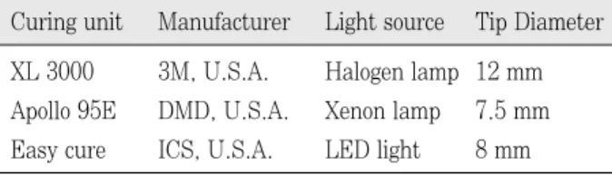
Table 2.Tested light curing unit
XL 3000 3M, U.S.A. Halogen lamp 12 mm Apollo 95E DMD, U.S.A. Xenon lamp 7.5 mm Easy cure ICS, U.S.A. LED light 8 mm Curing unit Manufacturer Light source Tip Diameter
Table 3.Light source, resin shade and irradiation time for tested group
Halogen XL 3000 10, 20, 40sec B1, A3 Plasma arc Apollo 95E 3, 6, 10sec B1, A3 LED Easy cure 10, 20, 40sec B1, A3 Light source Curing unit Time of i Tested rradiation shade Table 1.Composition of Esthet∙X
Matrix Filler
ethoxylated Bisphenol-A- dimethacrylate
Triethylene glycol dimet- hacrylate(TEGDMA) photoinitiation system diketone, Camphoroquin- one(CQ)
inorganic bariumalumino fluroborosilicate(BAFG) glass with nano-sized sili- con dioxide particles vol: 60%
weight: 77%
Fig. 1.Schematic drawing of specimen fabrication.
Light guide
1, 2, 3mm
Slide glass Composite
Emitting Diode)광원으로 Easy cure (ICS, U.S.A.)를 사용하였다(Table 2). 플라스마 아크 광원을 사용하는 조 사기는 1930mW/cm2의 출력11)으로 연속 조사하는 방식을 선택하였다. LED 광원으로 사용된 Easy cure는 두부에 7 개의 다이오드가 포함되어 있다.
2. 시편제작
직경 4.0 mm 두께 1, 2 및 3 mm의 알루미늄 주형을 이 용하여 각 군 당 5개의 시편을 제작하였다. Accufilm Ⅱ (CE, U.S.A.)의 검정색 면 위에 셀룰로이드 스트립과 주형 을 위치시킨 후 광중합형 복합레진을 충전하였다. 상면에 셀룰로이드 스트립을 얻은 후 1 mm 두께의 투명 슬라이드 글라스(Superior, Germany)로 압접하여 과잉 레진을 제 거하였다. 각각의 광조사기를 슬라이드 글라스에 접촉시킨 후 정해진 시간에 따라 중합하였다(Fig. 1, Table 3). 제작 된 시편은 24시간 동안 증류수에 보관하였다.
3. 깊이에 따른 비커스 경도 측정
1 mm 시편의 상부와 1, 2 및 3 mm 시편의 하부에 미세 경도기(Microhardness tester, Zwick, Germany)를 사용 하였다. 표면 각이 136�인 피라미드 형태의 다이아몬드 압 입체를 이용하여 3회씩 500 g의 하중을 10초간 적용한 후 정사각형 모양의 압입 자국에서 두 개의 꼭지점간의 거리를 200배로 확대하여 측정하였다.
4. 통계처리
광원, 조사시간 및 중합깊이에 따른 비커스 경도를 비교하 기 위해 two-way ANOVA를 사용하여 비교, 분석하였으며 Student-Newman-Keuls method로 사후 검정하였다.
동일 깊이에서 각 군간 비커스 경도값을 비교하기 위해 Kruskal-Wallis one way ANOVA를 사용한 후 Student- Newman-Keuls method로 사후 검정하였다.
Ⅲ. 연구 결과
1. 색상, 깊이 및 조사시간에 따른 비커스 경도
B1, A3 색상에서 광조사기와 조사시간에 따른 비커스 경 도는 다음과 같다(Table 4, 5, Fig. 2-4).
B1 색상에서는 20.43-70.66으로 나타났으며 Easy cure 를 40초 간 조사한 군의 표층에서 가장 높은 값을, Easy cure를 10초 간 조사한 군의 3 mm에서 가장 낮은 값을 나 타내었다. 동일한 광원에서 조사시간이 증가함에 따라 표면 경도는 증가하였다(p<0.05). 할로겐 광원 조사군의 1 mm 와 2 mm 간을 제외한 모든 군에서 깊이가 증가함에 따라 표면경도는 감소하였다(p<0.05). A3 색상에서 XL 3000을 40초 간 조사한 군에서는 모든 깊이에서 B1값과 유의한 차 이가 없었으나(p>0.05), XL 3000을 10초, 20초 간 조사 한 군과 Apollo 95E와 Easy cure를 이용한 군에서는 2 mm, 3 mm에서 B1값과 유의한 차이를 보였다(p<0.05).
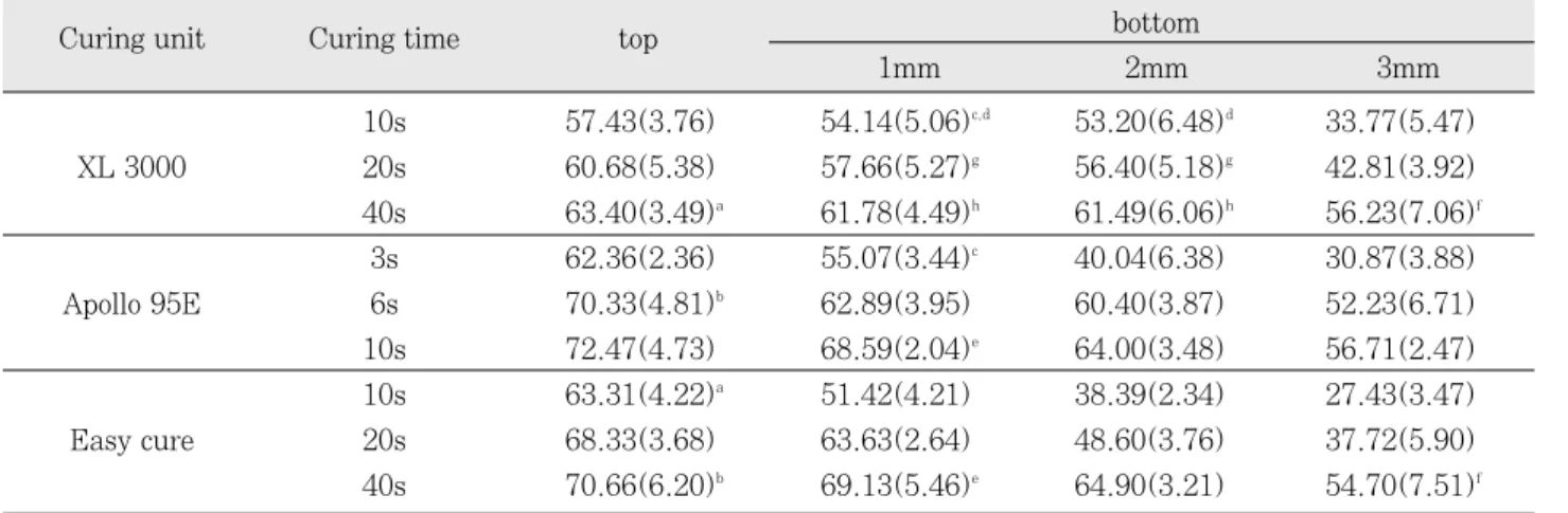
Table 4. Vicker's hardness number of B1 shade Mean(S.D.), n=15
10s 57.43(3.76) 54.14(5.06)c,d 53.20(6.48)d 33.77(5.47)
XL 3000 20s 60.68(5.38) 57.66(5.27)g 56.40(5.18)g 42.81(3.92)
40s 63.40(3.49)a 61.78(4.49)h 61.49(6.06)h 56.23(7.06)f
3s 62.36(2.36) 55.07(3.44)c 40.04(6.38) 30.87(3.88)
Apollo 95E 6s 70.33(4.81)b 62.89(3.95) 60.40(3.87) 52.23(6.71)
10s 72.47(4.73) 68.59(2.04)e 64.00(3.48) 56.71(2.47)
10s 63.31(4.22)a 51.42(4.21) 38.39(2.34) 27.43(3.47)
Easy cure 20s 68.33(3.68) 63.63(2.64) 48.60(3.76) 37.72(5.90)
40s 70.66(6.20)b 69.13(5.46)e 64.90(3.21) 54.70(7.51)f
The same letters were not significantly different.
Curing unit Curing time top bottom
1mm 2mm 3mm
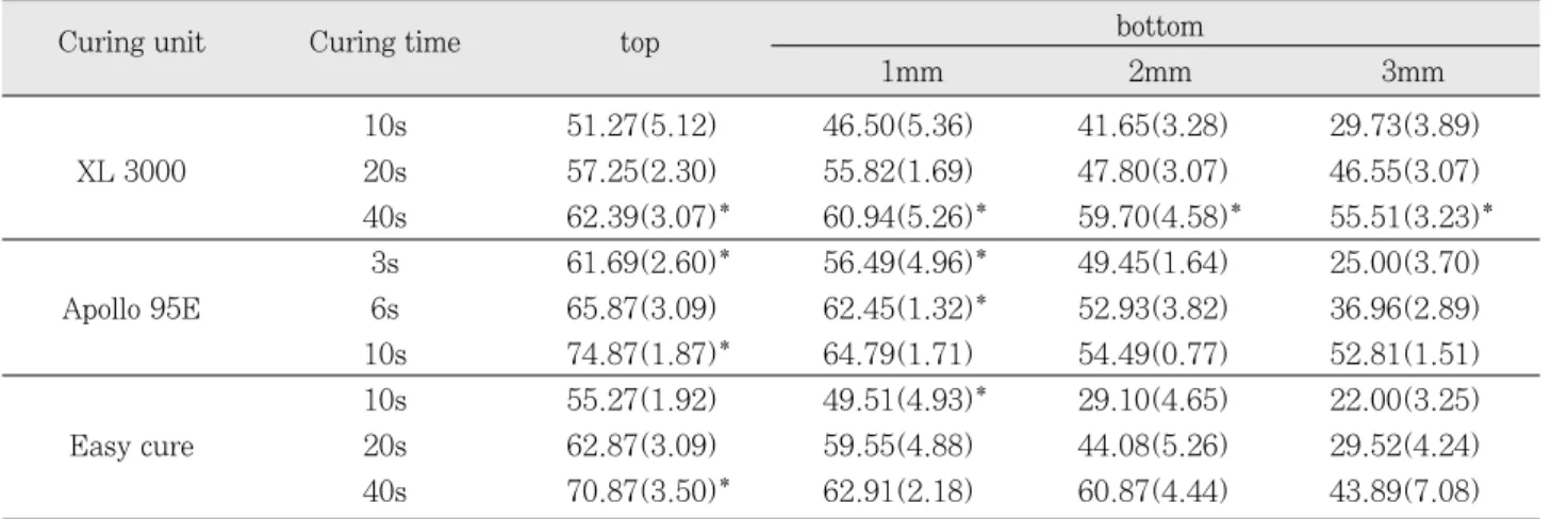
10s 51.27(5.12) 46.50(5.36) 41.65(3.28) 29.73(3.89)
XL 3000 20s 57.25(2.30) 55.82(1.69) 47.80(3.07) 46.55(3.07)
40s 62.39(3.07)* 60.94(5.26)* 59.70(4.58)* 55.51(3.23)*
3s 61.69(2.60)* 56.49(4.96)* 49.45(1.64) 25.00(3.70)
Apollo 95E 6s 65.87(3.09) 62.45(1.32)* 52.93(3.82) 36.96(2.89)
10s 74.87(1.87)* 64.79(1.71) 54.49(0.77) 52.81(1.51)
10s 55.27(1.92) 49.51(4.93)* 29.10(4.65) 22.00(3.25)
Easy cure 20s 62.87(3.09) 59.55(4.88) 44.08(5.26) 29.52(4.24)
40s 70.87(3.50)* 62.91(2.18) 60.87(4.44) 43.89(7.08)
* : No significant difference with B1 shade.
Fig. 2.Vicker's hardness number of composite cured by XL 3000
Fig. 3.Vicker's hardness number of composite cured by Apollo 95E.
Fig. 4.Vicker's hardness number of composite cured by Easy cure.
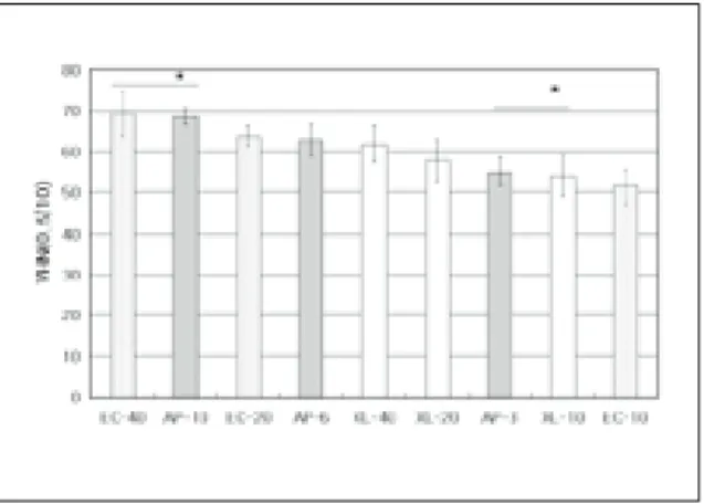
Fig. 5.Mean VHN of B1 shade groups on top surface.
* : No significant difference between groups.
2. B1 색상에서 깊이에 따른 비커스 경도는 다음과 같다 (Fig. 6-9).
모든 깊이에서 Apollo 95E를 10초 간 조사한 군이나
Easy cure를 40초 간 조사한 군에서 가장 높았으며 Easy cure나 XL 3000를 10초 간 조사한 군에서 가장 낮은 값을 보였다(p<0.05). 표층에서는 Easy cure를 40초 간 조사한 군과 Apollo 95E를 6초 간 조사한 군, XL 3000을 40초 간 조사한 군과 Easy cure를 10초 간 조사한 군간에 유의 한 차이가 없었다. 1 mm 하방에서는 Easy cure 40초 간 조사한 군과 Apollo 95E를 10초 간 조사한 군, Apollo 95E를 3초 간 조사한 군과 XL 3000을 10초 간 조사한 군 간에 유의한 차이가 없었다. 2 mm 하방에서는 모든 군 간 에 유의한 차이가 있었으며, 3 mm 하방에서는 XL 3000을 40초간 조사한 군과 Easy cure를 40 초 간 조사한 군 간에 유의한 차이가 없었다.
3. 비커스 경도비
B1 색상에서 깊이에 따른 표층과의 비커스 경도비는 Fig.
9와 같다.
Fig. 6.Mean VHN of B1 shade experimental groups on 1mm from the top surface.
Fig. 7.Mean VHN of B1 shdae experimental groups on 2mm from the top surface.
Fig. 8.Mean VHN of B1 shade experimental groups on 3mm from the top surface.
Fig. 9.Ratio of the VHN by depth in B1 shade. Fig. 10.Ratio of the VHN by depth in A3 shade.
Ⅳ. 총괄 및 고찰
중합광선은 수복물을 통과함에 따라 산란, 흡수되어 수복 물의 불완전한 중합을 초래하게 된다. 이는 수복물의 구강 내 용해도 증가12), 마모저항성의 감소1), 수복물의 파절13),
미세누출14,15)과 술 후 과민증1)등의 문제점을 야기한다. 복합
레진의 중합률에 영향을 미치는 인자로는 광강도1,16,17), 중합
시간18-21), 레진의 광투과성22), 필러의 크기23,24)와 함량25) 및
색상26,27)등이 있다. 복합레진의 중합률은 다양한 방법으로
측정될 수 있는데, FT-IR 분광 분석28), 표면경도, 수분 흡 수도, 레진 유리도 및 핵자기 공명 미세영상분석29)등을 측 정함으써 나타낼 수 있다4,30). 이 중 표면경도의 측정은 같은 종류의 재료에서 복합레진의 중합률을 나타내는 유용한 방 법으로, 복합레진의 표면경도는 중합률28), 충전재의 함량25) 및 크기24)에 의해 영향받으며, Shortal 등31)은 표면경도와 중합률은 색상과 밀접한 관계가 있다고 하였다. 또한 Peutzfeldt와 Asmussen4)은 복합레진 중합 후 잔류 이중 결합의 양은 표면경도과 관련이 깊다고 하였으며, Warren32)도 복합레진의 중합률을 표면경도를 이용하여 측 정하였다. 표면경도 측정법 중 하나로 비커스 경도는 피라 미드 형태의 압입체를 이용하여 일정 힘을 정해진 시간동안 가한 후 압입 자국의 직경을 측정하여 표면경도를 구하게 된다23,25,29).
광중합기에서 발생된 빛은 광중합형 복합레진의 중합개시 제인 camphoroquinone을 활성화시키며 이것은 삼차 아민 에서 자유 라디칼을 생성하여 중합반응을 야기한다2). 레진 내부의 광개시제를 활성화시키기 위해서는 충분한 광에너 지(광강도×조사시간)가 필요하며, 광강도와 중합시간은 레 진의 중합에 영향을 미치는 결정적인 인자로 보고되고 있
다16,17). Rueggeberg 등16)은 233 mW/cm2이상의 광강도를
추천하면서 400-578 mW/cm2의 광원에서 수복물이 2 mm일 경우 40초 이상을 중합할 것을 추천하였다. 본 실험 에서 할로겐 광원으로 사용된 XL 3000을 Radiometer (Curing radiometer, Demetron, U.S.A.)로 광강도 측정 결과 500 mW/cm2를 나타내었다.
Sharkey 등33)은 플라스마 아크 광원을 이용한 중합기에 서 짧은 조사시간은 수복물의 이상적인 강도를 얻는데 부족 하며, Peutzfeldt 등9)은 중합깊이를 비교한 실험에서 할로 겐 광중합기를 20초간 조사한 군이 플라스마 아크를 3초간 조사한 군에 비해 높은 값을 나타냈음을 보고하였다. Jandt 등27)은 할로겐 광중합기와 LED 광중합기를 이용하여 40초 간 중합한 후 중합깊이를 비교하여 할로겐 광조사기가 20% 정도 깊은 중합깊이를 나타냈음을 보고하였다. 또한
phoroquinone의 빛 흡수곡선과 유사한 파장 분포를 가지 고 있기 때문이라고 분석하였다. Kurachi 등34)은 두부에 두 개에서 여섯 개까지 LED를 포함하는 광중합기를 자체 제 작하여 전통적인 할로겐 광중합기와 비교 연구하였다. 실험 결과 6개의 LED를 포함하는 광중합기에서 가장 높은 광강 도를 가졌으나, 동일시간에 할로겐 광중합기로 중합한 군에 비해 낮은 비커스 경도를 보인다고 보고하였다. 본 연구 결 과 B1 색상에서 할로겐 광원을 40초 간 조사한 군의 비커 스 경도를 다른 광원과 비교 시 표층과 1 mm 에서는 플라 스마 아크 광원을 6초, 10초 조사한 군과, LED 광원을 40 초, 60초 간 조사한 군에 비하여 낮은 값을 보였고, 2 mm 에서는 플라스마 아크광원을 10초 간 조사한 군과 LED 광 원을 40초 간 조사한 군에 비해 낮은 값을, 3 mm 깊이에서 는 플라스마 아크 광원을 10초 간 조사한 군에 비해 낮은 값을 보였으며 LED 광원을 40초 간 조사한 군과는 유의한 차이가 없었다. 본 연구에서 플라스마 아크 광원으로 Apollo 95E를 사용하였으며 LED 광원으로 Easy cure를 사용하였다. 제조자는 2-3 mm 수복물을 중합하는데 각각 3초와 8초간 조사할 것을 추천하고 있으나, 실험결과는 이 들 중합기에서 조사시간의 연장이 필요함을 보여주고 있다.
중합깊이와 레진 색상과의 관계에 대하여 많은 연구가 있 어왔다. Ferracane 등30)은 색상과 무관하게 동일 제품의 중 합깊이는 같다고 보고하였으나, Kawaguchi 등22)은 레진 색상은 광투과도에 영향을 미치며, 동일 제품에서 밝은 색 은 광투과도가 더 높으므로 중합깊이도 이에 비례한다고 보 고하였으며, Harrington과 Wilson26), Shortall 등31)의 연 구에서도 A2 색상이 A3.5에 비해서 유의하게 높은 중합깊 이를 보임을 보고하였다. Kwaguchi 등22)은 수종의 레진에 서 광투과도와 중합깊이의 연관성에 대해 연구하였는데 실 험에서 사용된 레진의 광투과도는 0.042-0.263 이었으며, 레진 색상이 광투과도와 직접적인 연관이 있음을 보고하였 다. 또한, 그는 대부분 실험군에서 중합깊이와 광투과도 사 이에 명백한 비례관계가 있음을 보고하였다. Shortall 등31) 은 5종의 레진에서 A2, A3.5 및 C2 색상에 따른 중합깊이 를 penetrometer26)를 이용하여 비교한 결과 모든 제품에서 A2가 A3.5 색상에 비해 유의하게 높은 중합깊이를 보임을 보고하였다. 본 실험결과 A3 색상에서 할로겐 광원을 40초 간 조사한 군에서는 모든 깊이에서 B1값과 유의한 차이가 없었으나, 할로겐 광원을 10초, 20초 간 조사한 군과 플라 스마 아크 광원과 LED 광원을 이용한 군의 2 mm, 3 mm 에서 B1값에 비해 유의하게 낮은 표면경도를 보였다.
중합광선은 레진을 통과함에 따라 흡수, 산란되어 광강도 가 감소하게 되며 이는 표층과 바닥에서의 경도차이를 야기
하게 된다28). 이를 보상하기 위해 Pilo와 Cardash24)는 표층 과 바닥에서의 경도비는 0.8 이상이 되도록 할 것을 추천하 였다. 연구 결과 2 mm 깊이에서 비커스 경도비를 보면 B1 색상에 있어서 할로겐 광원을 사용한 모든 군에서 0.8이상 을 나타냈으나, 플라스마 아크 광원을 3초, LED 광원을 10초, 20초 간 조사한 군에서는 0.8 이하를 나타내었으며, A3 색상에 있어서 할로겐 광원을 이용한 보든 군에서 0.8 이상을 나타내었으나. 플라스마 아크 광원을 10초, LED 광원을 10초, 20초 간 조사한 군에서는 0.8 이하를 나타내 었다. 전반적으로 조사시간이 증가함에 따라 각 깊이에서 비커스 경도비가 증가하는 양상을 나타내었으며, 이는 조사 시간을 증가시킴으로서 하방의 불완전한 중합을 예방할 수 있음을 보여준다. 색상에 따른 비커스 경도비에도 차이를 보였으며, B1 색상에서는 평균 0.87, A3에서는 0.83으로 나타났다. 이는 어두운 색일수록 깊이가 증가함에 따라 중 합률이 크게 감소함을 보여준다.
B1, A3 색상에서 할로겐 광원으로 40초 간 조사한 군의 2 mm 하방의 비커스 경도를 기준으로 비교시 B1 색상에 서 할로겐 광원은 61.46 이었으며, 이는 플라스마 아크 광 원의 경우 10초, LED 광원의 경우 40초 간 조사해야 더 높 은 값을 나타내었으며, A3 색상에서 할로겐 광원은 59.70 이었으며 플라스마 아크 광원의 경우 10초 간 조사한 군에 서도 보다 낮은 값을 보였으며 LED 광원의 경우 40초간 조사하여야 더 높은 값을 얻을 수 있었다.
Ⅴ. 결 론
본 실험에서는 할로겐 광원으로 XL 3000, 플라스마 아크 광원으로 Apollo 95E, LED 광원으로 Easy cure를 사용 하여 복합레진을 광중합 시 중합 광원에 따른 중합 깊이를 비커스 경도를 이용하여 측정하여 다음과 같은 결과를 얻었 다.
1. 모든 광원에서 조사시간이 증가함에 따라 중합깊이는 증가하였다(p<0.05).
2. 할로겐 광원 조사군의 1 mm와 2 mm 간을 제외한 모 든 군에서 깊이가 증가함에 따라 비커스 경도는 감소하 였다(p<0.05).
3. 할로겐 광원으로 10초 및 20초간 조사한 군에서 A3 색상 복합레진의 표면경도는 B1 색상에 비해 보든 깊 이에서 낮았으나(p<0.05), 40초간 조사한 군은 색상 에 따른 차이를 보이지 않았다.
4. Plasma arc와 LED 광원 조사군은 표층과 1 mm 깊 이에서는 색상에 따라 비커스 경도의 차이를 보이지 않 았으나, 2 및 3 mm 깊이에서는 색상간에 유의한 차이 를 보였다(p<0.05).
참고문헌
1. Stephen C, Bayne MS, Harald O, Heymann HO, Edward J, Swift JR. : Update on dental composite restorations. J Am Dent Assoc 125:687-701, 1994.
2. Mills RW, Jandt KD : Blue LEDs for curing polymer- based dental filling materials. LEOS Newsletter 12:9- 10, 1998.
3. Shintani H, Inoue T, Yamaki M : Analysis of cam- phorquinone in visible light cured composite resins.
Dent Mat 1:14-126, 1985.
4. Peutzfeldt A, Asmussen E : Hardness of restorative resins: effect of camphorquinone, amine, and inhibitor.
ACTA Odontol Scand 47:229-231, 1989.
5. Fan PL, Wozniak WT, Reyes WD, Stanford JW : Irradiance of visible light-curing units and voltage variation effects. J Am Dent Assoc 115:442-445, 1987.
6. Rueggeberg FA, Caughman WF : Factors affecting light transmission of single-use, plastic light-curing tips. Oper Dent 23:179-184, 1998.
7. Pouls JG, Styner DL : Curing lights: Changes in intensity output with use over time. General Dent January-February 70-73, 1997.
8. Pilo R, Oelgiesser D, Cardash HS : A survey of inten- sity and potential for depth of cure among light-curing units in clinical use. J Dent 27:235-241, 1999.
9. Peutzfeldt A, Sahafi A, Asmussen E : Characterization of resin composites polymerized with plasma arc curing units. Dent Mater 19:330-336, 2000.
10. Munksgaard EC, Peutzfeldt A, Asmussen E : Elution of TEGDMA and BisGMA from a resin and a resin composite cured with halogen or plasma light. Eur J Oral Sci 108:341-345, 2000.
11. Clinical research associates newsletter volume 23, Issue 5-6, May-June, 1999.
12. Pearson GJ, Longman CM : Water sorption and solu- bility of resin-based materials following inadequate polymerization by a visible-light curing system. J Oral Rehabil 16:57-61, 1989.
13. Eakle WS : Fracture resistance of teeth restored with class II bonded composite resin. J Dent Res 65:149- 153, 1986.
14. Youngson CC, Glyn Jhones JC, Fox K, Smith IS, Wood DJ, Gale M : A fluid filtration and clearing techneque to assess microleakage associated with three dentine bonding systems. J Dent 27:223-233, 1999.
15. Tsunekawa M, Setcos JC, U.S.A.mi Y, Iwaku M, Marshall SJ : A new light-activated adhesive compos- ite: shear bond strength and microleakage. Dent Mater 8:234-237, 1992.
16. Rueggeberg FA, Caughman, WF, Curtis JW : Effect of light intensity and exposure duration on cure of resin composites. Oper Dent 19:26-32, 1994.
17. Pires JAF, Cvitko E, Denehy G, Swift EJ Jr : Effect of curing tip distance on light intersity and composite resin microhardness. Quintessence int 24:517-521, 1993.
18. Davidson-Kaban SS, Davidson CL, Feilzer AJ, Gee AJ, Erdilek N : The effect of curing light variations on bulk curing and wall-to-wall quality of two types and vari- ous shades of resin composites. Dent Mater 13:344-52, 1997.
19. Vargas MA, Cobb DS, Schmit JL : Polymerization of composite resins: Argon laser vs conventional light.
21. Tanoue N, Matsumura H, Atsuta M : Curing depth of prosthetic composite materials polymerized with their proprietary photo-curing units. J Oral Rehabil 26:594- 599, 1999.
22. Kawaguchi M, Fukushima T, Miyazaki M : The rela- tionship between cure depth and transmission coeffe- cient of visible-light-activated resin composites. J Dent Res 73:516-521, 1994.
23. Wassell RW, McCabe JF, Walls AW : Subsurface deformation associated with hardness measurements of composites. Dent Mater 8:218-223, 1992.
24. Pilo R, Cardash HS : Post-irradition polymerization of different anterior and posterior visible light-activated resin composites. Dent Mater 8:299-304, 1992.
25. Chung KH, Greener EH : Correlation between degree of conversion, filler concentration and mechanical properities of posterior composite resins. J Oral Rehabil 17:487-494, 1990.
26. Harrington E, Wilson HJ : Depth of cure of radiation- activated materials: Effect of mould material and cavi- ty size. J Dent 21:305-311, 1993.
27. Jandt KD, Mills RW, Flackwell GB, Ashworth SH : Depth of cure and compressive strength of dental com- posites cured with blue light emitting diodes(LEDs).
Hunter G, Pananalkis D, Abel EW : Determination of the depth of cure for VLC composites by nuclear mag- netic restoration microimaging. Dent Mater 10:128- 133, 1994.
30. Ferracane JL, Aday P, Matsumoto H, Marker VA : Relationship between shade and depth of cure for light-activated dental composite resins. Dent Mater 2:80-84, 1986.
31. Shortall AC, Wilson HJ, Harrington E : Depth of cure of radiation-activated composite restoratives-influence of shade and opacity. J Oral Rehabil 22:337-342, 1995.
32. Warren K : An investigation into the microhardness of a light cured composite when cured through varying thicknesses of porcelain. J Oral Rehabil 17:327-334, 1990.
33. Sharkey S, Ray N, Burke F, Ziada H, Hannigan A : Surface hardness of light-activated resin composites cured by two different visible-light sources: An in vitro study. Quintessence int 32:401-405, 2001.
34. Kurachi C, Tuboy AM, Magalhaes DV, Bagnato VS : Hardness evaluation a dental composite polymerize with experimental LED-based devices. Dent Mater 17:309-315, 2001.