Introduction
That is rare but crucial extra-intestinal complications dur- ing an exacerbation in patients with inflammatory bowel dis- ease (IBD)–ulcerative colitis (UC) and Crohn’s disease.1)2) Car- diac complications occur particularly with UC,2) and few cases reported patients with various manifestation from fulminant myocarditis to mild form.3-5) Control of the underlying diseas- es is often associated with resolution of myocarditis and pre- vention of recurrence.6) Here we report a young female patient with fulminant myocarditis induced by UC treated with ex- tracorporeal life support and infliximab.
Case
A 28-year-old woman was consulted for dyspnea and elevat- ed cardiac biomarker (CK-MB: 11.4 ng/mL, troponin I: 0.512 ng/mL) from division of gastroenterology to cardiology. Sinus tachycardia and non-specific ST change was noted on her elec- trocardiography (ECG) and mild cardiomegaly and pulmo- nary congestion were noted on chest X-ray (CXR). She was suffering from exacerbation of UC treated with corticosteroid.
Her blood pressure was 106/70 mm Hg and heart rate was
CASE REPORT J Cardiovasc Ultrasound 2016;24(2):163-167
138 bpm. Initial echocardiography showed dilated left ven- tricular (LV) dimension and decreased LV ejection fraction (EF:
33%) with preserved LV tissue velocities (Fig. 1, Supplemen- tary movie 1). We decided to apply extracorporeal membrane oxygenation (ECMO) for cardiac rest under impression of acute myocarditis because her coronary angiography showed normal coronary arteries (Fig. 2) in combination with global hy- pokinesia of LV. Follow up echocardiography performed the following day (Fig. 3, Supplementary movie 2) showed much depressed LV systolic function (EF: 10%) and myocardial tis- sue velocities (E’: 10 → 3 cm/s, S’: 6 → 2 cm/s). The 7th day after ECMO, massive hematochezia was occurred even with maintenance of corticosteroid (methylprednisolone 33 mg).
Emergent sigmoidoscopy revealed diffuse maroon colored blood oozing from inflamed mucosa of transverse colon with- out discernable specific bleeding focus. Increased LV wall thickness (8 → 13 mm) and still depressed LV performance (EF: 16%, E’: 2 cm/s, S’: 3 cm/s) detected by echocardiogra- phy (Fig. 4, Supplementary movie 3), wherein we added inf- liximab (215 mg) infusion for uncontrolled inflammation.
Two days later, removal of ECMO was possible because she sta-
Successfully Treated Acute Fulminant Myocarditis Induced by Ulcerative
Colitis with Extracorporeal Life Support and Infliximab
Han-Kyul Kim, MD1,Kun Il Kim, MD, PhD2,Sung Won Jung, MD, PhD3, Hee-Sun Mun, MD1, Jung Rae Cho, MD1,Namho Lee, MD, PhD1, and Min-Kyung Kang, MD, PhD1
Departments of 1Cardiology, 2Cardiothoracic Surgery, 3Gastroenterology, Kangnam Sacred Heart Hospital, Hallym University Medical Center, Seoul, Korea
We report a case of successfully treated acute fulminant myocarditis induced by ulcerative colitis with extracorporeal life support and infliximab. Myocarditis is a rare but crucial complication during an exacerbation of inflammatory bowel disease. In our case, we applied extracorporeal membrane oxygenation (ECMO) for cardiac rest under impression of acute myocarditis associated with ulcerative colitis, and added infliximab for uncontrolled inflammation by corticosteroid. As a result, our patient was completely recovered with successful weaning of ECMO.
KEY WORDS: Myocarditis · Ulcerative colitis · Extracorporeal membrane oxygenation.
• Received: October 20, 2015 • Revised: December 12, 2015 • Accepted: May 10, 2016
• Address for Correspondence: Min-Kyung Kang, Department of Cardiology, Kangnam Sacred Heart Hospital, Hallym University Medical Center, 1 Singil-ro, Yeongdeungpo-gu, Seoul 07441, Korea Tel: +82-2-2820-5294, Fax: +82-2-2846-4669, E-mail: [email protected]
• This is an Open Access article distributed under the terms of the Creative Commons Attribution Non-Commercial License (http://creativecommons.org/licenses/by-nc/3.0) which permits unrestricted non-commercial use, distribution, and reproduction in any medium, provided the original work is properly cited.
bilized without further bleeding and echocardiography (Fig. 5, Supplementary movie 4) showed improved LV systolic func- tion (EF: 16 → 60%) and myocardial tissue velocities (E’: 3 → 5 cm/s, S’: 2 → 7 cm/s). Last echocardiography (Fig. 6, Supple- mentary movie 5) showed normal LV wall thickness (11 → 8 mm), EF (65%), and myocardial tissue velocities (E’: 12 cm/s, S’: 10 cm/s). In addition, cardiac biomarkers (CK-MB: 11.4 → 8.4 → 5.8 → 1.4 ng/mL, troponin I: 0.512 → 0.298 → 0.099 → 0.025 ng/mL), ECG, and CXR were all normalized (Fig. 7).
Now she is stable and followed regularly on outpatient basis
with maintenance of low dose steroid.
Discussion
UC is known to be a severe disease with many extra-colonic manifestations, such as skin involvement, rheumatic prob- lems, and ocular complications.2) Here we present a young fe- male patient with UC and fatal cardiac complication that were not resolved with corticosteroids.
In our patient, acute myocarditis was developed presenting dyspnea and elevated cardiac biomarkers during exacerbation of
Fig. 1. Initial echocardiography shows dilated left ventricle by 2D (A) and decreased left ventricular (LV) ejection fraction about 33% by M-mode (B).
Velocity time integral from LV out flow track showed 12 cm (C) but relatively preserved LV myocardial tissue velocities (D).
A
C
B
D
Fig. 2. Coronary aniography shows normal left (A) and right (B) coronary arteries.
A B
UC in the maintenance of corticosteroid. We rapidly decided to apply ECMO for cardiac rest even her LV EF was not severely depressed and also with preserved LV myocardial tissue veloci-
ties. As a result, our decision was not so impetuous because echocardiography performed the very next day revealed severely depressed LV EF and myocardial tissue velocities. Unexpected
Fig. 3. Follow up echocardiography after extracorporeal life support shows further decreased left ventricular ejection fraction and velocity time integral (A, B, and C) and depressed myocardial tissue velocities (D).
A
C
B
D
Fig. 4. Increased left ventricular wall thickness was detected after ECMO procedure (A and B) and still depressed velocity time integral from left ventricular out flow track and myocardial tissue velocities (C and D). ECMO: extracorporeal membrane oxygenation.
D B
C A
dium suggesting inflammatory phase of acute myocarditis.7) Therefore we apply infliximab to control underlying bowel in- flammation and acute management for UC. Follow up echocar- diography after infusion infliximab, result showed improved LV abrupt hematochezia was occurred despite of maintenance of
corticosteroid (methylprednisolone 33 mg). Not only sigmoid- oscopy showed uncontrolled inflammation of transverse colon, echocardiography also revealed increased thickness of myocar-
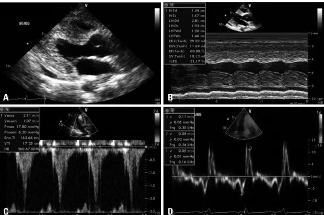
Fig. 5. The echocardiography after infusion of infliximab showed still increased left ventricular (LV) wall thickness but improved LV systolic function (A and B) and velocity time integral from LV out flow track and myocardial tissue velocities (C and D).
D B
C A
Fig. 6. The last echocardiographic study showed completely recovered myocardial function and resolved myocardial thickness. Normalized left ventricular (LV) wall thickness and improved LV systolic funciton (A and B) and normalized velocity time integral from LV out flow track and myocardial tissue velocities (C and D).
A B
D C
systolic function (EF: 16 → 60%) and myocardial tissue veloci- ties (E’: 3 → 5 cm/s, S’: 2 → 7 cm/s). Infliximab is a chimeric monoclonal antibody against tumor necrosis factor alpha used to treat autoimmune diseases. Although this agent has various adverse effects, some life-threatening, common to drugs in the class of immunosuppressants, patients with UC with treatment of infliximab was shown effective inflammation control.8)
In conclusion, we report a young female patient with fulmi- nant myocarditis associated with UC successfully treated with ECMO and infliximab. Serial follow up of echocardiography and early decision of applying ECMO were particularly essen- tial for management in our patient. Control of underlying dis- ease might be helpful to improve cardiac function, in case of acute myocarditis due to underlying IBD.
Supplementary movie legends
Movie 1. Initial echocardiography–dilated left ventricular (LV) dimension and decreased LV ejection fraction (ejection fraction: 33%) with preserved LV tissue velocities.
Movie 2. Second echocardiography–much depressed left ventricular systolic function (ejection fraction: 10%) and myo- cardial tissue velocities (E’: 10 → 3 cm/s, S’: 6 → 2 cm/s).
Movie 3. 7th echocardiography–increased left ventricular (LV) wall thickness (8 → 13 mm) and still depressed LV per- formance (ejection fraction: 16%, E’: 2 cm/s, S’: 3 cm/s).
Movie 4. 9th echocardiography–improved left ventricular
systolic function (ejection fraction: 16 → 60%) and myocardi- al tissue velocities (E’: 3 → 5 cm/s, S’: 2 → 7 cm/s).
Movie 5. Last echocardiography–normal left ventricular wall thickness (11 → 8 mm), ejection fraction (65%), and myocardial tissue velocities (E’: 12 cm/s, S’: 10 cm/s).
References
1. Mowat NA, Bennett PN, Finlayson JK, Brunt PW, Lancaster WM.
Myopericarditis complicating ulcerative colitis. Br Heart J 1974;36:724-7.
2. Becker SA, Wishnitzer R, Botwin S, Eliraz A, Bass DD. Myopericar- ditis associated with inflammatory bowel disease. J Clin Gastroenterol 1981;
3:267-70.
3. Freeman HJ, Salh B. Recurrent myopericarditis with extensive ulcerative colitis. Can J Cardiol 2010;26:549-50.
4. Varnavas VC, Reinsch N, Perrey M, Nensa F, Schlosser T, Baba HA, Gerken G, Erbel R, Janosi RA, Katsounas A. Recurrent lymphocytic myo- carditis in a young male with ulcerative colitis. Eur J Med Res 2014;19:11.
5. Gruenhagen B, Alraies MC, Vakil KP, March SK. Ulcerative colitis-in- duced myocarditis. BMJ Case Rep 2014 May 22 [Epub]. http://dx.doi.
org/10.1136/bcr-2014-204818.
6. Kristensen KS, Høegholm A, Bohr L, Friis S. Fatal myocarditis asso- ciated with mesalazine. Lancet 1990;335:605.
7. Kindermann I, Barth C, Mahfoud F, Ukena C, Lenski M, Yilmaz A, Klingel K, Kandolf R, Sechtem U, Cooper LT, Böhm M. Update on myocarditis. J Am Coll Cardiol 2012;59:779-92.
8. Iwasa R, Yamada A, Sono K, Furukawa R, Takeuchi K, Suzuki Y. C- reactive protein level at 2 weeks following initiation of infliximab induction therapy predicts outcomes in patients with ulcerative colitis: a 3 year follow-up study. BMC Gastroenterol 2015;15:103.
Fig. 7. Improvement of initial sinus tachycardia with non-specific ST changes on ECG (A) to normal sinus rhythm (B) and initial cardiomegaly and pulmonary congestion on chest X-ray (C) to normal (D). ECG: electrocardiography.
D B
C A