• 검색 결과가 없습니다.

Changes of Cytokine and Chemokine mRNA Expression in Whole Blood Cells from Active Pulmonary Tuberculosis Patients after T-Cell Mitogen and Mycobacterium tuberculosis Specific Antigen Stimulation

N/A
N/A
Protected

Academic year: 2021

Share "Changes of Cytokine and Chemokine mRNA Expression in Whole Blood Cells from Active Pulmonary Tuberculosis Patients after T-Cell Mitogen and Mycobacterium tuberculosis Specific Antigen Stimulation"

Copied!
6
0
0

로드 중.... (전체 텍스트 보기)

전체 글

(1)

Biomedical Science Letters 2014, 20(3): 162~167 http://dx.doi.org/10.15616/BSL.2014.20.3.162 eISSN : 2288-7415

Changes of Cytokine and Chemokine mRNA Expression in Whole Blood Cells from Active Pulmonary Tuberculosis Patients after T-Cell Mitogen and

Mycobacterium tuberculosis Specific Antigen Stimulation

Sunghyun Kim 1,2,§ , Sangjung Park 3,§ , and Hyeyoung Lee 1,†

1

Department of Biomedical Laboratory Science, College of Health Sciences, Yonsei University, Wonju 220-710, Korea

2

Institute for Life Science and Biotechnology, Yonsei University, Seoul 120-749, Korea

3

Department of Clinical Laboratory Science, College of Medical Science, Daegu Haany University, Gyeongsan 712-715, Korea

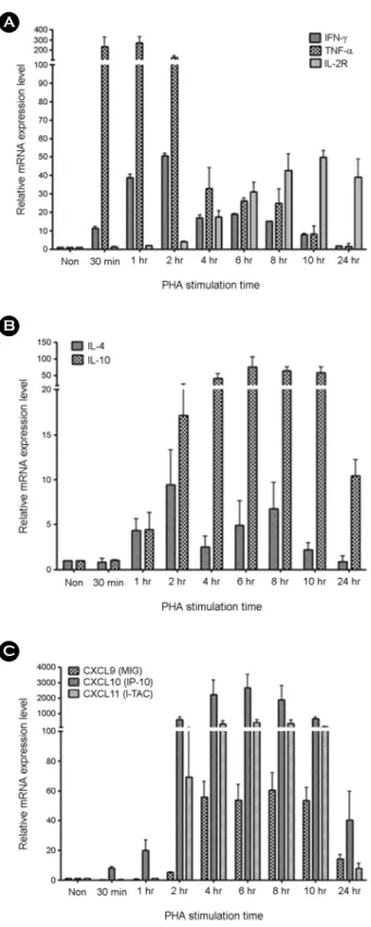
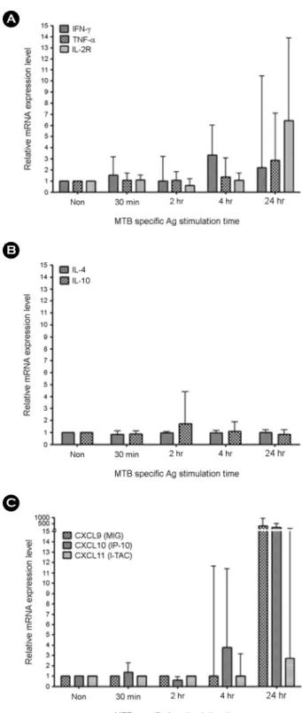
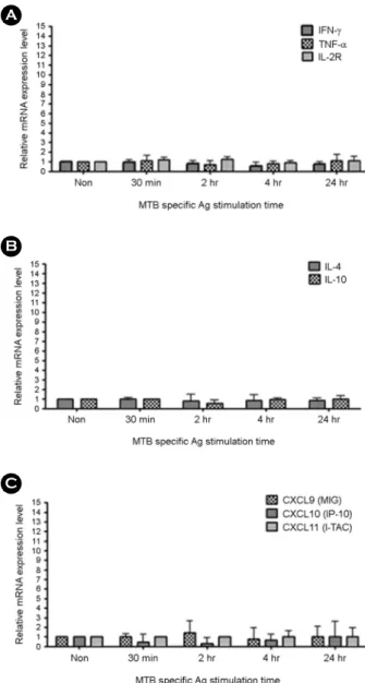
Tuberculosis (TB) is one of the major global health problems and it has been estimated that in 5~10% of Mycobacterium tuberculosis (MTB)-infected individuals, the infection progresses to an active disease. Numerous cytokines and chemokines regulate immunological responses at cellular level including stimulation and recruitment of wide range of cells in immunity and inflammation. In the present study, the mRNA expression levels of eight host immune markers containing of IFN-γ, TNF-α, IL-2R, IL-4, IL-10, CXCL9, CXCL10, and CXCL11 in whole blood cells from active pulmonary TB patients were measured after T-cell mitogen (PHA) and MTB specific antigens (ESAT-6, CFP-10, and TB7.7). Among the TH1-type factors, IFN-γ mRNA expression was peaked at 4 h, TNF-α and IL-2R mRNA expression was significantly high at the late time points (24 h) in active TB patients, TH2-type cytokine (IL4 and IL10) mRNA expression levels in both active TB and healthy controls samples did not changed significantly, and the mRNA expression of the three IFN-γ-induced chemokines (CXCL9, CXCL10, and CXCL11) were peaked at the late time points (24 h) in active TB patients after MTB specific antigen stimulation. In conclusion, the mRNA expression patterns of the TB-related immune markers in response to the T-cell mitogen (PHA) differed from those in response to MTB specific antigens and these findings may helpful for understanding the relationship between MTB infection and host immune markers in a transcripts level.

Key Words: Active pulmonary tuberculosis, Cytokines, Chemokines, T-cell mitogen, Mycobacterium tuberculosis specific antigens

Tuberculosis (TB) is mainly caused by Mycobacterium tuberculosis (MTB) and it remains a major infectious disease with approximately 9.3 million new TB cases annually and 1.8 million deaths per year. Forty thousand new cases of TB and 3,000 deaths occur in the Republic of Korea every

year (Dye, 1999; Lew, 1995). Most MTB infections are asymptomatic however about 10% of all infections even- tually progress to active TB disease (Comstock, 1974).

Typically, about 90% of active cases are pulmonary TB disease and 15~20% of active cases are extrapulmonary TB disease including tuberculous pleurisy, meningitis, lymphadenitis, and urogenital TB (Golden, 2005). The 6 kDa early secretory antigenic target (ESAT-6) and 10 kDa culture filtrate antigen (CFP-10) of MTB form a hetero- dimeric complex and both genes are expressed from region of difference 1 (RD1) region (Renshaw, 2005). These secretory antigens (proteins) contribute to the virulence of

*

Received: June 25, 2014 / Revised: September 12, 2014 Accepted: September 17, 2014

§

Equal contributors

Corresponding author: Hyeyoung Lee. Department of Biomedical Laboratory Science, College of Health Sciences, Yonsei University, 1 Yonseidae-gil, Wonju 220-710, Korea.

Tel: +82-33-760-2740, Fax: +82-33-760-2561 e-mail: [email protected]

C

The Korean Society for Biomedical Laboratory Sciences. All rights reserved.

Brief Communication

(2)

MTB and have a potential of T-cell antigen (Pathak, 2007;

Meher, 2006). TB7.7 is encoded by a phage-inserted region (phiRv2) which is highly specific for MTB (Behr, 1999;

Brosch, 2002; Cole, 1998). Phytohaemagglutinin (PHA) is a lectin found in plants and which is very useful as a mito- gen to trigger T-cell division and activation (Hamelryck, 1996). Cytokines and chemokines regulate immunological responses at cellular level including stimulation and recruit- ment of wide range of cells in immunity and inflammation.

Cytokines have pleiotropic actions, cascade effect, and antagonistic action and chemokines have homeostatic action, roles in promoting angiogenesis or guide cells to tissues, and inflammatory effect (Adane and Markos, 2013). Numerous cytokines and regulatory factors have been implicated in the pathogenesis of TB (Ulrichs, 1998; Dlugovitzky, 1999;

Azzurri, 2005; Okamoto, 2005). Other studies have shown that IFN-γ, TNF-α, and IL-2 expression profiles of CD4+

T-cells were highly observed in active TB disease (Harari, 2011) and IL-4 and IL-10 levels were also closely linked to active TB (Walzl, 2011). CXCL9 (monokine induced by interferon gamma, MIG), CXCL10 (interferon gamma inducible protein 10, IP-10), and CXCL11 (interferon- inducible T cell alpha chemoattractant, I-TAC) are inflam- matory chemokines which is induced by pro-inflammatory cytokines and secreted by monocytes or macrophages. These chemokines have been found in a various TB diseases (Kaplan, 1987; Okamoto, 2005; Juffermans, 1999). In the present study, relative mRNA expressions of IFN-γ, TNF-α, IL-2R, IL-4, IL-10, CXCL9, CXCL10, and CXCL11 in whole blood cells from active pulmonary TB patients and healthy donors (non-TB) were measured after PHA and MTB specific antigen stimulation time dependently and the results were compared.

The study was approved by the Institutional Ethics Committee of Yonsei University Severance Hospital (approval number 4-2010-0527) and Yonsei University Wonju Campus (approval number 2012-3) and all partici- pants provided written informed consent. MTB culturing confirmed individuals from respiratory specimens were considered as active pulmonary TB group. Individuals with HIV infection, end-stage renal disease, or leukemia/

lymphoma, and those who had received anti-TB therapy

for more than 2 weeks or immunosuppressive therapy,

including anti-cancer chemotherapy for malignant disease,

within 3 months of enrollment, were excluded from the

study. Enrolled healthy (non-TB) control group contains

young healthy adults (20s) with no symptoms of active TB,

no known history of contact with TB patients. T-cell

mitogen stimulation was performed using the commercial

mitogen tubes, QFT-IT test Mitogen tube (Cellestis, Carnegie,

Australia) which contains anticoagulant and PHA. One mL

of peripheral whole blood samples were collected into

mitogen tubes and incubated at 37℃ for 30 min, 1, 2, 4, 6,

8, 10, and 24 h. Plasma was removed by centrifugation at

2,000 x g for 5 min, mitogen stimulated whole blood cell

pellets were treated with RNA/DNA Stabilization Reagent

for Blood/Bone Marrow solution (Roche Diagnostics,

Mannheim, Germany) and stored at -80℃ until for total

RNA isolation. MTB-specific antigen stimulation was per-

formed using the commercial antigen tubes, QFT-IT test

MTB specific antigen tube (Cellestis, Carnegie, Australia)

which contains anticoagulant and ESAT-6, CFP-10 and

TB7.7. One mL of peripheral whole blood samples were

collected into antigen tubes and incubated at 37℃ for 30

min, 2, 4, and 24 h. Plasma was removed by centrifugation

at 2,000 x g for 5 min, mitogen stimulated whole blood cell

pellets were treated with RNA/DNA Stabilization Reagent

for Blood/Bone Marrow solution (Roche Diagnostics,

Mannheim, Germany) and stored at -80℃ until for total

RNA isolation. MagNA Pure LC 2.0 instrument and MagNA

Pure LC RNA Isolation Kit-High Performance (Roche

Diagnostics, Mannheim, Germany) were was used for

isolating total RNA from T-cell mitogen and MTB specific

antigen stimulated whole blood cells. Complementary DNA

(cDNA) was synthesized by M-MLV Reverse Transcriptase

kit and random hexamers (Invitrogen, Carlsbad, CA, USA)

with manufacturer's recommendations. Briefly, 10 μL of

extracted total RNA was added to master mixture containing

1 μL of 10 mM dNTP mix, 0.25 μg of random hexamers,

and 5 μL of DEPC-treated water in PCR tubes, and the

reaction mixture was incubated at 65℃ for 5 min and

quickly chilled on ice. Subsequently the mixture of 4 μL of

5× First-Strand Buffer, 2 μL of 0.1 M dithiothreitol (DTT),

and 1 μL of (M-MLV) reverse transcriptase (RT) was added

(3)

to the previous reaction mixture in PCR tubes and the second reaction was performed at the condition of 10 min at 25℃, 50 min at 37℃, and 15 min at 70℃. Real-time RT-PCR was performed using the TaqMan assay with ABI 7500 FAST instrument (Applied Biosystems, Foster City, CA, USA). Targets for real-time RT-PCR were IFN-γ, TNF-α, IL-2R, IL-4, IL-10, CXCL9, CXCL10, CXCL11, and glyceraldehyde 3-phosphate dehydrogenase (GAPDH).

The real-time PCR premix contained 10 μL of PreMix EX Taq (TaKaRa Bio Inc., Shiga, Japan), 10 pmol sense and antisense primers, 5 pmol TaqMan probes, and 3 μL of cDNA in a final volume 20 μL. The thermal cycling con- ditions were 10 min at 95℃, followed by 40 cycles of 10 s at 95℃, 30 s at 60℃. The standard curves for the target and references genes indicated the efficacy of the method (>

90%). The relative quantitation was automatically analyzed using 7500 Software version 2.0.4 (Applied Biosystems, Foster City, CA, USA) with Quantitation-Comparative threshold cycle (Ct) protocol which was based on the 2-DDCT method; relative expression was calculated as the ratio between the mean Ct values of the target genes and endogenous reference gene in each stimulated sample in relation to a unstimulated sample (Kim, 2013).

To determine the initiation time point of mRNA expression levels of TH1-type factors (IFN-γ, TNF-α, and IL-2R), TH2-type cytokines (IL-4 and IL-10), and IFN-γ-induced chemokines (CXCL9, CXCL10, and CXCL11) after T-cell mitogen stimulation, peripheral whole blood were stimulated with PHA and blood cell pellets were harvested at 30 min and 1, 2, 4, 6, 8, 10, and 24 h after incubation. In the TH1-type factor group, IFN-γ mRNA expression was detected at 30 min, it increased up to 2 h, and then it decreased until 24 h in a time-dependent manner. The TNF-α mRNA expression level was the highest in the early time points (30 min and 1 h), remained constant up to 2 h and then significantly reduced from 4~24 h. The IL-2R mRNA expression level increased from 30 min to 8 h time dependently and was constant between 8 and 24 h (Fig. 1-A).

In summary, in samples treated with the T-cell mitogen (PHA), the IFN-γ mRNA expression level peaked at 2 h, the TNF-α mRNA expression level was significantly high in the early time points, and the IL-2R mRNA expression

Fig. 1. The changes of target gene mRNA expression level after

stimulation of T-cell mitogen in time dependent manner. Whole

blood was stimulated with T-cell mitogen (PHA) for indicated

time period (0, 30 min, 1, 2, 4, 6, 8, 10, and 24 h). The target gene

(A. TH1-type factors; IFN-γ, TNF-α and IL-2R, B. TH2-type cyto-

kines; IL-4 and IL-10, C. IFN-γ induced chemokines; CXCL9

(MIG), CXCL10 (IP-10) and CXCL11 (I-TAC)) mRNA expression

levels of whole blood cells were analyzed by real-time RT-PCR.

(4)

level increased in the late time points (after 8 h) in a time-dependent manner. All these changes occurred after activation of other TH1-type cytokines (IFN-γ and TNF-α).

In the TH2-type cytokine group, IL-4 mRNA expression was detected at 1 h, increased up to 2 h, decreased at 4 h, increased again at 6~8 h and decreased until 24 h time dependently. IL-10 mRNA expression was detected at 1 h, increased until 4 h, was constant between 4 and 10 h and decreased at 24 h (Fig. 1-B). In summary, IL-4 mRNA expression peaked at 2 time points. IL-10 mRNA expression remained constant for a longer time (2~10 h) than that of other targets genes. In the IFN-γ-induced chemokine group, CXCL9 and CXCL11 mRNA expression increased at 2 h, was constant between 4 and 10 h and decreased at 24 h.

The CXCL10 mRNA expression increased from 30 min to 2 h, was constant between 2 and 10 h, and decreased at 24 h (Fig. 1-C). In summary, the mRNA expression of the three IFN-γ-induced chemokine (CXCL9, CXCL10, and CXCL11) remained constant for a longer time (2~10 h) with a significantly higher expression level than those of other groups (TH-1 type factors and TH-2 type cytokines) when stimulated with PHA. To determine the changes of mRNA expression levels of eight target genes (IFN-γ, TNF-α, IL-2R, IL-4, IL-10, CXCL9, CXCL10, and CXCL- 11) after MTB specific antigen stimulation, peripheral blood was collected from active TB patients and healthy controls in two QFT-IT tubes (MTB specific antigen; ESAT-6, CFP-10, TB7.7 and nil). Blood cell pellets were harvested at 30 min and 2, 4, and 24 h after incubation for total RNA isolation. Plasma samples were also collected at the same points to measure the plasma target protein levels. Target genes were grouped as. In the TH1-type factor group in active TB patients' samples, IFN-γ mRNA expression was detected at 30 min, increased up to 4 h, and decreased until 24 h in a time-dependent manner. TNF-α and IL-2R mRNA expression was constant until 4 h and peaked at 24 h (Fig.

2-A). However, the mRNA expression of IFN-γ, TNF-α, and IL-2R did not respond to MTB specific antigens in healthy controls samples (Fig. 3-A). In active TB patients' samples treated with MTB specific antigens, IFN-γ mRNA expression peaked at 4 h, and TNF-α and IL-2R mRNA expression was significantly high at the late time points (24 h).

Fig. 2. The changes of target gene mRNA expression level after

stimulation of MTB specific Antigen in time dependent manner

in samples from active TB patients. Whole blood from active

TB patients was stimulated with MTB specific antigens (ESAT-6,

CFP-10 and TB7.7) for indicated time period (0, 30 min, 2, 4 and

24 h). The target gene (A. TH1-type factors; IFN-γ, TNF-α and

IL-2R, B. TH2-type cytokines; IL-4 and IL-10, and C. IFN-γ

induced chemokines; CXCL9 (MIG), CXCL10 (IP-10) and CXCL-

11 (I-TAC)) mRNA expression levels of whole blood cells were

analyzed by real-time RT-PCR.

(5)

In the TH2-type cytokine group in active TB patients' samples, IL-4 and IL-10 mRNA expression was constant from the early time point to the late time point (Fig. 2-B). It did not respond to MTB specific antigens in healthy controls samples (Fig. 3-B). These data reveal that TH2-type cyto-

kine mRNA expression levels in both active TB and healthy controls samples did not change significantly after MTB specific antigen stimulation. In the IFN-γ-induced chemo- kine group, CXCL9 mRNA expression was constant until 4 h and increased significantly at 24 h. CXCL10 mRNA expression increased at 4 h and was significantly high at 24 h. CXCL11 mRNA expression was constant until 4 h and increased slightly at 24 h (Fig. 2-C). However, the mRNA expression of CXCL9, CXCL10, and CXCL11 did not respond to MTB specific antigens in healthy control samples (Fig. 3-C). In summary, the mRNA expression of the three IFN-γ-induced chemokines (CXCL9, CXCL10, and CXCL- 11) peaked at the late time points (24 h). In particular, CXCL9 and CXCL10 mRNA expression at 24 h was several hundred times higher than those of other targets in response to MTB specific antigens. CXCL10 mRNA was expressed earlier than CXCL9 and CXCL11 mRNA. In conclusion, the mRNA expression patterns of the TB-related immune markers in response to the T-cell mitogen (PHA) differed from those in response to MTB specific antigens.

Because the concentration of the peptide antigen was much higher in the T-cell mitogen control than in the MTB- specific antigen control, overall target mRNA expression was initiated at an early time point in the T-cell mitogen control. Additionally, while PHA first causes mitosis specifi- cally in T-cells among the immune cells, MTB specific antigens simultaneously stimulate various immune cells in whole blood cells. There were no significant changes of cytokine and chemokine mRNA expression by MTB specific antigens stimulation in healthy controls comparing to the active TB patients because the healthy controls involved in the present study did not have active TB symptoms and signs, and did not have any previous TB history. These results may contribute to a better understanding of the relationship between MTB specific antigens and host immune markers in a transcript level. In addition, these also provide essential information for development of next generation TB immunodiagnostic testing.

Acknowledgements

This study was supported by a grant of the Korea Healthcare technology R&D Project, Ministry of Health &

Fig. 3. The changes of target gene mRNA expression level after

stimulation of MTB specific antigens in time dependent manner

in samples from healthy controls. Whole blood from healthy

controls was stimulated with MTB specific antigens (ESAT-6,

CFP-10 and TB7.7) for indicated time period (0, 30 min, 2, 4 and

24 h). The target gene (TH1-type factors; IFN-γ, TNF-α and IL-2R,

TH2-type cytokines; IL-4 and IL-10, and IFN-γ induced chemo-

kines; CXCL9 (MIG), CXCL10 (IP-10) and CXCL11 (I-TAC))

mRNA expression levels of whole blood cells were analyzed by

real-time RT-PCR.

(6)

Welfare, Republic of Korea (grant HI10C1708 [A101750]

to H. L.). This work was supported (in part) by the Yonsei University Research Fund of 2013.

REFERENCES

Adane M, Markos A. Cytokines and Chemokines as Biomarkers of Tuberculosis. J Mycobac Dis. 2013. 3: 1-4.

Azzurri A, Sow OY, Amedei A, Bah B, Diallo S. IFN-gamma inducible protein 10 and pentraxin 3 plasma levels are tools for monitoring inflammation and disease activity in Myco- bacterium tuberculosis infection. Microbes Infect. 2005. 7:

1-8.

Behr MA, Wilson MA, Gill WP, Salamon H, Schoolnik GK, Rane S, Small PM. Comparative genomics of BCG vaccines by whole genome DNA microarray. Science. 1999. 284: 1520 -1523.

Brosch R, Gordon SV, Marmiesse M, Brodin P, Buchrieser C, Eiglmeier K, Garnier T, Gutierrez C, Hewinson G, Kremer K, Parsons LM, Pym AS, Samper S, Soolingen D van, Cole ST. A new evolutionary scenario for the Mycobacterium tuberculosis complex. Proc Natl Acad Sci USA. 2002. 99: 3684-3689.

Cole ST, Brosch R, Parkhill J, Garnier T, Churcher C, Harris D, Gordon VS, Eiglmeier K, Gas S, Barry III CE, Tekaia F, Badcock K, Basham D, Brown D, Chillingworth T, Connor R, Davies R, Devlin K, Feltwell T, Gentles S, Hamlin N, Holroyd S, Hornsby T, Jagels K. Deciphering the biology of Mycobacterium tuberculosis from the complete genome sequence. Nature. 1998. 393: 537-544.

Comstock GW, Livesay VT, Woolpert SF. The prognosis of a positive tuberculin reaction in childhood and adolescence.

Am J Epidemiol. 1974. 99: 131-138.

Dlugovitzky D, Bay ML, Rateni L, Urizar L, Rondelli CF. In vitro synthesis of interferon-gamma, interleukin-4, transforming growth factor-beta and interleukin-1 beta by peripheral blood mononuclear cells from tuberculosis patients: relationship with the severity of pulmonary involvement. Scand J Immunol.

1999. 49: 210-217.

Dye CS, Schelle P, Dolin V, Pathania, Raviglione MC. Global burden of tuberculosis: estimated incidence, prevalence, and mortality by country. JAMA. 1999. 282: 677-686.

Golden MP, Vikram HR. Extrapulmonary tuberculosis: an over- view. Am Fam Physician. 2005. 72: 1761-1768.

Hamelryck TW, Dao-Thi MH, Poortmans F, Chrispeels MJ, Wyns L, Loris R. The crystallographic structure of phytohemagglutinin

-L. J of Biol Chem. 1996. 271: 20479-20485.

Harari A, Rozot V, Enders FB, Perreau M, Stalder JM. Dominant TNF-alpha+ Mycobacterium tuberculosis-specific CD4+ T cell responses discriminate between latent infection and active disease. Nat Med. 2011. 17: 372-376.

Juffermans NP, Verbon A, van Deventer SJ. Elevated chemokine concentrations in sera of human immunodeficiency virus (HIV)-seropositive and HIV-seronegative patients with tuber- culosis: a possible role for mycobacterial lipoarabinomannan.

Infect Immun. 1999. 67: 4295-4297.

Kaplan G, Luster AD, Hancock G, Cohn ZA. The expression of a gamma interferon-induced protein (IP-10) in delayed immune responses in human skin. J Exp Med. 1987. 166: 1098-1108.

Kim S, Kim YK, Lee H, Cho J-E, Kim HY, Uh Y, Kim YM, Kim H, Cho S-N, Jeon B-Y, Lee H. Interferon gamma mRNA quantitative real-time polymerase chain reaction for the diagnosis of latent tuberculosis: a novel interferon gamma release assay. Diagn Microbiol and Infect Dis. 2013. 75: 68-72.

Lew WT, Lee EG, Kwon DW, Kim SJ, Hong YP, Kim JB. The fate of intractable tuberculosis cases under national tuberculosis programme. Tuberc and Resp Dis. 1995. 42: 11-18.

Meher AK, Bal NC, Chary KV, Arora A. Mycobacterium tuber- culosis H37Rv ESAT-6-CFP-10 compleand biochemical stability". FEBS J. 2006. 273: 1445-1462.

Okamoto M, Kawabe T, Iwasaki Y. Evaluation of interferon- gamma, interferon-gamma-inducing cytokines, and interferon- gamma-inducible chemokines in tuberculous pleural effusions.

J Lab Clin Med. 2005. 145: 88-93.

Pathak SK, Basu S, Basu KK, Banerjee A, Pathak S, Bhattacharyya A, Kaisho T, Kundu M, Basu J. Direct extracellular interaction between the early secreted antigen ESAT-6 of Mycobacterium tuberculosis and TLR2 inhibits TLR signaling in macrophages.

Nat Immunol. 2007. 8: 610-618.

Renshaw PS, Lightbody KL, Veverka V, Muskett FW, Kelly G, Frenkiel TA, Gordon SV, Hewinson RG, Burke B, Norman J, Williamson RA, Carr MD. Structure and function of the complex formed by the tuberculosis virulence factors CFP-10 and ESAT-6. EMBO J. 2005. 24: 2491-2498.

Ulrichs T, Munk ME, Mollenkopf H, Behr-Perst S, Colangeli R.

Differential T cell responses to Mycobacterium tuberculosis ESAT6 in tuberculosis patients and healthy controls. Eur J Immunol. 1998. 28: 3949-3958.

Walzl G, Ronacher K, Hanekom W, Scriba TJ, Zumla A. Immuno- logical biomarkers of tuberculosis. Nat Rev Immunol. 2011.

11: 343-354.

수치

Fig. 1. The changes of  target gene mRNA expression level after  stimulation of  T-cell mitogen in time dependent manner
Fig. 2. The changes of  target gene mRNA expression level after stimulation of  MTB specific Antigen in time dependent manner in  samples  from  active  TB  patients
Fig. 3. The changes of  target gene mRNA expression level after stimulation of  MTB specific antigens in time dependent manner in  samples  from  healthy  controls

참조

관련 문서

In this way, it is possible to shorten the time of accident restoration and maintenance by minimizing the expression error of the high-strength facial

This study was conducted under the hypothesis that a group art therapy program may increase emotional expression and decrease life event stress in patients with

Results : The expression of p21 was increased in boderline serous tumor and serous cystadenocarcinoma in contrast to benign serous tumors. The expression of

Activated H-Ras expression in human fibroblast cell lines increases the activity of Ku80 to bind injuried DNA, reduces γ-H2AX expression by UV irradiation,

Effect of caloric restriction on the expression of PGC-1 and PPARs mRNA in liver of Otsuka Long-Evans Tokushima Fatty Rats.. Long-lived growth hormone

4 > Effects of Taro on COX-2 expression and iNOS expression(hot water) in human thyroid cancer cells. The cells were pretreated for 48hours with either

We have already looked at this problem from one standpoint for which we found the value of the potential was doubled at the surface and the expression for the

In the present study, consistent to that report, up-regulation of VEGF mRNA was observed in the colon cancer cell with the acquired resistance to 5-FU and this