ABSTRACT
Purpose:
Thioacetamide (TAA) produces reactive oxygen species (ROS) in the liver, and the generated ROS induces liver injury through inflammatory reactions. The current study was undertaken to investigate the hepatoprotective effect of Artemisiae Capillaris Herba water extract (AC), imparted via its antioxidant activity, in an animal model of TAA-induced liver injury.
Methods:
Animal experiments were conducted in 5 groups: normal, control (TAA 200 mg/kg), SM (TAA 200 mg/kg + silymarin 100 mg/kg), ACL (TAA 200 mg/kg + AC 100 mg/kg), ACH (TAA 200 mg/kg + AC 200mg/kg). TAA (intraperitoneal) and treatment compounds (per oral) were administered for 3 days. Serum levels of ammonia concentration and myeloperoxidase (MPO) activity were subsequently measured. Liver tissues were subjected to western blot analysis for measuring the oxidative stress (NADPH oxidase), anti-oxidative activity (Nrf2, heme oxygenase-1 [HO-1], superoxide dismutase [SOD], catalase, and GPx-1/2), tissue inhibitors of metalloproteinase (TIMP)-1, and matrix metalloproteinases (MMPs) protein expressions.
Results:
Serum ammonia levels and MPO activity were significantly increased in the TAA- induced control group, whereas groups administered AC treatment showed markedly reduced levels. Western blot analysis revealed significantly increased NOX2 and p22
phoxexpressions, (oxidative stress-related factors) in the TAA-induced control group. These levels were determined to be significantly decreased after AC exposure. Moreover, antioxidant- related factors including Nrf2, HO-1, SOD, catalase, and GPx-1/2 were significantly decreased in the control group and increased in the AC treated groups. In addition, MMP expressions were significantly suppressed in the AC treatment group due to increased levels of TIMP-1.
Conclusion:
Taken together, these data indicate that exposure to AC reduces the oxidative stress by inhibiting the expression of NADPH oxidase (NOX2 and p22
phox) through the Nrf2 signaling pathway. We therefore propose the potential of AC for the prevention and treatment of TAA-induced liver injury.
Keywords: thioacetamide, liver injury, Artemisiae Capillaris, oxidative stress, anti-oxidant
Research Article
Received: Mar 29, 2021 Revised: May 3, 2021 Accepted: May 11, 2021 Correspondence to Seong-Soo Roh
Department of Herbology, College of Korean Medicine, Daegu Haany University, 136 Sincheondong-ro, Suseong-gu, Daegu 42158, Korea.
Tel: +82-53-770-2350 E-mail: [email protected]
© 2021 The Korean Nutrition Society This is an Open Access article distributed under the terms of the Creative Commons Attribution Non-Commercial License (http://
creativecommons.org/licenses/by-nc/3.0/) which permits unrestricted non-commercial use, distribution, and reproduction in any medium, provided the original work is properly cited.
ORCID iDs Min Ju Kim
https://orcid.org/0000-0001-5901-3242 Jin A Lee
https://orcid.org/0000-0002-5615-4557 Mi-Rae Shin
https://orcid.org/0000-0002-4365-6988 Hae-Jin Park
https://orcid.org/0000-0002-4283-0809 Seong-Soo Roh
https://orcid.org/0000-0002-4162-6849 Funding
This work was supported by the National Research Foundation of Korea (NRF) grant funded by the Korea government (MSIT) (No.2018R1A5A2025272).
Protective effect of Artemisiae
Capillaris Herba water extract on liver injury induced by thioacetamide
Min Ju Kim
1, Jin A Lee
1, Mi-Rae Shin
1, Hae-Jin Park
2, and Seong-Soo Roh
11Department of Herbology, College of Korean Medicine, Daegu Haany University, Daegu 42158, Korea
2DHU Bio Convergence Testing Center, Gyeongsan 38610, Korea
인진호 열수 추출물이 thioacetamide 에
의해 유발된 간손상에 미치는 간보호 효과
김민주
1, 이진아
1, 신미래
1, 박해진
2, 노성수
11대구한의대학교 한의과대학 본초약리학교실
2대구한의대학교 DHU 바이오융복합시험센터
Conflict of Interest
There are no financial or other issues that might lead to conflict of interest.
서론
최근 과다한 음주
,불규칙한 식사
,흡연 등과 같은 문제로 인하여 간기능의 손상과 심할 경우 간질환으로 발병하는 경우가 일어나고 있다
[1].이러한 다양한 원인들로 발병되는 간질환은 염증 반응의 반복으로 간염이 유발되고 더욱이 간섬유화
,간경변 및 간암으로 진행되어 인체
에 치명적인 영향을 주는 것으로 알려져 있다
[2].간질환에 대한 연구 중
thioacetamide (TAA)로 유발된 간손상 모델에 대한 연구가 많이 진행되고 있다
.강력한 간독성제로 알려진
TAA에 의해 생성된 독성 대사산물은 간의 고분자에 결합하여 활성산소종
(reactive oxygen species, ROS)을 생성하고
,글루타티온을 고갈시켜 간세포의 손상을 유도한다고 알려져 있다
[3].본 연구에서는 이러한
TAA를 사용하여 간손상 개선에 대한 연구를 진행하였다
.국화과
(Asteraceae)에 속하는
400여종의 식물 중 하나인 사철쑥
(Artemisia capillaris THUNB)은 다년생 식물로 우리나라에서는 강가 또는 냇가 근처의 모래 땅에서 자라나며
,겨울에도 잘 죽지 않고 후년에 줄기에서 싹이 다시 나오는 특징을 지니고 있으며
,이러한 사철쑥의 지 상부를 건조하여 사용하는 것을 인진호
(茵蔯蒿; Artemisiae Capillaris Herba)라고 한다
[4,5].인진호는 약간의 찬 성질을 가지고 있으며 무독하고 맛은 쓰다
[6].주요성분으로는 항산화
와 항염증 효능이 있다고 알려진
chlorogenic acid, scopoletin, cineol및
choline등이 있다고 알
려져 있다
[7,8].인진호에 대한 연구로는 발암물질인
dimethylnitrosamine으로 유발된 동물모
델에서 항 간섬유화 효과
,항 골다공증 효과 및 전뇌허혈
(forebrain ischemia)에 대한 신경보
호 효과 등이 있으나
, TAA로 유발된 간손상에 대한 간보호 효과에 대해서는 알려진 바가 없
다
[9-11].따라서 본 연구에서는
TAA로 유발된 간손상 동물 모델에서 인진호 열수 추출물의
간보호 효과에 대하여 연구를 실시하였으며
,이에 유의한 결과를 얻었기에 보고하는 바이다
.연구방법
실험재료
본 실험에 사용된
potassium persulfate, TAA, phenylmethylsulfonyl fluoride (PMSF)는
Sigma Aldrich Co., Ltd. (St. Louis, MO, USA)에서 구입하여 사용하였으며
, protease inhibitor mixture, ethylenediaminetetraacetic acid (EDTA)는
Wako Pure Chemical Industries, Ltd. (Osaka, Japan)에 서 구입하였다
. Enhanced chemiluminescence (ECL) Western Blotting Detection Reagents는
GE Healthcare (Bloomington, IN, USA)로부터 구입하여 사용하였다
. 1차 항체인
β-actin, histone, gp91-phox (NOX2), p22phox, Nrf2, heme oxygenase-1 (HO-1), superoxide dismutase (SOD), cata- lase, GPx-1/2, tissue inhibitors of metalloproteinase (TIMP)-1, matrix metalloproteinase (MMP)-1, MMP-2, MMP-8은
Santa Cruz Biotechnology (Dallas, TX, USA)에서 구입하였으며
, 2차 항체인
mouse immunoglobulin G (IgG) antibody와
rabbit IgG antibody는
GeneTex, Inc. (San Antonio,TX, USA)
에서 구입하여 사용하였다
.시료추출
본 실험에 사용된 인진호는 옹기한약국
(Daegu, Korea)에서 구매하였으며
,증류수
1 L에 인진
호
100 g을 열탕추출기
(DWT-1800T; Daewoong Pharmaceutical Co., Ltd., Seoul, Korea)에 넣고
2시간 가열 추출하여 얻은 추출액을 농축한 후 동결건조기
(FD5508; Ilshin Biobase Co., Ltd.,Yangju, Korea)
를 이용하여 완전 건조시켜 가루형태
(수율
, 10%)로 만들었으며
,실험에 사용
하기 전까지
−80°C에서 냉동보관 하였다
.실험동물
본 실험에 사용된 수컷
200 g내외의
6주령
Sprague-Dawley rat (DBL Co., Ltd., Eumseong, Korea)은 실험 당일까지 온도
22 ± 2°C,습도
55 ± 5%가 유지되며
conventional system으로 명암주기
12
시간이 잘 유지되는 사육 환경에서 물과 고형 사료
(Zeigler Bros, Inc., PA, USA)를 자유롭게
공급받으며 일주일간 사육실 적응기를 가진 후 본 실험에 투입되었다
.또한
,본 실험은 대구한의대학교 동물실험윤리 위원회
(Institutional Animal Care and Use Committee)에서 승인
(DHU2020-082)
을 받아 시행되었으며
,동물관리 규정을 준수하였다
.급성간손상동물모델
실험동물은 각 군당
8마리씩
5군으로 실험을 진행하였다
;정상군
(Normal),간손상을 위한
TAA만을 투여한 대조군
(Control), silymarin을
100 mg/kg/day로 경구투여한
SM군
(SM),인진 호 열수 추출물을
100 mg/kg/day (성인
60 kg일 경우
, 967.74 mg/60 kg/day)로 경구투여한
ACL군
(ACL),인진호 열수 추출물을
200 mg/kg/day (성인
60 kg일 경우
, 1,935.48 mg/60 kg/day)로 경구투여한
ACH군
(ACH).모든 실험동물은 체중을 측정하였으며
,정상군을 제외한 나머지 군은
1일
1회
TAA를
200 mg/kg/day로 복강 투여하여 간손상을 유발하였다
[12]. TAA와 인진 호 열수 추출물 투여를
3일간 진행한 후
, 4일째에 마취하여 복대정맥에서 혈액을 채취하였으 며
,간 조직을 적출하였다
.혈액은
4,000 rpm, 4°C으로
10분간 원심 분리 후 상층액인 혈청을 분리하였으며
,간 조직과 혈청은 실험에 사용하기 전까지
−80°C에 보관하였다
.혈청내암모니아함량과
myeloperoxidase (MPO)
활성분석혈청 내 암모니아 함량과
MPO활성은
ammonia assay kit (Abcam, Cambridge, UK)와
MPO colorimetric activity assay kit (BioVision, Milpitas, CA, USA)의 프로토콜에 따라 측정하였다
.Western blotting
간 조직의
western blotting분석을 위해 간 조직에
buffer A (1.5 M sucrose, pH 7.4/100 mM Tris- HCl, pH 7.5/5 mM Tris–HCl, protease inhibitor cocktail, 0.1 M DTT, 15 mM CaCl2, 2 mM MgCl2)를 넣고 조직 분쇄기
(BioSpec Product, Bartlesville, OK, USA)를 사용하여 분쇄하고
ice위에서
30분간 정치한 다음
, 10% NP-40를 넣고 잘 혼합하여
4°C, 12,000 rpm으로
2분간 원심 분리하
여 세포질이 함유된 상층액을 분리하였다
.남아있는
pellet을
10% NP-40가 포함된
buffer A로 두 번 세척한 다음
buffer C (0.3 mM NaCl, 50 mM KCl, 10% glycerol, 50 mM HEPES, 0.1 mM EDTA, 1 mM DTT, 0.1 mM PMSF)를 넣고
10분 간격으로
3번
vortex하여
4°C, 12,000 rpm으로
10분간 원심 분리하여 핵을 함유하고 있는 상층액을 분리하였다
.분리된 세포질과 핵 시료
는
-80°C에서 보관되었다
.세포질 내의
β-actin, NOX2, p22phox, HO-1, SOD, catalase, GPx-1/2, TIMP-1, MMP-1, MMP-2, MMP-8와 핵 내의
Histone, Nrf2단백질 발현을 측정하기 위해
12 µg의 단백질을
8%–12% SDS-polyacrylamide gel에 전기영동 후
, acrylamide gel을
nitrocellulose membrane으로 이동시켰다
.그 다음
,각각의
1차 항체
(phosphate buffered saline with Tween 20 [PBST]를 사용하여
1:1,000으로 희석
)를 처리하여
4°C에서
overnight후
PBST로 세척하였
으며
, 1차 항체에 맞는
2차 항체
(PBST를 사용하여
1:3,000으로 희석
)로 상온에서
2시간 반응
후
, PBST로 세척하였다
.그리고
membrane을
ECL을 사용해
Sensi-Q2000 Chemidoc (LugenSci Co. Ltd, Seoul, Korea)
에서 감광시켜 단백질 발현을 확인하였다
.또한
ATTO Densitograph Software (ATTO Corporation, Tokyo, Japan)프로그램으로
band를 정량하였으며
,각군들의 단 백질 수준을 정상군의 단백질 수준
(정상군의 발현비율을
1로 기준
)으로 나누어 상대적 비로 나타내었다
.병리조직분석
10% neutral buffered formalin
에 간 조직을
1일 고정한 다음 정제된 알코올을 사용해 탈수시키
고
,파라핀을 사용해
block을 제작하였다
.그 후
, microtome으로 조직을
5 µm의 두께로 절편 하여
hematoxylin & eosin (H&E)염색을 실시하였으며
,광학현미경
(DSCHX50V; Sony, Tokyo,Japan)
을 사용하여 간 조직의 병변을 확인하였다
.통계분석
모든 수치는 평균
±표준편차로 표기하였으며
,군간 유의수준은
SPSS (version 25.0; IBM, Armonk, NY, USA)에서
one-way analysis of variance test후
, least-significant differences (LSD) test로 사후검정을 실시하여
p < 0.05에서 검증하였다
.결과
실험동물의체중변화
실험동물의 체중 변화량
(fold of normal)을 측정한 결과
,정상군
1.00 ± 0.05에 비하여 대조군
−2.53 ± 0.30 (p < 0.001)
으로 유의하게 감소하였으며
,간 보호 유효 약물 및 인진호 추출물 투
여군은
SM군
−1.92 ± 0.08 (p < 0.01), ACL군
−2.00 ± 0.12 (p < 0.05), ACH군
−1.82 ± 0.10 (p < 0.01)으로 인진호 투여군이 대조군과 비교하여 농도의존적으로 유의하게 증가하는 결과를 나타 냈다
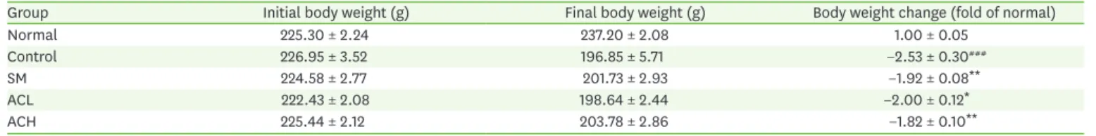
(Table 1).혈청내암모니아함량과
MPO
활성분석혈청 내 암모니아 함량을 측정한 결과
,정상군에 비해 대조군에서
50.5% (p < 0.001)유의하게 증가하였다
. SM및 인진호 추출물 투여군들은 대조군에 비해
SM군
16.3% (p < 0.05), ACL군
28.3% (p < 0.01)감소하였으며
,특히
ACH군은 대조군 대비
36.4% (p < 0.001)감소하여 정상
군 수준으로 나타났다
.혈청 내
MPO활성을 측정한 결과
,대조군은 정상군에 비해
p < 0.001Table 1. Initial and final body weight, body weight change
Group Initial body weight (g) Final body weight (g) Body weight change (fold of normal)
Normal 225.30 ± 2.24 237.20 ± 2.08 1.00 ± 0.05
Control 226.95 ± 3.52 196.85 ± 5.71 −2.53 ± 0.30###
SM 224.58 ± 2.77 201.73 ± 2.93 −1.92 ± 0.08**
ACL 222.43 ± 2.08 198.64 ± 2.44 −2.00 ± 0.12*
ACH 225.44 ± 2.12 203.78 ± 2.86 −1.82 ± 0.10**
All data are expressed as means ± SD (n = 8 rats per group).
Groups are represented as below: Normal, normal rats; Control, TAA-induced rats; SM, TAA-induced + silymarin 100 mg/kg body weight rats; ACL, TAA-induced + Artemisiae Capillaris Herba water extract 100 mg/kg body weight rats; ACH, TAA-induced + Artemisiae Capillaris Herba water extract 200 mg/kg body weight rats.
###p < 0.001 vs. Normal, *p < 0.05 and **p < 0.01 vs. Control.
수준으로 유의하게 증가하였으며
,이러한 대조군과 비교했을 때
SM군은
35.8% (p < 0.01)감 소하였다
.또한
,인진호 투여군인
ACL군과
ACH군은 대조군에 비하여 각각
27.8% (p < 0.05), 51.4% (p < 0.001)감소함으로 농도의존적인 경향이 나타났다
(Fig. 1).간조직내
NADPH oxidase
발현분석간 조직 내 산화적 스트레스를 측정하기 위해
western blotting으로
NADPH oxidase (NOX2, p22phox)의 발현을 확인하였다
.그 결과
, NOX2발현은 정상군에 비해 대조군의 발현이
29.0%(p < 0.001)
유의하게 증가하였다
.대조군과 비교하여 각각
SM군은
11.6% (p < 0.05), ACL군은
19.4% (p < 0.01), ACH군은
22.5% (p < 0.001)로
NOX2의 발현이 유의하게 감소하였으며
,특히
ACH
군은 정상군 수준까지 감소하는 것을 확인할 수 있었다
. p22phox의 발현 측정 결과
,정상
군과 비교해 대조군에서
20.0% (p < 0.01)유의하게 증가하였으며
,이러한 대조군에 비해 간 보호 약물 및 추출물 투여군들은 각각
SM군
11.7% (p < 0.05), ACL군
14.2% (p < 0.01), ACH군
15.0% (p < 0.01)로 유의하게 감소하였다
(Fig. 2).0 5 10 15 20
Normal Control SM ACL ACH Normal Control SM ACL ACH
Ammonia concentration (nmol/µL) MPO activity (mU/mL)
0 200 100 300 400
###
*
***
** *
###
**
***
A B
Fig. 1. Ammonia concentration and MPO activity in serum. Ammonia concentration (A) and MPO activity (B). TAA (200 mg/kg of body weight, I.P) and drug treatment compounds (P.O) were administered for 3 days. Groups are represented as below: Normal, normal rats; Control, TAA induce rats; SM, TAA-induced + silymarin 100 mg/kg body weight rats; ACL, TAA-induced + Artemisiae Capillaris Herba water extract 100 mg/kg body weight rats; ACH TAA-induced + Artemisiae Capillaris Herba water extract 200 mg/kg body weight rats. All data are expressed as means ± SD (n = 8 rats per group).
MPO, myeloperoxidase.
###p < 0.001 vs. Normal, *p < 0.05, **p < 0.01, and ***p < 0.001 vs. Control.
0 0.5 1.0 1.5
Normal Control SM ACL ACH Normal Control SM ACL ACH
Fold of normal Fold of normal
0 0.5 1.0
### 1.5
* ** *** **
##
* **
β-actin p22phox NOX2
β-actin
A B
Fig. 2. Expressions of NOX2 and p22phox protein in liver tissue. The protein expression level of NOX2 (A) and p22phox (B) were detected by western blotting. TAA (200 mg/kg of body weight, I.P) and drug treatment compounds (P.O) were administered for 3 days. Groups are represented as below: Normal, normal rats; Control, TAA induce rats; SM, TAA- induced + silymarin 100 mg/kg body weight rats; ACL, TAA-induced + Artemisiae Capillaris Herba water extract 100 mg/kg body weight rats; ACH TAA-induced + Artemisiae Capillaris Herba water extract 200 mg/kg body weight rats.
All data are expressed as means ± SD (n = 8 rats per group).
##p < 0.01 and ###p < 0.001 vs. Normal, *p < 0.05, **p < 0.01, and ***p < 0.001 vs. Control.
간조직내항산화관련인자발현분석
간 조직 내 항산화 관련 전사 인자로 알려진
Nrf2의 발현을 측정한 결과
,정상군에 비해 대조
군에서
22.0% (p < 0.001)유의하게 감소하였으며
,대조군과 비교해
SM군
15.4% (p < 0.05), ACL군
16.7% (p < 0.05)로 증가했으면서
, ACH군은 증가율은
29.5% (p < 0.001)로 정상군 수준 까지 증가하였다
. HO-1, SOD, Catalase및
GPx-1/2발현 측정결과
,모든 인자에서 정상군 대비 대조군이 유의하게 감소하였으며
(p < 0.001),간 보호 약물 및 추출물 투여군들은 대조군에 비해 증가하는 경향을 보였다
.특히
ACH군은
GPx-1/2에서 대조군 보다
46.4% (p < 0.001)증
가하여 정상군 수준으로 나타났다
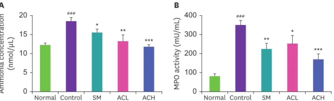
(Fig. 3).간조직내
TIMP-1
과MMPs
발현분석간 조직 내
MMPs발현을 측정한 결과
,모두 정상군에 비하여 대조군이 유의하게 증가하는 것
을 확인하였으며
(p < 0.001), MMP-2와
MMP-8의 발현이
ACH군에서 대조군에 비해 정상군
수준까지 유의하게 감소하는 것을 확인할 수 있었다
(p < 0.001).이러한
MMPs를 억제하는
TIMP-1
의 발현 측정결과
,정상군에 비해 대조군에서
45.0%유의하게 감소하였으며
,대조군
과 비교해
SM군은
30.9% (p < 0.01), ACL군은
38.2% (p < 0.001), ACH군은
58.2% (p < 0.001)증
가하는 것을 확인할 수 있었다
(Fig. 4).Normal Control SM ACL ACH
Normal Control SM ACL ACH
Normal Control SM ACL ACH
Fold of normal Fold of normal Fold of normal
0 0.4 0.8 1.2
0 0.4 0.8 1.2
0 0.4 0.8
** 1.2
### *
**
###
** **
###
***
**
Catalase β-actin
GPx-1/2 β-actin SOD
β-actin
C D E
0 0.4 0.8 1.2
Normal Control SM ACL ACH Normal Control SM ACL ACH
Fold of normal Fold of normal
0 0.4 0.8 1.2
### * ***
* **
###
* **
β-actin HO-1 Nrf2
Histone
A B
Fig. 3. Expressions of anti-oxidant protein in liver tissue. The protein expression level of Nrf2 (A), HO-1 (B), SOD (C), Catalase (D), and GPx-1/2 (E) were detected by western blotting. TAA (200 mg/kg of body weight, I.P) and drug treatment compounds (P.O) were administered for 3 days. Groups are represented as below:
Normal, normal rats; Control, TAA induce rats; SM, TAA-induced + silymarin 100 mg/kg body weight rats; ACL, TAA-induced + Artemisiae Capillaris Herba water extract 100 mg/kg body weight rats; ACH TAA-induced + Artemisiae Capillaris Herba water extract 200 mg/kg body weight rats. All data are expressed as means
± SD (n = 8 rats per group).
SOD, superoxide dismutase.
###p < 0.001 vs. Normal, *p < 0.05, **p < 0.01, and ***p < 0.001 vs. Control.
간조직
H&E
염색간 조직을
H&E염색하여 관찰한 결과
,정상군에 비해
TAA를 주입한 대조군에서 중심정맥 주
변을 기준으로 간 손상 병변과 염증세포의 침윤을 관찰할 수 있었다
.대조군과 비교하여 인 진호 추출물 투여군에서 염증세포의 침윤이 줄어든 것을 확인할 수 있었다
(Fig. 5).고찰
간은 우리 몸에서 대사기능에 매우 중요한 역할을 하는 기관으로
,전세계적으로 다양한 형태
의 간질환의 이환율과 사망율이 증가하고 있는 추세이다
[13].간에서 해독작용이 일어날 때
많은 양의 활성산소종
(ROS)이 생성되는데
,과도한
ROS로 인한 산화적 스트레스는 간에서 염증반응을 발생시켜 간염을 일으키게 되고
,간염이 만성적으로 지속되게 되면
,간섬유화와
간경변증 및 간암 등으로 진행되어 심하면 사망까지 이르게 되는 것으로 알려져 있다
[14].최
근에는 이러한 산화적 스트레스를 감소시키는 항산화 물질에 대한 연구가 많이 진행되고 있 다
.그렇기에 본 연구에서는
TAA의 산화적 스트레스에 의한 간손상 동물모델에서 인진호 열 수 추출물의 간보호 효과를 알아보았다
.0 0.5 1.0 1.5
Normal Control SM ACL ACH Normal Control SM ACL ACH
Fold of normal Fold of normal
0 0.5 1.0 1.5
### ** *** ***
*
###
*** *
β-actin MMP-1 TIMP-1
β-actin
A B
0 0.5 1.0 1.5
Normal Control SM ACL ACH Normal Control SM ACL ACH
Fold of normal Fold of normal
0 0.5 1.0 1.5
###
*** ** *** **
###
* ***
β-actin MMP-8 MMP-2
β-actin
C D
Fig. 4. Expressions of TIMP-1 and MMPs protein in liver tissue. The protein expression level of TIMP-1 (A), MMP-1 (B), MMP-2 (C), and MMP-8 (D) were detected by western blotting. TAA (200 mg/kg of body weight, I.P) and drug treatment compounds (P.O) were administered for 3 days. Groups are represented as below: Normal, normal rats; Control, TAA induce rats; SM, TAA-induced + silymarin 100 mg/kg body weight rats; ACL, TAA-induced + Artemisiae Capillaris Herba water extract 100 mg/kg body weight rats; ACH TAA-induced + Artemisiae Capillaris Herba water extract 200 mg/kg body weight rats. All data are expressed as means ± SD (n = 8 rats per group).
TIMP, tissue inhibitors of metalloproteinase; MMP, matrix metalloproteinase.
###p < 0.001 vs. Normal, *p < 0.05, **p < 0.01, and ***p < 0.001 vs. Control.
동물실험 후
,혈중 암모니아 함량과
MPO활성을 측정하였다
.간세포 효소
(transaminase)에 의해 장과 간 사이의 혈관인 간문맥을 지나 간으로 이동한 암모니아는
TAA를 주입할 경우 질
소 화합물인 요소
(urea)에 의해 원할한 대사활동을 하지 못하게 되어 혈중 암모니아 수치가
올라가게 된다
[15]. MPO는 호중구 과립구에 많이 있는 혈단백질로 많은 연구에서 호중구 침
윤의 지표로 많이 사용되며
, TAA에 의한 간손상 동물모델에서
MPO활성이 증가한다고 알려
져 있다
[3].이와 같은 암모니아 함량과
MPO활성을 혈청 내에서 측정한 결과
,정상군에 비
해
TAA로 간손상이 유발된 대조군에서 유의하게 증가하였으며
,대조군과 비교해 인진호 열 수 추출물 투여 시 농도의존적으로 유의하게 감소시키는 것을 확인할 수 있었다
.인진호 열수 추출물이 산화적 스트레스에 미치는 영향에 대해 알아보기 위해 간 조직 내에서 산화적 스트레스와 관련된 인자의 발현을 측정하였다
. NADPH oxidase (NOX)는 여러 자극들
에 의해 반응하여
ROS를 생성하는 효소이며
[16,17],간 내에서
NOX시스템에 의하여 발생하
는 산화적 스트레스는 간손상 등의 여러 간질환들의 진행에 중요한 역할을 한다고 알려져 있
다
[18].간 조직 내에서
NOX2와
p22phox의 발현을 측정한 결과
,정상군에 비해 대조군에서 유
의하게 발현이 증가한 것을 확인하였으며
,인진호 열수 추출물에서 대조군 보다 유의하게 감 소하는 것을 확인하였다
.우리 몸 안에서 산화적 스트레스가 일어나게 되면 세포는 산화
-환원 반응의 항상성을 유지하
기 위해서 항산화 작용을 하는
Nrf2와 기타 스트레스 반응 경로를 활성화 시킨다
[19].활성화
된
Nrf2는
antioxidant response element와 결합하여 주요 항산화 효소들을 활성화시키게 된다
[20].
본 연구에서는 간 조직 내 항산화와 관련된
Nrf2, HO-1, SOD, Catalase및
GPx-1/2의 단백 질 발현을 측정하였으며
,정상군과 비교해
TAA로 인해 산화적 스트레스가 일어난 대조군에 서 유의하게 감소하였고 대조군 대비 인진호 열수 투여군에서 농도의존적으로 유의하게 증
A B C
E D
Fig. 5. Pathological observation (hematoxylin & eosin) in liver tissue. Normal (A), Control (B), SM (C), ACL (D), and ACH (E). TAA (200 mg/kg of body weight, I.P) and drug treatment compounds (P.O) were administered for 3 days.
Groups are represented as below: Normal, normal rats; Control, TAA induce rats; SM, TAA-induced + silymarin 100 mg/kg body weight rats; ACL, TAA-induced + Artemisiae Capillaris Herba water extract 100 mg/kg body weight rats; ACH TAA-induced + Artemisiae Capillaris Herba water extract 200 mg/kg body weight rats (×200, scale bar 100 µm).
가하였다
.이를 통해 인진호 열수 추출물이 항산화 작용을 통해
TAA로 유발된 간손상 모델에 서 산화적 스트레스를 일으키는
ROS의 생성을 억제할 것으로 판단된다
.MMPs
는 세포외 기질
(extracellular matrix)의 대부분의 구성 요소를 분해할 수 있는 효소로
MMPs
의 활성은
TIMPs에 의해 조절된다
[21,22].또한
, MMPs는 간염
,지방간 및 간섬유증 등 의 간질환에서 간의 재생 과정에 관여하며
,급성 간손상에서 산화적 스트레스에 의해 발현이 증가한다고 알려져 있다
[23].그렇기에 본 연구에서는 간 조직 내에서
MMPs와
TIMP-1의 단 백질 발현을 측정하였다
.그 결과
, MMPs는 대조군 대비 인진호 열수 추출물 투여군에서 유 의하게 발현이 감소하였으며
, TIMP-1은 대조군과 비교해 인진호 열수 추출물 투여군에서 유 의하게 발현이 증가한 것을 확인할 수 있었다
.종합적으로 본 연구에서는 위와 같은 결과를 통해
TAA로 유발된 간손상에서 인진호 열수 추 출물이 항산화 작용을 통해 산화적 스트레스를 억제함으로써 간보호에 효과적인 것으로 판 단된다
.요약
본 연구는
TAA복강투여로 유발된 간 손상 동물모델에서 인진호 열수 추출물의 간보호 효
능을 평가하였으며 다음과 같은 결론을 얻었다
. TAA로 인해 줄어드는 체중은 인진호 열수 추출물을 투여한 군에서 유의하게 증가하였으며
,간손상에 의해 증가한 혈중 암모니아 함
량과
MPO활성은 인진호 열수 추출물 투여군에서 유의하게 감소하였다
.간 조직의
westernblotting
결과
,인진호 열수 추출물 투여가 산화적 스트레스 관련 인자들의 발현을 유의적으
로 감소시키고
,항산화 관련 인자들의 발현을 유의하게 증가시켰으며
, MMPs의 발현은 감소
시키고
TIMP-1의 발현은 증가시킴을 확인할 수 있었다
.따라서 인진호 열수 추출물은
TAA로
유발된 간손상 동물모델에서 항산화 작용을 통해 산화적 스트레스를 억제하여 간보호 효과 를 보이는 것으로 판단된다
.REFERENCES
1. Park JY, Park CM, Kim JJ, Song YS. Hepatoprotective activity of Dandelion (Taraxacum officinale) water extract against D-galactosamine-induced hepatitis in rats. J Korean Soc Food Sci Nutr 2008; 37(2): 177-183.
CROSSREF
2. Lee UE, Friedman SL. Mechanisms of hepatic fibrogenesis. Best Pract Res Clin Gastroenterol 2011; 25(2):
195-206.
PUBMED | CROSSREF
3. Özdemir-Kumral ZN, Erkek BE, Karakuş B, Almacı M, Fathi R, Yüksel M, et al. Potential effect of 1,25 Dihydroxyvitamin D3 on thioacetamide-induced hepatotoxicity in rats. J Surg Res 2019; 243: 165-172.
PUBMED | CROSSREF
4. Han JM, Kim HG, Choi MK, Lee JS, Lee JS, Wang JH, et al. Artemisia capillaris extract protects against bile duct ligation-induced liver fibrosis in rats. Exp Toxicol Pathol 2013; 65(6): 837-844.
PUBMED | CROSSREF
5. Kim JS, Kim KL. Anti-oxidative and anti-inflammatory effects of Artemisiae Capillaris Extract. Korean J Aesthet Cosmetol 2015; 13(6): 805-812.
6. Ham I, Jung SW, Lee KJ, Park KH, Choi HY. Effect of the aerial part of Artemisia capillaris, and A.
iwayomogi on the hyperlipidemia of rats induced by Triton WR-1339. Korea J Herbol 2005; 20(1): 45-52.
7. Kim HT, Kim JW, Lim MK, Jin TW, Yeo SG, Jang KH, et al. Cytotoxic effect of Artemisia capillaris extracts on the cancer cells on in vitro. J Vet Clin 2007; 24(3): 367-371.
8. Yu F, Qian H, Zhang J, Sun J, Ma Z. Simultaneous quantification of eight organic acid components in Artemisia capillaris Thunb (Yinchen) extract using high-performance liquid chromatography coupled with diode array detection and high-resolution mass spectrometry. J Food Drug Anal 2018; 26(2): 788-795.
PUBMED | CROSSREF
9. Kim KS, Yang HJ, Lee JY, Na YC, Kwon SY, Kim YC, et al. Effects of β-sitosterol derived from Artemisia capillaris on the activated human hepatic stellate cells and dimethylnitrosamine-induced mouse liver fibrosis. BMC Complement Altern Med 2014; 14(1): 363-372.
PUBMED | CROSSREF
10. Lee SH, Lee JY, Kwon YI, Jang HD. Anti-osteoclastic activity of Artemisia capillaris Thunb. extract depends upon attenuation of osteoclast differentiation and bone tesorption-associated acidification due to chlorogenic acid, hyperoside, and scoparone. Int J Mol Sci 2017; 18(2): 322-335.
CROSSREF
11. Kwon H, Jung JW, Lee YC, Ryu JH, Kim DH. Neuroprotective effect of the ethanol extract of Artemisia capillaris on transient forebrain ischemia in mice via nicotinic cholinergic receptor. Chin J Nat Med 2018;
16(6): 428-435.
PUBMED | CROSSREF
12. Luo M, Dong L, Li J, Wang Y, Shang B. Protective effects of pentoxifylline on acute liver injury induced by thioacetamide in rats. Int J Clin Exp Pathol 2015; 8(8): 8990-8996.
PUBMED
13. El Awdan SA, Amin MM, Hassan A. Cilostazol attenuates indices of liver damage induced by
thioacetamide in albino rats through regulating inflammatory cytokines and apoptotic biomarkers. Eur J Pharmacol 2018; 822: 168-176.
PUBMED | CROSSREF
14. Kim KJ, Shin MR, Kim SH, Kim SJ, Lee AR, Kwon O, et al. Protective effect of Tongyuhwalhyeol-tang on liver injury in thioacetamide-induced rat. Korea J Herbol 2018; 33(1): 37-46.
15. Brusilow SW. Hyperammonemic encephalopathy. Medicine (Baltimore) 2002; 81(3): 240-249.
PUBMED | CROSSREF
16. De Minicis S, Bataller R, Brenner DA. NADPH oxidase in the liver: defensive, offensive, or fibrogenic?
Gastroenterology 2006; 131(1): 272-275.
PUBMED | CROSSREF
17. Cheng G, Cao Z, Xu X, van Meir EG, Lambeth JD. Homologs of gp91phox: cloning and tissue expression of Nox3, Nox4, and Nox5. Gene 2001; 269(1-2): 131-140.
PUBMED | CROSSREF
18. Paik YH, Kim J, Aoyama T, De Minicis S, Bataller R, Brenner DA. Role of NADPH oxidases in liver fibrosis.
Antioxid Redox Signal 2014; 20(17): 2854-2872.
PUBMED | CROSSREF
19. Kasai S, Shimizu S, Tatara Y, Mimura J, Itoh K. Regulation of Nrf2 by mitochondrial reactive oxygen species in physiology and pathology. Biomolecules 2020; 10(2): 320-340.
PUBMED | CROSSREF
20. Lee JA, Park HJ, Kim SH, Kim MJ, Kim KJ, Shin MR, et al. Evaluation of Evodiae Fructus extract on the chronic acid reflux esophagitis in rats. Korea J Herbol 2019; 34(2): 15-23.
21. Kessenbrock K, Plaks V, Werb Z. Matrix metalloproteinases: regulators of the tumor microenvironment.
Cell 2010; 141(1): 52-67.
PUBMED | CROSSREF
22. Visse R, Nagase H. Matrix metalloproteinases and tissue inhibitors of metalloproteinases: structure, function, and biochemistry. Circ Res 2003; 92(8): 827-839.
PUBMED | CROSSREF
23. Naim A, Pan Q, Baig MS. Matrix metalloproteinases (MMPs) in liver diseases. J Clin Exp Hepatol 2017;
7(4): 367-372.
PUBMED | CROSSREF