ISSN 2234-3806 • eISSN 2234-3814
242 www.annlabmed.org http://dx.doi.org/10.3343/alm.2015.35.2.242 Ann Lab Med 2015;35:242-245
http://dx.doi.org/10.3343/alm.2015.35.2.242
Brief Communications
Clinical Microbiology
Molecular Characteristics of Noroviruses Genogroup I and Genogroup II Detected in Patients With Acute Gastroenteritis
Heejin Ham, M.S., Seah Oh, M.S., Hyunjung Seung, M.S., and Sukju Jo, M.S.
Department Microbiology, Seoul Metropolitan Government Research Institute of Public Health and Environment, Gwacheon, Korea
Noroviruses are the leading cause of epidemic gastroenteritis, including foodborne out- break, in Korea. The prevalence of human noroviruses was studied in diarrheal stool sam- ples of patients with acute gastroenteritis by conventional duplex reverse transcription (RT)-PCR. Diarrheal stool samples were collected from 1,685 patients from the local hos- pitals in Seoul. The prevalence of the noroviruses was 22.8% (222/972 patients) in 2012 and 11.2% (80/713 patients) in 2013, with a total of 17.9% (302/1,685 patients). Geno- typing was performed on 302 norovirus-positive stool samples to reveal 5.6% prevalence of genogroup I (GI) (17/302) and 94.4% prevalence of genogroup II (GII) (285/302). The patients with norovirus-associated acute gastroenteritis mostly showed prevalence of GII norovirus, especially GII.4 (64.6%; 195/302).
Key Words: Norovirus, Acute gastroenteritis, Prevalence, GI, GII, GII.4
Received: July 2, 2014
Revision received: September 4, 2014 Accepted: December 16, 2014 Corresponding author: Heejin Ham Microbiology Department, Seoul Metropolitan Government Research Institute of Public Health and Environment, 30 Janggunmaeul 3-gil, Gwacheon 427-070, Korea
Tel: +82-2-570-3426 Fax: +82-2-570-3275 E-mail: [email protected]
© The Korean Society for Laboratory Medicine This is an Open Access article distributed under the terms of the Creative Commons Attribution Non-Commercial License (http://creativecom- mons.org/licenses/by-nc/3.0) which permits unrestricted non-commercial use, distribution, and reproduction in any medium, provided the original work is properly cited.
Monitoring of diarrheal patients in Korea has revealed a declin- ing trend of bacterial infections and a rising trend of viral infec- tions. Gastroenteritis caused by noroviruses is becoming increas- ingly common and, owing to its contagious nature, it is easily transmitted [1-5]. Hence, noroviruses have been identified as the most common causative agent [6]. In the present study, we investigated the prevalence of common human norovirus in pa- tients with acute gastroenteritis.
Diarrheal stool samples from 1,685 patients suffering from acute gastroenteritis were collected from six local hospitals in Seoul (SW hospital, SG hospital, AM center, NP hospital, EJ hospital, and NM center) between 2012 and 2013. The samples were tested for the presence of norovirus. Briefly, 1 g of feces was mixed with 9 mL of sterile 0.1 M phosphate-buffered saline (PBS; pH 7.4; Sigma, St. Louis, MO, USA) and centrifuged at 1,660 g for 30 min at 4˚C. The supernatant was collected and
used for fragmentation at 4˚C. At the AM center, 0.5-1 mL of the rectal swab suspensions were collected and mixed with PBS for further processing. The 1,685 diarrheal stool samples (direct rectal swabs or diarrheal feces of patients) from six hospitals were transported at 4˚C within 8 hr of collection to the Seoul Metropolitan Research Institute of Public Health and Environ- ment, where they were stored at -70˚C until further analysis.
For the detection of noroviruses, one-step reverse transcrip- tion (RT)-PCR, followed by electrophoresis, was performed [7].
RNA was extracted by using the Viral RNA Mini Kit (QIAgen, Hilden, Germany) [8]. One-step duplex RT-PCR was performed by using the RT/PCR Diagnostic Norovirus Kit (Bioneer, Dae- jeon, Korea) and the GI-FIM, GI-RIM, and GI-F2 primer pairs for GI detection; the GII-FIM, GII-RIM, and GII-F3 primer pairs for GII detection; -and primers hybridized within the open reading frame 3 regions (5´-CTGCCCGAATTYGTAAATAAATGATGAT-3´,
Ham H, et al.
Noroviruses in gastroenteritis patients
http://dx.doi.org/10.3343/alm.2015.35.2.242 www.annlabmed.org 243
5´-CCAACCCARCCATTRTACATYTG-3´, and 5´-ATGATGATGGC- GTCTAAGGACGC-3´ respectively) for GI detection and another 3 regions (5´-GGGAGGGCGATCGCAATCT-3´, 5´-CCRCCIG- CATRICCRTTRTACAT-3´, and 5´-TTGTGAATGAAGATGGCGTC- GART-3´, respectively) for GII detection [1-5]. Positive results were defined as the presence of a 314-bp band for norovirus GI and a 313-bp band for norovirus GII [1-5].
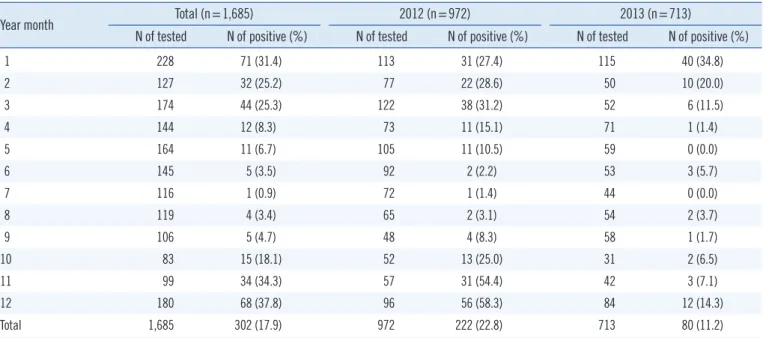
Our analysis showed that the norovirus detection rate was 22.8% (222/972 patients) in 2012 and 11.2% (80/713 pa- tients) in 2013, with a total rate of 17.9% (302/1,685 patients) (Table 1). In subsequent studies, monthly surveillance of norovi- rus prevalence over a period of 2 yr (2012-2013) revealed the highest frequency between October and March.
Genotyping of 302 (17.9%) norovirus-positive samples re- vealed 5.6% (17/302) prevalence of GI- and 94.4% (285/302) prevalence of GII. Notably, in norovirus-associated acute gastro- enteritis patients, the prevalence of GII genotype norovirus was the highest, especially of GII.4 (64.6%;195/302) (Table 2).
In the Hallym University Hospital, the epidemicity of norovirus started in October 2007, peaked in November 2007, and lasted until January in 2008; again, in the same hospital, the epidemic- ity started in December 2009, peaked in January 2010, and lasted until February 2010 [6]. Kong et al. [8] reported the high- est detection rate of norovirus in November 2005 (12.3%, 28/227) and in March 2006 (35.3%, 79/224), followed by that in March 2007 (45.9%, 62/135) in Incheon. Ham et al. [4] re- ported the highest peak in January (35.6%, 366/1,028), and the Table 1. The recorded incidences of norovirus infection and the number of detected noroviruses from patients with acute gastroenteritis during 2012-2013 in Seoul via periodic surveillance
Year month Total (n=1,685) 2012 (n=972) 2013 (n=713)
N of tested N of positive (%) N of tested N of positive (%) N of tested N of positive (%)
1 228 71 (31.4) 113 31 (27.4) 115 40 (34.8)
2 127 32 (25.2) 77 22 (28.6) 50 10 (20.0)
3 174 44 (25.3) 122 38 (31.2) 52 6 (11.5)
4 144 12 (8.3) 73 11 (15.1) 71 1 (1.4)
5 164 11 (6.7) 105 11 (10.5) 59 0 (0.0)
6 145 5 (3.5) 92 2 (2.2) 53 3 (5.7)
7 116 1 (0.9) 72 1 (1.4) 44 0 (0.0)
8 119 4 (3.4) 65 2 (3.1) 54 2 (3.7)
9 106 5 (4.7) 48 4 (8.3) 58 1 (1.7)
10 83 15 (18.1) 52 13 (25.0) 31 2 (6.5)
11 99 34 (34.3) 57 31 (54.4) 42 3 (7.1)
12 180 68 (37.8) 96 56 (58.3) 84 12 (14.3)
Total 1,685 302 (17.9) 972 222 (22.8) 713 80 (11.2)
Table 2. The distribution of norovirus genotypes identified in Seoul Genotype N of positive samples Positive rate (%)
GI GI.1 1 0.3
GI GI.2 3 1.0
GI GI.3 1 0.3
GI GI.4 4 1.3
GI GI.7 2 0.7
GI GI.8 3 1.0
GI GI.10 1 0.3
GI GI.13 1 0.3
GI GI.14 1 0.3
GII GII.1 10 3.3
GII GII.2 16 5.3
GII GII.3 20 6.6
GII GII.4 195 64.6
GII GII.5 1 0.3
GII GII.6 19 6.3
GII GII.7 1 0.3
GII GII.8 2 0.7
GII GII.11 8 2.7
GII GII.16 2 0.7
GII GII.17 1 0.3
GII non-typable 7 2.3
GII mixed typable 3 1.0
Total 302 100
Ham H, et al.
Noroviruses in gastroenteritis patients
244 www.annlabmed.org http://dx.doi.org/10.3343/alm.2015.35.2.242 lowest in July (2.4%, 20/818) from 2007 to 2011, followed by
that in January, February, and November from 2010 to 2013, in Seoul [5]. In addition, the Korea Centers for Disease Control and Prevention (KCDC) reported that norovirus prevalence- peaks from November to March in Korea [9]. In this study, the preva- lence of noroviruses was found to be the highest between Octo- ber and March in the years 2012 and 2013.
Of the 11,202 acute gastroenteritis patients whose stool sam- ples were examined from 2007 to 2011 in Seoul, 16.6% (1,861/
11,202) were found to be positive for noroviruses by RT-PCR in the Hallym University Hospital. Of the 1,861 noroviruses, 95.5%
were detected as GII and 3.9% were detected as GI [4]. Ap- proximately 6,618 acute gastroenteritis patients were recorded between 2005 and 2007 in Incheon; of the 6,618 stool samples tested, 10.7% (708/6,618) were determined to be infected with noroviruses by RT-PCR. Of the 708 noroviruses, GI constituted 0.6% and GII constituted 99.4% (69.7% GII.4 and 17.2%
GII.3) [8]. Of the 1,718 acute gastroenteritis patients recorded between 2012 and 2013 in the Hallym University Hospital, 14.8% (254/1718) were positive for human norovirus as deter- mined by using the norovirus antigen test kit. Of the 254 norovi- rus cases detected, 2.2% was attributable to GI and 97.8% to GII. Of the 97.8% GII noroviruses detected, 63.7% was attribut- able to GII.4, constituted by 60.4% GII.4 Sydney and 3.3% GII.4 Non-Sydney (Den Haag 2006b) [10]. Yu et al. [11] reported that norovirus GI accounted for 4% of the detected norovirus cases, while norovirus GII accounted for 92%, of which 70%
was attributable to GII.4, between 1999 and 2011 in China. Bok et al. [12] stated that norovirus GI accounted for 3.4% of the detected noroviruses and GII accounted for 95.8%. Of the GII- positive cases, 16% of the cases were attributed to GII.4; these cases were over a 34-yr period between 1974 and 1991 in the Washington DC region in the USA.
Consequently, most of the gastroenteritis patients were related to GII norovirus infection, especially noroviruses GII.4. Our results on the prevalence of noroviruses were in accordance with previ- ous results; for instance, the prevalence of GI (5.6%) and GII (94.4%) was similar to that in previous studies (0.6-3.9% and 92.0-99.4%, respectively) [4, 8, 10-12], while the prevalence of GII.4 (64.6%) was similar to that in other studies (63.7-70.0%) [8, 10, 11], but different from that in Bok et al. (16.0%) [12].
In addition, in Hallym University Hospital, 13.0% (1,463/
11,219) of the stool samples in gastroenteritis patients were de- termined to be positive for noroviruses from 2007 to 2010 by using the norovirus ELISA kit [6]. The KCDC reported that, out of 18,908 diarrheal stool samples, 8.5% (1,608) were found to
be positive for human norovirus in 2013 [9]. In 4,484 stool sam- ples obtained from 461 patients of food poisoning during on out- break from 2010 to 2013 in Seoul, 9.77% (438/4,484) were de- termined to be positive for noroviruses by RT-PCR; of the 438 noroviruses, 75.92% was positive for norovirus GII, and 54.05%
for norovirus GII.4 [5].
All stool samples were obtained from outpatients of six hospi- tals, including one primary hospital (AM center), three second- ary hospitals (SW hospital, NP hospital, and NM center), and two tertiary hospitals (SG and EJ hospitals). The norovirus de- tection rate was different among the three types of hospitals.
The order of norovirus detection rate in gastroenteritis patients was primary (14.6%) < secondary (20.5%) < tertiary (26.8%) in 2012 and secondary (9.0%) < tertiary (12.0%) < primary (12.6%) in 2013. However, no differences were noted in the norovirus positive rates between stool and rectal swab samples.
In conclusion, noroviruses were found to be prevalent in pa- tients with gastroenteritis, with a higher occurrence between Oc- tober and March over the 12-month period between 2012 and 2013. Our analysis of diarrheal stool samples collected during 2012 to 2013 from local hospitals in Seoul revealed norovirus detection in 17.9% of the gastroenteritis patients (302/1,685), of whom, 5.6% (17/302) was constituted by norovirus GI and 94.4% (285/302) by norovirus GII (64.6% GII.4).
Authors’ Disclosures of Potential Conflicts of Interest
No potential conflicts of interest relevant to this article were re- ported.
Acknowledgments
We are grateful to the EnterNet-Korea Project of the Korean CDC for supporting this study.
REFERENCES
1. Lee JI. Molecular characterization of enteric viruses isolated from acute gastroenteritis patients. Chung Buk University, 2008. Ph.D. Thesis.
2. Park SH, Kim EJ, Oh SA, Kim CK, Choi SS, Cho SJ, et al. Viral agents as- sociated with acute gastroenteritis in Seoul, Korea. Clin Lab 2011;57:
59-65.
3. Lee JI, Park SH, Kim MS, Oh YH, Yu IS, Choi BH, et al. Surveillance of acute gastroenteritis in Seoul, Korea, during May 2004 and June 2007.
J Bacteriol Virol 2009;39:363-71.
4. Ham HJ, Oh SA, Kim CK, Jang JI, Cho SJ, Choi SM. Molecular characteris- tics of human noroviruses genogroup I and genogroup II detected in acute gastroenteritis patients in Seoul. J Environ Health Sci 2012;38:57-65.
Ham H, et al.
Noroviruses in gastroenteritis patients
http://dx.doi.org/10.3343/alm.2015.35.2.242 www.annlabmed.org 245
5. Oh SA, Park SH, Ham HJ, Seung HJ, Jang JI, Suh SW, et al. Molecular characterization of norovirus and rotavirus in outbreak of acute gastro- enteritis in Seoul. J Bacteriol Virol 2013;43:307-16.
6. Park DJ, Kim JS, Park JY, Kim HS, Song WK, Kim HS, et al. Epidemio- logical analysis of norovirus infection between March 2007 and Febru- ary 2010. Korean J Lab Med 2010;30:647-53.
7. Korea Centers for Disease Control and Prevention (KCDC). K-Calcinet.
National norovirus laboratory surveillance, 2013. http://www.google.
co.kr/url?sa=t&rct=j&q=&esrc=s&frm=1&source=web&cd=1&ved=0C BwQFjAA&url=http%3A%2F%2Fwww.cdc.go.kr%2FCDC%2Fcms%2F cmsFileDownload.jsp%3Ffid%3D31%26cid%3D22015%26fieldName
%3Dattach1%26index%3D1&ei=nUCKVMOWDZLz8gWdnoHwDw&us g=AFQjCNEAE32JB_qUoBija4o14zIve9jOog&bvm=bv.81828268,d.
dGc&cad=rjt.
8. Kong YW, Oh BY, Kim HY, Lee MY, Kim YH, Go JM, et al. Molecular epi-
demiologic investigation of norovirus infections in Incheon city, Korea, from 2005 to 2007. J Bacteriol Virol 2008;38:249-57.
9. Korea Centers for Disease Control and Prevention (KCDC). EnterNet Korea. Yearly Report 2013;41-52. Laboratory surveillance of viral acute gastroenteritis in Korea, 2012. http://www.cdc.go.kr/CDC/notice/CdcK- rInfo0301.jsp?menuIds=HOME001-MNU0004-MNU0036-MNU 0037&cid=21904 (Updated on Nov 2013).
10. Kim HS, Hyun JW, Kim HS, Kim JS, Song WK, Lee KM. Emergence of GII.4 Sydney norovirus in South Korea during the winter of 2012-2013.
J Microbiol Biotechnol 2013;23:1641-3.
11. Yu Y, Yan S, Li B, Pan Y, Wang Y. Genetic diversity distribution of human norovirus in China (1999-2011). Biomed Res Int 2014;2014: 196169.
12. Bok K, Abente EJ, Realpe-Quintero M, Mitra T, Sosnovtsev SV, Kapikian AZ, et al. Evolutionary dynamics of GII.4 noroviruses over a 34-year pe- riod. J Virol 2009;83:11890-901.