ISSN 2234-3806 • eISSN 2234-3814
76 www.annlabmed.org https://doi.org/10.3343/alm.2020.40.1.76 Ann Lab Med 2020;40:76-79
https://doi.org/10.3343/alm.2020.40.1.76
Letter to the Editor
Diagnostic Hematology
First Case of Double T-Cell Receptor Alpha/Delta Rearrangements of t(11;14) and inv(14) and
Subsequent JAK2 Rearrangement in a Patient With T-cell Acute Lymphoblastic Leukemia
Chang Ahn Seol , M.D., Ph.D.1, Young-Uk Cho , M.D., Ph.D.1, Seongsoo Jang , M.D., Ph.D.1, Chan-Jeoung Park , M.D., Ph.D.1, Jung-Hee Lee , M.D., Ph.D.2, and Eul-Ju Seo , M.D., Ph.D.1
Departments of 1Laboratory Medicine and 2Internal Medicine, University of Ulsan College of Medicine and Asan Medical Center, Seoul, Korea
Dear Editor,
Primary cytogenetic abnormalities of T-cell ALL (T-ALL) frequently involve rearrangements between oncogenic transcription factor genes and T-cell receptor (TCR) loci. These are usually mutually exclusive and associated with specific genetic subgroups [1, 2].
We report the first case, to our knowledge, of T-ALL showing dis- tinct and rare abnormalities of sequential TCR alpha/delta (TRA/
D) locus rearrangements associated with t(11;14)(p13;q11.2), inv(14)(q11.2q32), and clonal evolution of JAK2 rearrangement.
This report was approved by Asan Medical Center Institutional Review Board, Seoul, Korea (S2019-1375-0001).
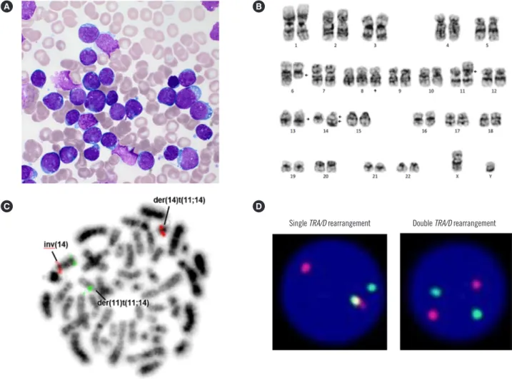
A 21-years-old man was admitted to Asan Medical Center in September 2017, with chest discomfort, petechiae, and oral bleeding. The study period was six months from admission. On admission, his hemogram showed a white blood cell count of 55×109/L with 68% lymphoblasts, 141 g/L Hb, and 71×109/L platelets. Computed tomography revealed a mediastinal mass, and a bone marrow (BM) examination showed that 90% of nu- cleated cells were lymphoblasts (Fig. 1A). The immunopheno- type of the lymphoblasts by four-color flow cytometric analysis (BD FACSCanto II; BD Biosciences, San Jose, CA, USA) was
positive for CD2, CD7, CD8, terminal deoxynucleotidyl transfer- ase, and cytoplasmic CD3. The karyotype of the BM cells was 47,XY,del(6)(q13q23),+8,t(11;14)(p13;q11.2),inv(14)(q11.2q32) [13]/46,XY[22] (Fig. 1B). Metaphase fluorescence in situ hy- bridization (FISH) analysis using the Vysis LSI TRA/D Dual Color Break Apart Rearrangement Probe (Abbott Laboratories, Abbott Park, IL, USA) showed double TRA/D rearrangements associ- ated with t(11;14) and inv(14) (Fig. 1C), while interphase FISH revealed a single TRA/D rearrangement in 12.5% of the cells and double TRA/D rearrangements in 74.0% of the cells (Fig. 1D).
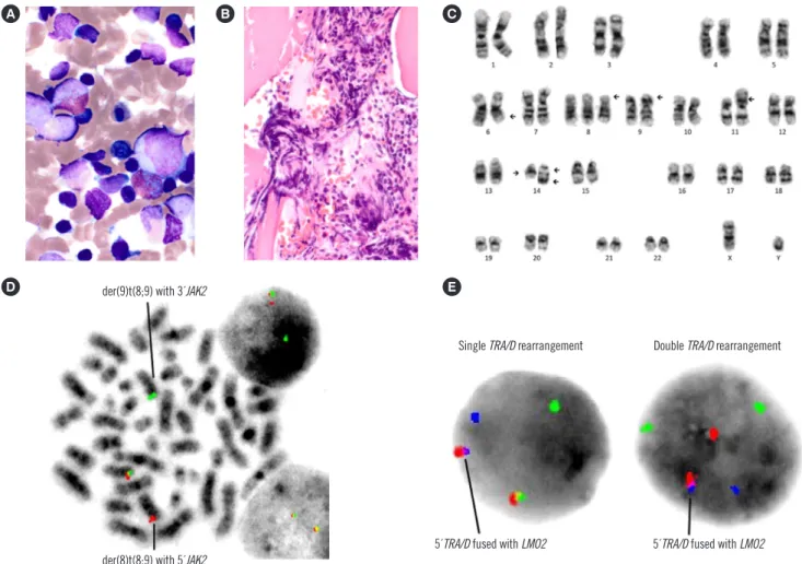
The patient received two cycles of cyclophosphamide, vincris- tine, doxorubicin, and dexamethasone (hyper-CVAD) for a month and achieved partial remission on day 47. Three weeks later, he was readmitted with disease progression. The follow-up BM ex- amination showed 57.4% lymphoblasts, increased eosinophils (4%), and focal fibrosis (MF-1) (Fig. 2A and 2B). The BM karyo- type was 47,XY,del(6)(q13q23),+8,t(8;9)(p22;p24),t(11;14)(p13;
q11.2),inv(14)(q11.2q32)[20]/46,XY[10] including clonal evolu- tion of t(8;9)(p22;p24) (Fig. 2C). The t(8;9)(p22;p24) compo- nent showed a JAK2 rearrangement by FISH analysis using the JAK2 Break Apart FISH Probe (Empire Genomics LLC, Buffalo,
Received: February 26, 2019 Revision received: June 3, 2019 Accepted: July 11, 2019
Corresponding author: Eul-Ju Seo, M.D., Ph.D.
Department of Laboratory Medicine, University of Ulsan College of Medicine and Asan Medical Center, 88 Olympic-ro 43-gil, Songpa-gu, Seoul 05505, Korea
Tel: +82-2-3010-4507, Fax: +82-2-478-0884, E-mail: [email protected]
© Korean Society for Laboratory Medicine
This is an Open Access article distributed under the terms of the Creative Commons Attribution Non-Commercial License (http://creativecommons.org/licenses/by-nc/4.0) which permits unrestricted non-commercial use, distribution, and reproduction in any medium, provided the original work is properly cited.
1 / 1 CROSSMARK_logo_3_Test
2017-03-16 https://crossmark-cdn.crossref.org/widget/v2.0/logos/CROSSMARK_Color_square.svg
Seol CA, et al.
Double TRA/D and JAK2 rearrangements in T-ALL
https://doi.org/10.3343/alm.2020.40.1.76 www.annlabmed.org 77
NY, USA) (Fig. 2D). Metaphase and interphase FISH at diagno- sis was negative for JAK2 rearrangement. Reverse transcriptase -PCR analysis using several primer sets for hotspot breakpoints of PCM1 (exons 23, 26, 36) and JAK2 (exons 3, 9, 11, 17) failed to detect the PCM1-JAK2 fusion transcript; these are most com- monly associated with t(8;9)(p22;p24) [3, 4]. Sequencing of NO- TCH1 revealed an in-frame deletion of the heterodimerization (HD) domain (NM_017617.5:c.4732_4734delGTG (p.V1578del)) and a nonsense mutation in the proline/glutamic acid/serine/
threonine (PEST) domain (NM_017617.5:c.5707C>T (p.Q2503*)).
We further investigated malignant clones, using a bacterial artifi- cial chromosome FISH probe for RP11-162F6 (Aqua, 11p13), which covers the LMO2 gene. FISH analysis using the RP11- 162F6 and TRA/D probes showed that LMO2 was fused with 5´TRA/D in all cells with a single TRA/D rearrangement as well
as double TRA/D rearrangements (Fig. 2E). This indicated that t(11;14) was probably the primary event, rather than inv(14), as a single TRA/D rearrangement was associated with LMO2 rear- rangement.
The patient was refractory to chemotherapy regimens. He died four months later, after disease progression.
The t(11;14)(p13;q11.2) component is associated with LMO2 on 11p13 and TRA/D on 14q11.2 and is found in 6% of T-ALL patients [1]. Nevertheless, inv(14)(q11.2q32) is very rare in T- ALL. In cases of T-ALL with inv(14)(q11.2q32), possible coexis- tence with rearrangements of other TCR loci, such as add(7) (q34), t(11;14)(p13;q11.2) and t(8;14)(q24;q11.2), has been reported, indicating that inv(14)(q11.2q32) could constitute a combined cytogenetic abnormality causing T-ALL [5, 6]. BCL11B and TRD may be associated with inv(14)(q11.2q32) [7].
Fig. 1. BM examination, karyotyping, and FISH analysis at diagnosis. (A) Lymphoblasts on the BM aspirate smear (Wright-Giemsa,
×1,000). (B) BM karyogram. (C, D) Metaphase and interphase FISH analyses with TRA/D Dual Color Break Apart Rearrangement Probe for 14q11.2 (5´TRA/D: SpectrumRed, 3´TRA/D: SpectrumGreen).
Abbreviations: BM, Bone marrow; FISH, fluorescence in situ hybridization; inv, inversion; der, derivative chromosome; TRA/D, T-cell receptor alpha/delta.
A B
C D
Single TRA/D rearrangement Double TRA/D rearrangement
Seol CA, et al.
Double TRA/D and JAK2 rearrangements in T-ALL
78 www.annlabmed.org https://doi.org/10.3343/alm.2020.40.1.76 Most cases in a provisional category of myeloid/lymphoid neo-
plasms associated with t(8;9)(p22;p24.1) and PCM1-JAK2 are considered myeloid neoplasms, and only a few cases are B-cell ALL (B-ALL) or T-cell lymphoma [8]. A recent study reported that hematopoietic neoplasms with JAK2 rearrangement are ex- tremely rare, and most are associated with PCM1-JAK2 rear- rangement [9]. Our patient, who had a JAK2 rearrangement of t(8;9)(p22;p24.1) as a clonal evolution, shared the characteris- tics of the entity with PCM1-JAK2 rearrangement such as eosin- ophilia and fibrosis on follow-up BM examination. A JAK2 inhib- itor, such as ruxolitinib, would be effective for treating patients with ALL involving JAK2 rearrangement [10].
The t(11;14)(p13;q11.2) component was probably the primary cytogenetic event, with inv(14) and t(8;9) occurring successively.
Clonal evolutions, dual NOTCH1 mutations, and additional chro-
mosomal abnormalities may contribute to a rapidly progressive clinical course. Follow-up cytogenetic evaluations and support- ing tests, including FISH, are important to detect cytogenetic clonal evolution.
Author Contributions
All authors equally contributed to this study. All authors have accepted their responsibility for the entire content of this manu- script and approved submission.
Conflict of Interest
None declared.
Fig. 2. BM examination, karyotyping, and FISH analysis at follow-up. (A) Eosinophils (Wright-Giemsa, ×1,000) and (B) focal fibrosis (HE,
×400). (C) BM karyogram. (D) Metaphase and interphase FISH analyses with JAK2 Break Apart FISH Probe for 9p24.1 (5´JAK2: Red, 3´JAK2: Green). (E) Interphase FISH analysis with RP11-162F6 (LMO2) for 11p13 (Aqua) and TRA/D Dual Color Break Apart Rearrange- ment Probe for 14q11.2 (5´TRA/D: SpectrumRed, 3´TRA/D: SpectrumGreen).
Abbreviations: BM, Bone marrow; HE, hematoxylin and eosin; FISH, fluorescence in situ hybridization; der, derivative chromosome; TRA/D, T-cell receptor alpha/delta.
A B C
D E
Single TRA/D rearrangement der(9)t(8;9) with 3´JAK2
der(8)t(8;9) with 5´JAK2 5´TRA/D fused with LMO2 5´TRA/D fused with LMO2
Double TRA/D rearrangement
Seol CA, et al.
Double TRA/D and JAK2 rearrangements in T-ALL
https://doi.org/10.3343/alm.2020.40.1.76 www.annlabmed.org 79
ORCID
Chang Ahn Seol https://orcid.org/0000-0001-8470-7633 Young-Uk Cho https://orcid.org/0000-0002-4403-8989 Seongsoo Jang https://orcid.org/0000-0002-0045-1747 Chan-Jeoung Park https://orcid.org/0000-0003-4396-8348 Jung-Hee Lee https://orcid.org/0000-0002-3127-0068 Eul-Ju Seo https://orcid.org/0000-0002-8247-3746
REFERENCES
1. Belver L and Ferrando A. The genetics and mechanisms of T cell acute lymphoblastic leukaemia. Nat Rev Cancer 2016;16:494-507.
2. Chen B, Jiang L, Zhong ML, Li JF, Li BS, Peng LJ, et al. Identification of fusion genes and characterization of transcriptome features in T-cell acute lymphoblastic leukemia. Proc Natl Acad Sci U S A 2018;115:
373-8.
3. Reiter A, Walz C, Watmore A, Schoch C, Blau I, Schlegelberger B, et al.
The t(8;9)(p22;p24) is a recurrent abnormality in chronic and acute leukemia that fuses PCM1 to JAK2. Cancer Res 2005;65:2662-7.
4. Murati A, Gelsi-Boyer V, Adélaïde J, Perot C, Talmant P, Giraudier S, et al. PCM1-JAK2 fusion in myeloproliferative disorders and acute ery- throid leukemia with t(8;9) translocation. Leukemia 2005;19:1692-6.
5. Liu Y, Easton J, Shao Y, Maciaszek J, Wang Z, Wilkinson MR, et al. The genomic landscape of pediatric and young adult T-lineage acute lym- phoblastic leukemia. Nat Genet 2017;49:1211-8.
6. Wilkinson A, Walker DA, Smith NM, Calvert A, Monk AJ, Parkin CA. In- version (14)(q11q32) in a patient with childhood T-cell acute lympho- blastic leukemia. Cancer Genet Cytogenet 1996;88:76-9.
7. Przybylski GK, Dik WA, Wanzeck J, Grabarczyk P, Majunke S, Martin-Su- bero JI, et al. Disruption of the BCL11B gene through inv(14)(q11.2q32.31) results in the expression of BCL11B-TRDC fusion transcripts and is as- sociated with the absence of wild-type BCL11B transcripts in T-ALL.
Leukemia 2005;19:201-8.
8. Swerdlow SH, Campo E, et al. eds. WHO classification of tumours of haematopoietic and lymphoid tissues. Revised 4th ed. Lyon: Interna- tional Agency for Research on Cancer; 2017:78-9.
9. Tang G, Sydney Sir Philip JK, Weinberg O, Tam W, Sadigh S, Lake JI, et al. Hematopoietic neoplasms with 9p24/JAK2 rearrangement: a multi- center study. Mod Pathol 2019;32:490-8.
10. Girardi T, Vicente C, Cools J, De Keersmaecker K. The genetics and molecular biology of T-ALL. Blood 2017;129:1113-23.