서 론
급성골수성백혈병(Acute myeloid leukemia, AML) 환자의 229
229
급성골수성백혈병 환자에서 다제내성 관련 유전자 발현과 c-Jun-N-Terminal Kinase 활성이 치료 결과에 미치는 영향
Treatment Outcome of Multidrug Resistance Related mRNA Expression and c-Jun-N-Terminal Kinase Activity in Patients with Acute Myeloid Leukemia
Jeong Hwa Do, M.D.
1, Seung Hwan Oh, M.D.
1, Eun Ju Song
1, Joo Seop Chung, M.D.
2, Chi Duk Kang, M.D.
3, and Eun Yup Lee, M.D.
1Departments of Laboratory Medicine1, Internal Medicine2, and Biochemistry3, Pusan National University School of Medicine, Busan, Korea 도정화1∙오승환1∙송은주1∙정주섭2∙강치덕3∙이은엽1
부산대학교 의학전문대학원 진단검사의학교실1, 내과학교실2, 생화학교실3
229 229
접 수: 2006년 6월 30일 접수번호:KJLM1962 수정본접수: 2007년 6월 20일
게재승인일: 2007년 6월 20일 교 신저 자: 이 은 엽
우 602-739 부산광역시 서구 아미동 1가 10 부산대학교 의학전문대학원 진단검사의학교실 전화: 051-240-7415, Fax: 051-247-6560 E-mail : [email protected]
Background :The multidrug resistance (mdr1), multidrug resistance associated protein (mrp1), and glutathione-s-transferase (gst) genes have been associated with treatment failure in acute myeloid leukemia (AML). c-jun N-terminal kinase (JNK) activity is increased in response to che- motherapeutic agent.
Methods :To investigate the significance of multidrug resistance (mdr) parameters and JNK activ- ity, bone marrow or peripheral blood cells from 52 patients with AML were analyzed. RT-PCR was performed for mdr1, mrp1, and gst gene expression. JNK expression and activity were measured using an immunoe- nzymatic kinase assay and a western blot method.
Results :High level expression of mdr1, mrp1, and gst mRNA was observed in 38.5%, 48.1%
and 54.3% of AML cases, respectively. The remission rate was significantly low in cases with an older age (>55 yr), a high WBC count, poor chromosomal abnormalities, a high level expression of mdr1 and mrp1. The WBC count and mdr1 mRNA expression were independent predictors for the out- come to induction chemotherapy. There was a shorter duration of overall survival in the patients with an older age, a high WBC count, chromosome aberrations, high level expressions of mdr1 and mrp1 mRNA, and JNK activation. The patient’s age, WBC count and chromosomal abnormalities were independent predictors for overall survivals. The majority (28/30) of AML cases did not show any levels of JNK activation except for two cases, which were associated with an extremely high WBC count, chromosomal aberration, high level expressions of mdr1, mrp1 and gst mRNA, and treatment resistance.
Conclusions :These data indicate the influences of mdr1 and mrp1 mRNA expression on the clinical outcome of AML to induction chemotherapy. But it will be necessary to investigate further whether blast cells of AML resistant to chemotherapy retain the capacity to activate JNK, and relate to MDR parameters. (Korean J Lab Med 2007;27:229-36)
Key Words : Drug Resistance, JNK, AML
대부분은 화학요법 치료 후 관해에 도달하지만 약 20-40%는 치 료에 실패하게 된다[1]. 원인은 약제내성이 주로 문제가 되는데, 다약제내성(multidrug resistance, mdr) P-당단백이 가장 잘 알 려져 있다. 다약제관련 내성단백(multidrug associated resistance protein, mrp)도 약물 수송 단백의 하나로 활성이 증가하면 약물 을 배출시켜서 약제내성에 관여하게 된다[2]. AML 치료제 an- thracyclines, anthracenediones, vinca alkaloids, epipodophyllo- toxins는 이런 기전으로 약제내성을 나타낸다. AML 환자에서 P- 당단백 발현, 활성, 또는mdr1 유전자 발현이 증가하면 치료 후 관해 유도가 어렵거나 관해 기간이 짧아지며[3-7], mrp1 유전자 가 과발현되어도 치료 실패율이 높다는 보고들이 있다[8, 9].
Glutathione-S-transferase (gst)의 촉매작용으로 형성된 glu- thathionyl S-복합물을 mrp 단백이 세포밖으로 배출시키므로 gst와 약제내성과의 관련성이 제시되고 있으나[10], mrp 단백에 의한 다약제내성의 역할은 아직 밝혀져 있지 않다.
신호전달에 관여하는 c-jun은 gst , mrp, gammaglutamyl cysteine synthase 발현을 조절하므로, c-jun이 관여하는 전사 과정이 다약제내성과 관련이 있을 수 있다[11]. c-jun이 관여하 는c-jun N-terminal kinase (JNK) 유전자를 소실시키면 산화 스트레스나 cisplatinum에 감수성이 관찰되며, P-당단백이나 mrp1 단백은 발현되지 않는다[12]. 그러나 adriamycin으로 종양 세포주를 처리하면 세포사멸과정에서 JNK 활성이 관찰되기도 한 다[13]. JNK 활성과 c-jun 과발현 다제내성 세포주 HL-60/ADR 에 dominant-negative c-jun 유전자를 유입시키면 mrp 기능이 없어지면서 daunorubicin에 높은 감수성을 보이며, JNK 활성이 증가한 AML 환자에서 치료 실패율이 높다는 보고가 있다[14].
AML에서 mdr/P-당단백 발현, 활성 및 유전자 발현이 관해 율에 영향을 미치는 예후인자의 하나로 제시되고 있으나, 생존예 측을 위한 예후인자로는 연구자들에 따라 차이가 있다[7, 9, 15- 17]. AML에서 mrp 발현과 활성의 예후인자로서의 가치는 보 고자들에 따라 의견의 차이가 크다[7, 16-18]. 신호전달에 관여 하는 JNK의 활성이 증가한 AML 환자들에서 치료 실패와 관련 성이 보고되었고, JNK 활성 증가가 AML에서 거의 관찰되지 않 지만 약제와 반응시키면 활성이 증가된다는 보고도 있다[14, 19].
본 연구는 AML 관해유도 치료를 받은 환자에서mdr1, mrp1 및gst 유전자 발현을 측정하여 이들 다약제내성 인자들이 치 료반응에 영향을 주는지를 평가하고, 동시에 신호전달에 관여하 는 JNK 발현과 활성을 측정하여 JNK 활성과 다약제내성 유전 자 발현과의 관련성에 대해 알아 보고자 하였다.
대상 및 방법
1. 대상1998년 1월부터 2003년 6월까지 AML로 진단되어 환자 또는
보호자로부터 서면 동의를 받아 관해유도 화학요법으로 치료를 받았던 환자를 대상으로 하였다. 본 연구는 유전자 검사에 따른 부산대학교병원 기관 생명윤리위원회의 승인을 받았다.
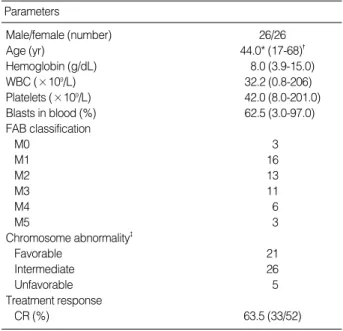
골수형태, 세포화학염색, 면역표현형, 세포유전학검사, FAB 기준에 따라 진단하였다. 이차성 AML과 재발 환자는 제외하였 다. 대상군 나이 중앙치는 44세였고, 남자 26명, 여자 26명이었 다. 혈색소 농도 평균은 8.0 g/dL, 백혈구 수 평균은 32.2×10⁹/ L, 혈소판 수 평균은 42.0×10⁹/L이었고, 혈액 아세포 평균은 62.5%였다. M0형 3명(5.8%), M1형 16명(30.8%), M2형 13명 (25.0%), M3형 11명(21.1%), M4형 6명(11.5%), M5형이 3명 (5.8%)이었다. 염색체 핵형은 예후[20]에 따라 양호군 21명(40.4
%), 불량군 5명(9.6%), 중간군 26명(50.0%)이었다. 환자의 33명 (63.5%)이 관해에 도달하였으며(Table 1), 전체 생존기간의 중 앙값은 12.0 (1.7-22.3)개월이었다.
2. 검사 방법
1) 세포 분리
골수 또는 말초혈액에서 백혈병세포를 분리하였다. 대조세포는 정상 골수세포, mdr1 음성대조는 CCRF-CEM 세포주를 사용하 였고, JNK는 K562 세포주로, mdr1 양성대조는 KG1 세포주를 사용하였으며 한국세포주은행(Seoul, Korea)에서 구입하였다.
*, median; �, the parenthesis means the range; �, AML patients were grouped into three different risk categories: t(8;21), inv(16), t(15;17) as favorable; -5/-7, 11q23 abnormalities, inv(3q), t(6:9), complex karyo- types as unfavorable. All other aberration and normal karyotypes were regarded as intermediate.
Abbreviation: CR, complete remission.
Parameters
Male/female (number) 26/26
Age (yr) 44.0* (17-68)�
Hemoglobin (g/dL) 8.0 (3.9-15.0)
WBC (×109/L) 32.2 (0.8-206)
Platelets (×109/L) 42.0 (8.0-201.0)
Blasts in blood (%) 62.5 (3.0-97.0)
FAB classification
M0 3
M1 16
M2 13
M3 11
M4 6
M5 3
Chromosome abnormality�
Favorable 21
Intermediate 26
Unfavorable 5
Treatment response
CR (%) 63.5 (33/52)
Table 1.Clinical and laboratory parameters of patients with acute myeloid leukemia
2) RNA 분리 및 역전사PCR
UltraspecTM-II RNA 분리 시약(Biotecx Laboratories, Hous- ton, TX, USA)을 사용하여 RNA를 분리하고 순도와 총량을 측 정하였다.
역전사반응은 M-MLV Reverse Transcriptase (Promega Corporation, Madison, WI, USA)와 Random Hexamer (Roche Molecular Systems, Indianapolis, IN, USA)를 시발체로 하여 cDNA를 합성하였다.
Mdr1, mrp1, gst , 2-microglobulin 유전자의 증폭은 iCy- clerTM Thermal Cycler (Bio Rad, Herlcules, CA, USA)에서 변성, 결합, 연장 반응 순으로 94℃ 40초, 55℃ 40초, 72℃ 1분 을 30회 반복 진행하였다. 유전자의 PCR 시발체의 염기서열 및 증폭산물 크기는 다음과 같다[20, 22].
Mdr1 (229 bp); GGAGAGATCCTCACCAAGCG, GTTGCCAACCATAGATGAAGG Mrp (326 bp); CGTGTACTCCAACGCTGAC,
CTGGACCGCTGACGCCCGTGAC Gst (256 bp); GGTGAATGACGGCGTGGAG,
GAGAGGAGGGGGAACGCAT
2-microglobulin; AGATGAGTATGCCTGCCGT, (261bp) TCAAACATGGAGACAGCACT PCR 반응산물은 전기영동 후 자외선 투시기로 2-microglob- ulin, mdr1, mrp1 및gst 유전자 발현을 확인하였다. 유전자 발현은 2-microglobulin 발현량을 기준으로 광학밀도 비(UVI- PRO, Cambridge, UK)를 측정하여, 반정량적으로 분석하였다.
3) JNK 발현 및JNK활성
JNK 발현: 차가운 용해 완충액으로 분리세포를 혼합하여 세 포용해액을 준비하였다. 12% sodium dodecyl sulfate (SDS) polyacrylamide 겔상에서 전기영동하여 니트로셀룰로스 막에 전 사한 후 JNK에 대한 일차 항체와 반응시켰다. Horseradish per- oxidase 결합 항 rabbit 이차 항체와 반응시킨 후 화학발광으로 발현량을 확인하였다.
JNK 활성 측정: c-Jun은 두 개의 인산화 부위 Ser 63과 Ser 73의 N-말단에 JNK와 친화성이 높은 결합부위를 가지므로 glu- tathione-sepharose bead에 결합되어 있는 N-terminal c-Jun 융합단백이 세포용해액 내에 있는 JNK를 끌어 당기는 성질을 이 용한 SAPK/JNK 활성 측정 키트(Cell Signaling Technology, Beverly, MA, USA)로 JNK 활성을 측정하였다. 차가운 PBS 세척 세포 용해액을 초음파기로 세포를 깨뜨려서 고속 원침하고 상층액에 glutathinone S-transferase-c-Jun 융합단백 beads를 넣고 침전물을 kinase 완충액으로 세척한 후 ATP 첨가 kinase 완충액과 반응시킨다. 3×SDS 완충액으로 반응을 종료하고, 12%
SDS-PAGE 겔 전기영동하여, 항 phospho-c-Jun 항체로 western blot하여 인산화된 c-Jun을 측정하여 JNK 활성을 분석하였다.
3. 관해 유도 치료
관해유도를 위해 cytarabine과 anthracycline계 약물[23]의 표 준 관해유도요법을 시행하였으며, 완전관해에 도달하면 공고 화학 요법을 시행하였고, M3 아형은 all-trans-retinoic acid 유지요법 을 추가하였다.
4. 통계
약제내성 인자mdr1, mrp 1, gst 유전자 발현, JNK 활성 증가에 따른 완전관해율 및 전체생존기간을 비교하였다. 완전관 해 판정은 National Cancer Institute 기준[24]을 따랐다. 치료 후 관해에 도달하지 못하거나 사망한 환자는 치료실패로 간주하 였다. 전체 생존기간은 진단일로부터 최종 추적일까지로 하였다.
약제내성 관련인자 발현에 따른 임상특징과 완전 관해율의 차이 는 Fish 정확확률, 또는 카이제곱 검정으로 비교하였다. 전체 생 존기간은 Kaplan-Meier법으로 구하고 Log-Rank 검사로 비교하 였다. 완전관해율에 영향을 주는 예후인자의 다변량분석은 Lo- gistic regression으로 구하였고, 전체 생존기간에 영향을 주는 예 후인자의 다변량분석은 Cox regression으로 구하였으며, 통계학적 유의 수준은P값이 0.05 미만으로 하였다.
결 과
1.mdr1
,mrp1
,gst
mRNA 발현 양상양성대조 세포주의mdr1, mrp1 및gst 유전자는 강한 발현 을 보였다. 음성대조 세포주에서 mdr1 유전자는 발현되지 않았 고mrp1과gst 유전자는 중등도 발현을 보였다. 정상 골수세 포 5예에서 시행한mdr1 유전자 발현은 음성이었고, mrp1 유전 자는 중등도(평균 밀도량 0.5), gst 유전자는 중등도 이상(평균밀 도량 0.9) 발현을 보였다(Fig. 1).
AML 환자에서mdr1 유전자 발현은 일부 환자에서 약하거나 중등도 발현을 보여 겔상mdr1 밴드가 관찰되면mdr1 발현 양 성으로 구분하였다. mrp1 유전자는 환자마다 다양하여 대부분 중 등도 또는 강한 발현을 보였고, gst 유전자는 중등도 내지는 강한 발현을 보였다(Fig. 1), mrp1과gst 유전자 발현은 정상 골수 유전자 발현의 광학밀도량의 평균 이상이면 양성으로 구분 하였다[7].
2.
mdr1
,mrp1
,gst
mRNA 발현 빈도AML 환자에서mdr1 유전자 발현 양성률은 38.5% (20/52) 이었다. mrp1 유전자 발현 양성률은 48.1% (25/52)이었다. gst 유전자 발현 양성률은 54.3% (25/46)였다. mdr1 유전자 발
현 환자 20명 중에서 14명이mrp1 유전자 과발현을 보여, 일치 율은 70%로 두 유전자 상호 간에 상관성이 있었다(r=0.651, P<
0.000). 그러나gst 와mdr1과 그리고mrp1 유전자 발현 간에 상관성은 없었다(Table 2).
3. JNK 단백 발현과JNK활성
JNK 항체로 시행한 western blot에서 p46 및 p54 JNK 단백 은 AML 환자 모두에서 발현되었다. 그러나 정상 골수세포에서 JNK 단백은 발현되지 않았다(Fig. 2). JNK 활성은 AML 환자 30명 중 2명(6.7%)에서 관찰되었다.
4. 임상 및 검사 소견과의 관련성
mdr1, mrp1, gst 유전자 발현에 따른 임상 및 검사 소견 차 Normal bone
marrow
2-MG mdr1 mrp1 gst
mdr1 (229 bp) mrp1 (326 bp) gst (256 bp)
2-microglobulin (261 bp)
M3 M2 M5 M1 M5 M0 M2 Negative cell line
Positive cell line
Fig. 1.RT-PCR results for mdr1, mrp1, gst , and 2-microglob- ulin mRNA (internal control) with KG1 and CCRF-CEM T-cell lines as mdr1 mRNA positive and negative control and normal bone marrow (A). Expression levels of mdr1, mrp1 and gst mRNA using RT-PCR in patients with acute myeloid leukemia (B).
Abbreviations: mdr1, multidrug resistance; mrp1, multidrug resistance associated protein; gst, glutathione-s-transferase.
*, P<0.05.
Abbreviations: See Fig. 1.
mrp1 gst
mdr1 0.651 (0.000)* -0.031 (0.839)
mrp1 0.071 (0.641)
Table 2.Correlation among mdr1, mrp1 and gst mRNA ex- pression
Parameters mdr1+ (%) P mrp1+ (%) P gst + (%) P
Positive N 20/52 (38.5) 25/52 (48.1) 25/46 (54.3)
Age (yrs)
<55 11/36 (30.6) (0.122) 14/36 (38.9) (0.071) 18/33 (54.5) (1.000)
55 9/16 (56.3) 11/16 (68.8) 7/13 (53.8)
Sex
Male 11/26 (42.3) 11/26 (42.3) (0.579) 15/23 (65.2) (0.236)
Female 9/26 (34.6) 14/26 (53.8) 10/23 (43.5)
WBC (/L)
<5×10⁹ 11/28 (39.3) (0.776) 11/28 (39.3) (0.254) 14/23 (60.9) (0.762)
5×10⁹ 9/22 (40.9) 13/22 (59.1) 11/21 (52.4)
CD34 (%)
<20 8/22 (36.4) (1.000) 13/22 (59.1) (0.578) 10/22 (45.5) (0.375)
20 12/30 (40.0) 12/30 (40.0) 15/24 (62.5)
FAB
M0 2/3 (66.7) (0.123) 1/3 (33.3) (0.154) 2/3 (66.7) (0.228)
M1 5/16 (31.3) 9/16 (56.3) 10/13 (76.9)
M2 8/13 (61.5) 7/13 (53.8) 5/11 (45.5)
M3 1/11 (9.1) 2/11 (18.2) 5/10 (50.0)
M4 3/5 (60.0) 3/5 (60.0) 3/6 (50.0)
M5 1/3 (33.3) 3/3 (100) 3/3 (100)
Chromosome
Good 6/21 (28.6) (0.105) 7/21 (33.3) (0.121) 10/19 (52.6) (0.469)
Intermediate 10/26 (38.5) 14/26 (53.8) 11/22 (50.0)
Poor 4/5 (80.0) 4/5 (80.0) 4/5 (80.0)
Treatment
Response 7/33 (21.2) (0.001*) 12/33 (36.4) (0.043*) 15/29 (51.7) (0.762)
Failure 13/19 (68.4) 13/19 (68.4) 10/17 (58.8)
Table 3.Mdr1, mrp1, gst mRNA expression and clinical and laboratory findings in patients with acute myeloid leukemia
*, P<0.05; +, expression or overexpression.
Abbreviations: See Fig. 1.
A
B
이를 Table 3에 나타내었다. mdr1, mrp1, gst 유전자 발현은 환자 나이, 성별, 백혈구 수, CD34 항원 유무, FAB형 및 염색체 핵형에 따른 차이는 없었다.
5. 치료 반응
나이 55세 이상, 높은 백혈구 수, 예후 불량 핵형에서 관해율이
유의하게 낮았으며(P<0.05 또는P<0.01), mdr1, mrp1 유전자 발현 양성 환자의 관해율도 유의하게 낮았다(P<0.05 또는 <0.01).
환자 성별, CD34 양성 유무, gst 유전자 발현, JNK 활성 유 무에 따른 관해율은 차이가 없었다. 다변량분석으로 백혈구 수와 mdr1 유전자 발현은 관해에 영향을 주는 예측인자였다(P<0.01 또는 0.05). 전체 생존기간은 나이, 백혈구 수, 염색체 핵 형에 따 라 차이가 있었고, mdr1, mrp1 유전자 발현 양성 환자의 전체 생존기간이 유의하게 짧았으나(P<0.01), gst 유전자 발현에 따른 차이는 없었다. 다변량분석으로 나이, 백혈구 수, 염색체 핵 형은 전체 생존기간에 영향을 주는 예측인자였다(P<0.01 또는 0.05) (Table 4).
6. JNK 활성 증가 환자의 임상 소견 및 특징
JNK 발현은 AML 환자에서 모두 관찰되었다. 그러나 JNK 활성 증가는 2명에서만 관찰되어 통계적으로 비교할 수 없었다.
JNK 활성이 증가된 환자의 특징을 보면, 67세 남자와 24세 여자 로서 FAB 아형은 각각 M1과 M4형이었다. 백혈구 수는 115.3×
Total JNK p46 phospho c-Jun p35
-actin p42
M3 M2 M5 M1 M5 M0 M2 M3 M4 P N
Fig. 2.Immunoblotting of cell lysates of AML patients to analyse c-Jun-N-terminal kinase (JNK) activity and -actin expression, and JNK activity (phospho c-Jun). JNK activity was undetec- table in leukemic samples except 2 cases (middle). P, K562; N, normal bone marrow. Total JNK, 46, 54 kDa; phospho c-Jun, 35 kDa; -actin; 42 kDa.
Parameters N of cases Chemotherapy
Remission (%) P
Overall survival
Median±SE (months) P
Age
<55 36 27 (75.0) 0.014* 16.0±3.3 0.001*
55 16 6 (37.5) 2.0±3.0
Sex
Male 26 15 (57.7) 0.565 60.0±3.1 0.117
Female 26 18 (69.2) 16.0±6.9
WBC (/L)
<5×10⁹ 28 23 (82.1) 0.004* 17.0±1.8 0.004*
5×109 22 9 (40.9) 3.0±0.5
CD34 (%)
<20 24 14 (58.3) 0.568 21.0±3.5 0.129
20 28 19 (67.9) 7.0±4.0
Chromosome
Good 21 19 (90.5) 0.002* 27.1±3.2 0.000*
Intermediate 26 13 (50.0) 8.3±1.6
Poor 5 1 (20.0) 2.4±0.4
mdr1
Negative 32 26 (81.3) 0.001* 19.9±2.8 0.001*
Positive 20 7 (35.0) 2.0±0.7
mp1
Negative 27 21 (77.8) 0.043* 17.0±5.7 0.003*
Positive 25 12 (48.0) 4.0±1.6
gst
Low 21 14 (66.7) 0.762 21.6±3.4 0.296
High 25 15 (60.0) 7.0±3.6
JNK activity
Negative 28 20 (71.4) 0.103 16.0±8.8 0.013*
Positive 2 0 (0.0) 2.0±0.0
Table 4.Chemotherapeutic response and overall survivals of AML patients according to clinical findings, and mdr1, mrp1, gst mRNA and JNK activity
*, P<0.05.
Abbreviations: See Fig. 1.
109/L, 181.3×10⁹/L로 과백혈구증가를 보였고, 모두 염색체 이 상(48,XY,+8,+9 및 46,XX,t[4;5][q33;p11.3])이 관찰되었다. 그 리고 둘 다mdr1, mrp1 유전자 발현 양성이었고 화학요법 치료 에 반응하지 않았다.
고 찰
Hunault 등[9]은 AML 환자의 40%에서mdr1 유전자 발현 양성이었고, 50세 이상, 불량 염색체, mdr1 유전자 과발현에서 관해유도 치료에 높은 내성과 짧은 전체 생존기간을 보이므로, mdr1 유전자 발현이 관해유도 예측을 위한 예후 인자의 하나라 고 하였다. Schaich 등[7]도 AML 환자에서mdr1 유전자 발현 양성은 25%에서 관찰되었고, mdr1 유전자 발현은 관해유도 치 료와 전체생존결과 예측을 위한 독립적 예후 인자라고 하였다. 본 연구에서도 AML 환자의mdr1 유전자는 38.5%에서 양성이었 으며, mdr1 유전자 발현 환자에서 관해율이 낮았고 전체 생존기 간도 짧아서mdr1 유전자 발현은 관해유도 치료의 독립적 예후 인자였다. Schaich 등[25]은 AML에서mdr1 유전자 발현 빈도 가 비정상염색체에서 정상염색체보다 더 높았으며, 비정상염색체 중에서는mdr1 유전자 양성군의 관해율이 음성군 보다 유의하게 낮았고, 예후 불량 염색체 환자에서mdr1 유전자 양성군은 관해에 도달하지 않았으나, 음성군은 정상염색체와 유사한 관해율을 보였 고, 복합염색체이지만 mdr1 유전자 음성군의 관해율이 63%나 되므로, 특정 염색체이상 환자들 사이에mdr1 유전자 발현이 차 이가 있다고 하였다. 그러므로 불량 염색체 군에서도mdr1 유전 자 발현 상태는 관해유도 치료에 결정적 인자가 될 수 있을 것이 라고 하였다. 본 연구에서 불량 염색체 군에서mdr1 유전자의 발현 빈도가 높았고, 염색체 예후 군에 따라 관해율과 전체생존 기간은 차이가 있었으며, 염색체는 전체 생존예측을 위한 예후인 자의 하나였으나 특정염색체에 따른mdr1 유전자 발현은 대상 환자가 적어서 구분하여 비교할 수는 없었다.
따라서 이상의 결과로 AML에서mdr1 유전자 발현은 위험도 에 기초한 치료계획 수립 시 중요한 예후 인자로서 적극적으로 고 려되어야 할 것이다[7, 25].
Mrp1은mdr1 유전자 발현이 없는 약제내성 세포주에서 과발 현되므로 mrp1 유전자가 약제내성과 관련이 있을 수 있다[2].
mrp1 유전자는 재발한 AML 환자에서 과발현 빈도가 높으며 과발현 mrp1 환자에서 관해유도 치료에 불응 빈도가 높으므로 mrp1유전자 발현이 치료 결과에 영향을 줄 수 있다[8]. Schaich 등[7]은mrp1 유전자 발현 양성 AML 환자의 무질병 생존기간 이 음성 환자보다 짧으며, mrp1 유전자 발현은 무질병생존 예측 을 위한 예후인자의 하나이므로, mrp1 유전자 과발현 환자의 강 력한 치료 계획수립에 이용할 수 있을 것이라고 하였다[7]. 본 연구에서mrp1 유전자 과발현 AML 환자의 관해율이 낮고 전 체 생존기간도 짧았으나 독립적인 예후인자는 아니었다. 본 연구
와 함께 아직까지mrp1 발현이 관해유도 및 생존에 미치는 영향 에 대하여는 보고자들에 따라 차이가 있는데 적은 대상 환자 수 또는 검출법의 차이일 수 있을 것이다[16, 18].
세포 내 존재하는gst 는 약물에 의해 생성되는 자유 라디칼 을 해독하는 효소로서 mrp 단백에 의해 배출되는 gluthathionyl S-conjugates를 합성함으로 약제내성에 관여하는 것으로 알려지 고 있다[11]. gst 유전자는 재발한 AML 환자에서 발현이 증 가하였다는 보고가 있지만[26], 치료결과에 미치는 영향에 대한 연구는 거의 없다. 본 연구에서도 AML 환자에서gst 유전자 과발현이 관해율 및 전체 생존기간에 직접 영향을 주지는 않았다.
신호전달에 관여하는 JNK는 세포 사멸을 유도하기도 하지만 항 세포 사멸(anti-apoptotic)에도 관여하는 것으로 알려져 있다 [27]. Lunghi 등[19]은 AML에서 세포 활성 유도에 관여하는 MAP kinase 경로를 밝히기 위해 26명의 일차성 AML 환자에 서 JNK 활성을 측정하였는데, 한 명의 AML 환자에서만 백혈병 세포의 JNK 활성이 증가하였고 대부분 환자는 활성이 전혀 관찰 되지 않거나 아주 약하게 관찰되었다. 그래서 백혈병 세포를 화학 스트레스 유발 물질 anisomycin과 반응시킨 후 측정하였는데 JNK 활성이 처리 전 보다 증가하였고, etoposide와 cytorabine과 반응 시켰을 때도 JNK 활성이 증가되어 약물 노출에 의한 세포사멸 과정에서 JNK 활성 증가와의 관련성을 지적하였다[19]. 본 연구 에서도 일차성 AML을 대상으로 하였는데 환자 중 JNK 활성은 2명에서만 증가하였다. Cripe 등[14]은 AML 환자의 50%에서 JNK 활성이 증가하였고, mrp 배출 기능도 함께 증가하므로 JNK 활성 증가와 약제내성과 관련이 있다고 하였으며, JNK 활성이 증 가된 환자 대부분은 과다백혈구증가를 동반하였으며, JNK 활성 증가와 치료실패와 관련성이 있다고 하였는데 대부분 이들은 이차 또는 재발 환자라고 하였다. 본 연구에서 JNK 활성이 증가한 2 명은 일차성 AML 환자들이었으며, M1과 M4 아형, 과다백혈구 증가, mdr1, mrp1 유전자발현 양성이었고 치료에 반응하지 않았 다. AML에서 JNK 활성 증가는 약제 다제내성, 과다증식, 이차 성 또는 재발과 같은 특정한 AML에서 관찰되므로 불량한 예후 인자로서의 가능성에 대한 연구가 더 필요할 것으로 생각된다.
요 약
배경 : 급성골수성백혈병(AML)에서 다약제내성 지표인 mul- tidrug resistance (mdr1), multidrug resistance associated pro- tein (mrp1), glutathione-s-transferase (gst) 발현은 치료 실패와 관련이 있으며, c-jun N-terminal kinase (JNK) 활성은 화학약제들에 반응하여 증가한다. 따라서 다제내성 인자와 신호전 달 JNK 활성의 임상적 의의를 규명하고자 AML 환자에서mdr1, mrp1, gst 유전자 발현과 JNK 활성을 측정하였다.
방법 : 52명의 AML 환자에서 다약제내성 유전자 발현은 RT- PCR을 이용하여 측정하였고, JNK 발현 및 활성은 immunoen-
zymatic kinase 측정과 western blot법을 이용하여 검출하였다.
결과 : mdr1, mrp1, gst 유전자 발현 빈도는 각각 38.5%, 48.1%, 54.3%이었다. 고령, 백혈구 증가, 불량염색체, mdr1과 mrp1 유전자 발현 환자에서 관해율이 유의하게 낮았고, 백혈구 수와mdr1 유전자 발현은 치료 반응의 예측 인자였다. 고령, 백 혈구 수, 불량염색체, mdr1, mrp1 유전자 발현, JNK 활성 증가 환자의 전 생존기간이 유의하게 짧았고, 나이, 백혈구 수, 염색체 는 전체 생존기간에 영향을 주는 예측 인자였다. JNK 활성은 대 부분 환자(28/30)에서 검출되지 않고 2명(6.7%)에서만 증가하였 다. JNK 활성이 증가한 2명의 환자는 과백혈구 증가, 비정상염 색체, mdr1, mrp1, gst 유전자 과발현을 동반하였으며 치료에 반응하지 않았다.
결론 : AML에서 다약제내성인자 발현은 관해유도 화학요법 에 따른 치료 결과 예측에 유용하지만, JNK 활성과 다약제내성 인자와의 관련성은 연구가 더 필요할 것으로 생각되었다.
참고문헌
1. Zittoun R, Mandelli F, Willemze R, de Witte T, Labar B, Resegotti L, et al. Autologous or allogenic bone marrow transplantation com- pared with intensive chemotherapy in acute myelogenous leukemia.
European Organization for Research and Treatment of Cancer (EO- RTC) and the Gruppo Italiano Malattie Ematologiche Maligne dell’- Adulto (GIMEMA) Leukemia Cooperative Groups. N Engl J Med 1995;332:217-23.
2. Cole SP, Bhardwaj G, Gerlach JH, Mackie JE, Grant CE, Almquist KC, et al. Overexpression of a transporter gene in a multidrug-resis- tant human lung cancer cell line. Science 1992;258:1650-4.
3. Del Poeta G, Stasi R, Aronica G, Venditti A, Cox MC, Bruno A, et al.
Clinical relevance of P-glycoprotein expression in de novo acute myeloid leukemia. Blood 1996;87:1997-2004.
4. Campos L, Guyotat D, Archimbaud E, Calmard-Oriol P, Tsuruo T, Troncy J, et al. Clinical significance of multidrug resistance P-glyco- protein expression on acute nonlymphoblastic leukemia cells at diag- nosis. Blood 1992;79:473-6.
5. Sato H, Preisler H, Day R, Raza A, Larson R, Browman G, et al. MDR1 transcript levels as an indication of resistant disease in acute myel- ogenous leukaemia. Br J Haematol 1990;75:340-5.
6. Baek JH, Park SW, Kim DH, Jung JT, Kwak DS, Park SH, et al. The frequency and clinical significance of multidrug resistance-1(MDR- 1) gene expression in acute myeloid leukemia. Korean J Hematol 2000;35:117-25. (백진호, 박성원, 김동환, 정진태, 곽동석, 박소향등. 급 성 골수성 백혈병에서 다약제내성(MDR-1) 유전자 발현의 빈도 및 임상 적의의. 대한혈액학회지 2002;35:117-25.)
7. Schaich M, Soucek S, Thiede C, Ehninger G, Illmer T, SHG AML 96
study Group. MDR1 and MRP1 gene expression are independent predictors for treatment outcome in adult acute myeloid leukaemia.
Br J Haematol 2005;128:324-32.
8. Zhou DC, Zittoun R, Marie JP. Expression of multidrug resistance- associated protein (MRP) and multidrug resistance (MDR1) genes in acute myeloid leukemia. Leukemia 1995;9:1661-6.
9. Hunault M, Zhou D, Delmer A, Ramond S, Viguie F, Cadiou M, et al. Multidrug resistance gene expression in acute myeloid leukemia:
major prognossis significance for in vivo drug resistance to induc- tion treatment. Ann Hematol 1997;74:65-71.
10. Zaman GJ, Lankelma J, van Tellingen O, Beijnen J, Dekker H, Pau- lusma C, et al. Role of glutathione in the export of compounds form cells by the multidrug-resistance-associated protein. Proc Natl Acad Sci USA 1995;92:7690-4.
11. Yamane Y, Furuichi M, Song R, Van NT, Mulcahy RT, Ishikawa T, et al. Expression of multidrug resistance protein/GS-X pump and gamma-glutamylcysteine synthetase genes is regulated by oxida- tive stress. J Biol Chem 1998;273:31075-85.
12. Toone WM, Kuge S, Samuels M, Morgan BA, Toda T, Jones N. Reg- ulation of the fission yeast transcription factor Pap1 by oxidative stress: requirement for the nuclear export factor Crm1 (Exportin) and the stress-activated MAP kinase Sty1/Spc1. Genes Dev 1998;
12:1453-63.
13. Osborn MT and Chambers TC. Role of the stress-activated/c-Jun NH2-terminal protein kinase pathway in the cellular response to adriamycin and other chemotherapeutic drugs. J Biol Chem 1996;
271:30950-5.
14. Cripe LD, Gelfanov VM, Smith EA, Spigel DR, Phillips CA, Gabig TG, et al. Role for c-jun N-terminal kinase in treatment-refractory acute myeloid leukemia (AML): signaling to multidrug-efflux and hyperproliferation. Leukemia 2002;16:799-812.
15. Lamy T, Goasguen JE, Mordelet E, Grulois I, Dauriac C, Drenou B, et al. P-glycoprotein (P-170) and CD34 expression in adult acute myeloid leukemia (AML). Leukemia 1994;8:1879-83.
16. Leith CP, Kopecky KJ, Chen IM, Eijdems L, Slovak, ML, McConnel TS, et al. Frequency and clinical significance of the expression of the multidrug resistance proteins MDR1/P-glycoprotein, MRP1 and LRP in acute myeloid leukemia: a Southewest Oncology Group Study. Blood 1999;94:1086-99.
17. Van den Heuvel-Eibrink MM, van der Holt B, te Boekhorst PA, Pieters R, Schoester M , Lowenberg B, et al. MDR1 expression in an independent prognostic factor for response and survival in de novo acute myeloid leukemia. Br J Hematol 1997;99:76-83.
18. Filipits M Suchomel RW, Zochbauer S, Brunner R, Lechner K, Pir- ker R. Multidrug resistance associated protein in caute myeloid leukemia: No impact on treatment outcome. Clin Cancer Res 1997;
3:1419-25.
19, Lunghi P, Tabilio A, Pinelli S, Valmadre G, Ridolo E, Albertini R, et al. Expression and activation of SHC/MAP kinase pathway in pri- mary acute myeloid leukemia blasts. Hematol J 2001;2:70-80.
20. Grimwade D, Walker H, Oliver F, Wheatley K, Harrison C, Harri- son G, et al. The importance of diagnostic cytogenetics on outcome in AML: analysis of 1,612 patients entered into the MRC AML 10 trial. The Medical Research Council Adult and Children’s Leukae- mia Working Parties. Blood 1998;92:2322-33.
21. Beck J, Handgretinger R, Dopfer R, Klingebiel T, Niethammer D, Gekeler V. Expression of mdr1, mrp, topoisomerase II alpha/beta, and cyclin A in primary or relapsed states of acute lymphoblastic leukaemias. Br J Haematol 1995;89:356-63.
22. Stammler G, Sauerbrey A, Volm M. Determination of DNA topoi- somerase II in newly diagnosed childhood acute lymphoblastic leukemia by immunocytochemistry and RT-PCR. Cancer Lett 1994;
84:141-7.
23. Vogler WR, Velez-Garcia E, Weiner RS, Flaum MA, Bartolucci AA,
Omura GA, et al. A phase III trial comparing idarubicin and dau- norubicin in combination with cytarabine in acute myelogenous leukemia: a Southeastern Cancer Study Group Study. J Clin Oncol 1992;10:1103-11.
24. Cheson BD, Cassileth PA, Head DR, Schiffer CA, Bennett JM, Bloom- field CD, et al. Report of the National Cancer Institute-sponsored workshop on definitions of diagnosis and response in acute myeloid leukemia. J Clin Oncol 1990;8:813-9.
25. Schaich M, Harbich-Brutscher E, Pascheberg U, Mohr B, Soucek S, Ehninger G, et al. Association of specific cytogenetic aberrations with mdr1 gene expression in acute myeloid leukemia and its impli- cation for treatment outcome. Haematologica 2002;87:455-64.
26. Beck J, Handgretinger R, Klingebiel T, Dopfer R, Schaich M, Ehn- inger G, et al. Expression of PKC isozyme and MDR-associated genes in primary and relapsed state AML. Leukemia 1996;10:426-33.
27. Libermann DA. Normal development, oncogenesis and programm- ed cell death. Oncogene 1998;17:1189-94.