J Korean Ophthalmol Soc 2016;57(10):1570-1576 ISSN 0378-6471 (Print)⋅ISSN 2092-9374 (Online)
http://dx.doi.org/10.3341/jkos.2016.57.10.1570
Original Article
여러 가지 기기를 이용하여 측정한 전방깊이와 중심각막두께의 비교
Comparison of Anterior Chamber Depth and Central Corneal Thickness Measured Using Different Devices
정재균1⋅길태영1⋅배지현1⋅신승주1⋅정성근2
Jae Gyun Jeung, MD1, Tae Young Gil, MD1, Gi Hyun Bae, MD1, Seong Joo Shin, MD1, Sung Kun Chung, MD PhD2
삼육서울병원 안과1, 가톨릭대학교 의과대학 성바오로병원 안과 및 시과학교실2 Department of Ophthalmology, Sahmyook Medical Center1, Seoul, Korea
Department of Ophthalmology and Visual Science, St. Paul’s Hospital, College of Medicine, The Catholic University of Korea2, Seoul, Korea
Purpose: To compare the accuracy and clinical usefulness of different devices by measuring anterior chamber depth (ACD) with three devices and central corneal thickness (CCT) with four devices.
Methods: In 180 eyes of 90 healthy subjects, ACD was measured using A-scan, Lenstar LS900®, Pentacam®, and CCT was measured using ultrasound pachymetry (USP), Lenstar LS900®, Pentacam®, and anterior segment optical coherence tomog- raphy (OCT).
Results: The average ACT measurements using Lenstar LS900®, A-scan, and Pentacam® were 3.27 ± 0.35 mm, 3.26 ± 0.36 mm, and 3.25 ± 0.36 mm, respectively. The measurements were significantly correlated (p < 0.001) but without statistically sig- nificant difference (p = 0.017). The Bland-Altman plots showed a low degree of agreement. The average CCT measurements us- ing Pentacam®, USP, Lenstar LS900®, and OCT were 553.31 ± 25.23 μm, 547.26 ± 23.83 μm, 541.38 ± 24.49 μm, and 531.40
± 22.33 μm, respectively. The measurements were significantly correlated (p < 0.001) and statistically significantly different (p <
0.05). The Bland-Altman plots showed a low degree of agreement.
Conclusions: ACD and CCT measured using different devices were highly correlated, but the ACD measurements were not stat- istically different; however, the CCT measurements were statistically different, and agreement was low between both measurements.
J Korean Ophthalmol Soc 2016;57(10):1570-1576
Keywords: Anterior chamber depth, A-scan, Central corneal thickness, Lenstar®, Pentacam®
■Received: 2016. 4. 28. ■ Revised: 2016. 7. 27.
■Accepted: 2016. 9. 20.
■Address reprint requests to Sung Kun Chung, MD, PhD Department of Ophthalmology, The Catholic University of Korea St. Paul’s Hospital, #180 Wangsan-ro, Dongdaemun-gu, Seoul 02559, Korea
Tel: 82-2-958-2151, Fax: 82-2-960-2150 E-mail: eyedoc@catholic.ac.kr
ⓒ2016 The Korean Ophthalmological Society
This is an Open Access article distributed under the terms of the Creative Commons Attribution Non-Commercial License (http://creativecommons.org/licenses/by-nc/3.0/) which permits unrestricted non-commercial use, distribution, and reproduction in any medium, provided the original work is properly cited.
각막두께 및 앞방깊이를 포함한 안구생체계측을 정확하 게 하는 것은 굴절교정수술, 백내장수술에 있어 매우 중요 하며, 최근에는 녹내장 분야에 있어서도 매우 중요한 요소
가 되고 있다. 과거부터 안구생체계측에 검사방법이 간단 하고 비용이 저렴한 초음파 각막두께측정계, A-scan 등을 이용하였지만 초음파를 이용한 방식은 안구에 기계가 접촉 하기 때문에 환자에게 불편감을 초래할 수 있고, 안구를 함 입함으로써 측정값의 오차에 대한 우려가 제기되어 왔다.1,2 이에 각막에 직접 접촉하지 않고 각막두께를 측정할 수 있는 방법들이 여럿 개발되었다. 현재 전안부 빛간섭단층촬영계 (anterior segment optical coherence tomography), slit-scanning 방식을 이용한 ORBscan II® (Bausch & Lomb, Rochester, NY, USA), 부분결합간섭계원리를 이용한 기기인 IOL Master® (Carl Zeiss Meditec, Dublin, CA, USA) 및 저간섭반사계를
Table 1. Comparison of mean anterior chamber depth among devices
A-scan Lenstar LS900® Pentacam® p-value*
Mean ACD (mm) 3.26 ± 0.36 3.27 ± 0.35 3.25 ± 0.36 0.170
Range (mm) 2.25-4.12 2.46-4.27 2.22-4.34
Values are presented as mean ± SD.
ACD = anterior chamber depth; SD = standard deviation.
*p-value is obtained from repeated measured analysis of variance (ANOVA) test.
Figure 1. Bland and Altman plots comparing the level of agreement
between the 3 instruments for anterior chamber depth (ACD, mm).(A) A-scan and Lentar LS900®, (B) Ascan and Pentacam®, (C) Lenstar® and Pentacam®. SD = standard deviation.
이용한 Lenstar LS900® (Haag-Streit, Koeniz, Switzerland), Scheimpflug camera를 이용한 Pentacam® (Oculus, Wetzlar, Germany), Galilei™ (Ziemer, Port, Switzerland), Sirius® (Costruzione Strumenti Oftalmici, Florence, Italy) 등을 포 함한 다양한 기기들이 개발되어 임상에서 사용되고 있다.
지금까지 국내외에서 이러한 비접촉 방법들과 초음파를 이 용한 접촉식 방법으로 측정한 값들의 차이를 보고한 연구들이 여럿 있었다.3-13 본 연구에서는 A-scan (Pacscan 300A, Sonomed Inc., Chicago, IL, USA), Lenstar LS900®과 Pentacam®을 이용하여 앞방깊이를 측정해 비교하고, 접촉식 각막두께측 정계인 초음파 각막두께측정계(Pacscan 300A, Sonomed Inc., Chicago, IL, USA), Lenstar LS900®, Pentacam®, 전안 부 빛간섭단층촬영계(RTVue-100, Optovue Inc., Freemont, CA, USA)를 이용하여 중심각막두께를 측정하였고, 기존의 연구들과 비교하여 각 기기 간 계측의 정확성 및 임상적 유 용성에 대해 알아보고자 하였다. A-scan과 초음파 각막두 께측정계는 동일한 기기를 사용하였다.
대상과 방법
90명의 안과적 수술력이나 외상력, 각막질환의 과거력이 없는 건강한 성인 지원자를 대상으로 앞방깊이와 중심각막 두께를 측정하여 분석하였다. 대상은 남자 90안, 여자 90안 이었으며, 환자의 평균연령은 56.4 ± 15.02세(20-92세)였다.
본 연구는 가톨릭중앙의료원 임상시험심사위원회의 승인 을 받았다(IRB No. PC15risi0043).
중심각막두께(central corneal thickness, CCT)는 각막의 중앙부 전면(각막상피)에서 각막의 중앙부 후면(각막내피) 까지의 거리로 정의하였고, 앞방깊이(anterior chamber depth, ACD)는 각막의 중앙부 전면(각막상피)에서 수정체 중앙부 전면(수정체 전낭)까지의 거리로 정의하였다.
Pentacam®은 각막형태검사를 시행하여 컴퓨터 화면에 각막중심과 동공중심이 중앙에 위치하고, 내외측과 상하측 이 대칭이 되는 시점에서 스캔을 하였다. Lenstar LS900®는 광원이 동공 중심에 오게 하여 4회 측정 후 평균값을 측정 치로 하였다. 초음파 각막두께측정 검사와 A-scan 검사는 가장 마지막에 시행하였으며, 검사 전 propacanine 0.5% (Alcaine®,
(mm) (mm)
A
B
C
(mm)
Table 2. Mean difference, 95% limit of agreement (LoA), and Pearson correlation of anterior chamber depth
Mean difference ± SD p-value* 95% LoA Pearson correlation p-value†
A-scan–Lenstar® (mm) -0.01 ± 0.22 1.000 -0.43 to 0.41 0.816 <0.001
A-scan–Pentacam® (mm) 0.01 ± 0.26 0.204 -0.49 to 0.51 0.753 <0.001
Lenstar®–Pentacam® (mm) 0.02 ± 0.16 1.000 -0.30 to 0.34 0.897 <0.001
Values are presented as mean ± SD unless otherwise indicated.
SD = standard deviation.
*p-value is obtained from Bonferroni post hoc analysis; †p-value is obtained from Pearson correlation.
Table 3. Comparison of mean central corneal thickness among devices
USP Lenstar® Pentacam® AS-OCT p-value*
Mean CCT (μm) 547.26 ± 23.83 541.38 ± 24.49 553.31 ± 25.23 531.40 ± 22.33 <0.05
Range (μm) 486-606 485-599 487-611 478-584
Values are presented as mean ± SD.
USP = ultrasound pachymetry; AS-OCT= anterior segment optical coherence tomography; CCT = central corneal thickness; SD = standard deviation.
*p-value is obtained from Friedman test.
Table 4. Mean difference, 95% limit of agreement (LoA), and Pearson correlation of central corneal thickness
Comparisons Mean difference ± SD p-value* 95% LoA Pearson correlation p-value†
USP–Lenstar® (μm) 5.9 ± 27.4 0.000 -21.5 to 33.3 0.833 <0.001
USP–Pentacam® (μm) -6.0 ± 31.1 0.000 -37.2 to 25.1 0.792 <0.001
USP–AS-OCT (μm) 15.9 ± 25.7 0.000 -9.9 to 41.6 0.840 <0.001
Lenstar®–Pentacam® (μm) -11.9 ± 26.3 0.000 -38.2 to 14.4 0.855 <0.001
Lenstar®–AS-OCT (μm) 10.0 ± 16.5 0.000 -6.6 to 26.5 0.939 <0.001
Pentacam®–AS-OCT (μm) 21.9 ± 25.6 0.000 -3.7 to 47.5 0.856 <0.001
Values are presented as mean ± SD unless otherwise indicated.
USP = ultrasound pachymetry; AS-OCT = anterior segment optical coherence tomography; SD = standard deviation.
*p-value is obtained from Wilcoxon-rank signed test; †p-value is obtained from Pearson correlation.
Alcon Laboratories Inc., Cleveland, OH, USA)로 각막을 마 취한 다음 소식자(probe)를 각막 중심에 수직으로 접촉하여 5회 측정한 후 평균값을 측정치로 하였다. 모든 검사 시 대 상자의 머리와 턱을 각각의 고정대에 고정하고 눈높이를 적절하게 위치시킨 후 검사하는 동안 시스템 중앙의 주시 광선을 보게 하고 눈을 움직이지 않도록 하였다. 검사 전에 눈을 깜박거리게 하여 눈물층을 균일하게 하였으며, 속눈 썹이 각막을 가리지 않도록 눈을 크게 뜨도록 지시한 후 안 구에 압박이 가해지지 않도록 조심스럽게 눈꺼풀을 벌리면 서 측정하였다.
통계적 분석은 SPSS v18.0 for Windows (SPSS Inc., Chicago, IL, USA)를 사용하였다. 세 가지 방법을 이용해 측정한 앞방깊이는 repeated measured analysis of variance (ANOVA)를 이용하여 비교하였고, 사후 분석은 Bonferroni post hoc analysis를 이용하였다. p<0.05일 경우 통계적으로 유의한 것으로 판정하였다. 네 가지 방법을 이용해 측정한 중심각막두께는 정규성을 만족하지 않아 friedman test를 이용하여 독립된 4군의 크기를 비교하였고, Wilcoxon 부호 순위검정을 통해 모든 측정치의 쌍이 통계적으로 유의한 차이가 있는지를 알아보았다. 각 검사 방법 간의 일치도는
Bland-Altman plot을 사용하여 분석하였고, Pearson corre- lation을 이용하여 각 방법 간의 상관계수를 구하였으며, p<0.05를 유의한 상관관계가 있다고 보았다.14,15
결 과
앞방깊이(평균 ± 표준편차)는 A-scan, Lenstar LS900®, Pentacam®에서 각각 3.26 ± 0.36 mm, 3.27 ± 0.35 mm, 3.25
± 0.36 mm로 Lenstar LS900®에서가장 깊게, Pentacam®에 서 가장 얕게 측정되었으나, 전체 기기 간 측정치의 유의한 차이는 보이지 않았다(p=0.17) (Table 1). Bland-Altman 일 치도 분석결과 A-scan-Lenstar LS900® 간의 95% 일치도 범위는 0.84 mm (-0.43~0.41), A-scan-Pentacam®은 1.00 mm (-0.49~0.51), Lenstar LS900®-Pentacam®은 0.64 mm (-0.30~0.34)로 Lenstar LS900®-Pentacam® 간의 일치도 범 위가 가장 작았고 A-scan-Pentacam® 간의 일치도 범위가 가장 컸다(Fig. 1). 각 측정치는 모두 통계적으로 유의하게 높은 양의 상관관계를 보였고(all p<0.001) 상관계수는 각 각 0.816, 0.753, 0.897이었다(Table 2).
중심각막두께(평균 ± 표준편차)는 초음파 각막두께 측정
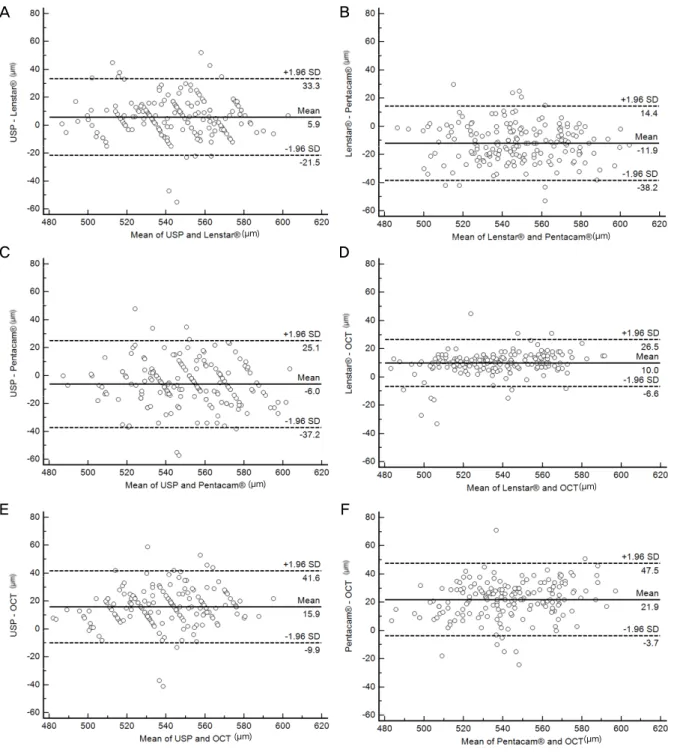
Figure 2. Bland and Altman plots comparing the level of agreement between the 4 instruments for central corneal thickness (CCT,
μm). (A) Ultrasound pachymetry (USP) and Lenstar LS900, (B) USP and Pentacam, (C) USP and anterior optical coherence tomog- raphy (OCT), (D) Lenstar and Pentacam, (E) Lenstar and anterior OCT, (F) Pentacam and anterior OCT. SD = standard deviation.계, Lenstar LS900®, Pentacam®, 전안부 빛간섭단층촬영계 에서 각각 547.26 ± 23.83 μm, 541.38 ± 24.49 μm, 553.31
± 25.23 μm, 531.40 ±22.33 μm였고, Pentacam®으로 측정 한 값이 가장 두꺼웠고, 전안부 빛간섭단층촬영계로 측정 한 값이 가장 얇게 나타났으며, 각 측정치 사이에 통계적으 로 유의한 차이를 보였다(p<0.05) (Table 3). 각 기기 간의 측정치 차이를 비교해 봐도 모든 군에서 통계적으로 의미
있는 차이를 보였다(p<0.05) (Table 4). Bland-Altman 일치 도 분석결과 초음파 각막두께측정계-Lenstar LS900® 간의 95% 일치도 범위는 54.8 μm (-21.5~33.3), 초음파 각막두 께 측정계-Pentacam®은 62.3 μm (-37.2~25.1), 초음파 각막두께 측정계-OCT는 51.5 μm (-9.9~41.6), Lenstar LS900®-Pentacam® 은 52.6 μm (-38.2~14.4), Lenstar LS900®-OCT는 33.1 μm (-6.6~26.5) Pentacam®-OCT는 51.2 μm (-3.7~47.5)로 Lenstar
A B
C D
E F
(μm)
(μm) (μm) (μm)
(μm) (μm)
LS900®-OCT 간의 일치도 범위가 가장 작았고, 초음파 각 막두께 측정계-Pentacam®의 일치도 범위가 가장 컸다(Fig. 2).
모든 측정치는 통계적으로 유의하게 높은 상관관계를 보였 고(all p<0.001), 상관계수는 각각 0.833, 0.792, 0.840, 0.855, 0.939, 0.856이었다(Table 4).
고 찰
전안부계측방법에는 여러 가지가 있지만 초음파를 이용 한 방법이 쉽게 사용할 수 있고 상대적으로 저렴한 검사비 용과 높은 정확도로 현재 널리 사용되고 있다. 하지만 검사 전 점안마취가 필요하고 소식자가 각막에 직접 접촉하여 측정하기 때문에 검사자에 따라 가해지는 압력의 차이나 측정 위치에 따라 측정값에 차이가 있을 수 있고, 각막 상 피의 손상, 감염의 전파 등의 위험이 있을 수 있다. 이러한 문제점들로 인해 최근 다양한 원리와 측정방법을 이용한 전안부 측정 기기들이 개발되어 사용되고 있는데 그중에 Pentacam®은 rotating Scheimpflug camera가 360° 회전하 며, 2초 내에 각막 전후면에 위치한 500개의 점을 포함하는 영상을 얻어 컴퓨터 재구성을 통해 전안부를 3차원으로 분 석할 수 있는 검사기구이며, 각막두께 및 각막 곡률치, 각막 지형도, 앞방깊이 측정뿐 아니라 전방각과 수정체의 혼탁과 두께의 정보를 추가로 얻을 수 있다.16 또 Lenstar LS900®는 820 μm의 파장의 광원으로 전간섭성반사(optical low-co- herence reflectometry) 원리를 이용하여 안축장, 전방 깊이, 각막 두께, 각막 곡률치, 동공 크기, 수정체 두께, 망막 두께 등의 측정이 가능한 안구 생체 계측 장비이며, 760 μm 파장 의 적외선 광선을 이용하며 부분결합간섭의 원리를 이용한 장비인 IOL Master®에 비해 투과력이 좋고, IOL Master®와 전안부 계측치 및 인공수정체 도수의 일치도도 매우 높다 고 알려져 있다.17 그리고 전안부 빛간섭단층촬영계는 비접 촉 영상기기로 조직 내에서 반사되는 빛의 시간 차이를 광 학적 간섭계를 통해 전안부 및 망막의 고해상도의 영상을 나타낼 수 있는 검사기구이다.18 하지만 오히려 이러한 다 양성이 기기별 측정 결과를 분석하는 데 혼란을 발생시킬 수 있다. 이에 본 연구에서는 앞방깊이와 중심각막두께에 관하여 접촉식 초음파를 이용한 방법과 비접촉식 광학적 방식을 이용하여 측정한 결과를 비교하고, 각 기기 간 계측 의 정확성 및 임상적 유용성에 대해 알아보고자 하였다.
본 연구에서 앞방깊이는 Lenstar LS900®, A-scan, Pentacam® 순서로 깊게 측정되었고, 모든 군에서 통계적으로 유의한 차 이는 보이지 않았다(p=0.841). Lenstar LS900®와 Pentacam® 의 측정치를 비교한 연구들 중 Huang et al3, OʼDonnell et al4, Uçakhan et al5에서는 각각 0.01 mm3, 0.03 mm4, 0.09 mm5
Pentacam®의 앞방깊이가 Lenstar LS900®보다 더 깊게 측 정되었으며 통계적인 차이가 있었고, 일치도에 관하여서는 기존 연구에서 95% 일치도 범위가 Pentacam®과 Lenstar LS900® 간에 0.16-0.28 mm로 중등도 이상이라고 보고하였 다. 본 연구에서는 Lenstar LS900®의 앞방깊이가 Pentacam® 보다 0.02 깊게 측정되어 기존의 연구들과 상반된 결과를 보였지만 차이가 적어 임상적으로 큰 의미는 없을 것으로 생각되며, 95% 일치도 범위는 0.64 mm 정도로 일치도가 높지 않았다.
Pentacam®과 A-scan의 측정치를 비교한 Elbaz et al6의 연구에서는 Pentacam®의 앞방깊이가 A-scan보다 0.1 mm 정도 깊게 측정되었고 통계적으로 유의한 차이가 있어 0.01 mm 정도의 차이가 났던 본 연구와 차이가 있었다. 95% 일 치도 범위는 0.60 mm (-0.197~0.404)로 일치도가 높지 않 다고 보고하였다. 본 연구에서도 95% 일치도 범위가 1.00 mm (-0.49~0.51)로 일치도가 높지 않았다.
Lenstar LS900®와 A-scan을 비교한 Tappeiner et al7의 연 구에서는 Lenstar LS900®가 A-scan에 비해 0.05 mm 정도 깊게 측정되었으나 유의한 차이는 없었고, Gursoy et al8의 연구에서는 Lenstar LS900®가 0.27 mm 정도 깊게 측정되 어 유의한 차이를 보였다. 95% 일치도 범위는 두 연구 모 두에서 모두 1.0 mm 이상으로 보고하였다. 본 연구에서는 Lenstar LS900®가 0.01 mm 정도 깊게 측정되어 유의한 차 이가 없었지만, 95% 일치도 범위가 0.84 mm로 앞선 연구 들과 마찬가지로 일치도가 높지 않았다.
본 연구에서 중심각막두께는 Pentacam®, 초음파 각막두 께측정계, Lenstar LS900®, 전안부 빛간섭단층촬영계 순서로 두껍게 측정되었고, 측정치들 사이에 유의한 차이가 존재하 였다. 기존의 Pentacam®, Lenstar LS900®, specular micro- scopy와 초음파 각막두께 측정계를 비교한 Borrego-Sanz et al9의 연구에서는 Pentacam®이 565.36 μm, 초음파 각막두 께측정계가 549.61 μm, lenstar LS900®가543.05 μm 순으로 두꺼웠고. 95% 일치도 범위는 Pentacam®-Lenstar LS900® 50.79 (-3.09-47.7) Pentacam®-초음파 각막두께측정계 56.52 (-12.51-44.01) Lenstar LS900®-초음파 각막두께측정계 30.8 (-21.95-8.85)였으며, 높은 상관관계를 가지지만 일치도가 높 지 않다고 보고하였다. 본 연구에서도 Pentacam® 553.31 μm, 초음파 각막두께 측정계 547.2 μm, Lenstar LS900® 541.38 μm, 순으로 두껍게 측정되었고, 95% 일치도 범위는 초음 파 각막두께 측정계-Lenstar LS900® 54.8 μm (-21.5~33.3), 초음파 각막두께 측정계-Pentacam® 62.3 μm (-37.2~25.1), Lenstar LS900®-Pentacam® 52.6 μm (-38.2~14.4)로 앞 연구와 마찬가지로 높은 상관관계를 가지지만 일치도가 높지 않았다.
반면에 Tai et al10의 연구에서는 Pentacam®이 538.4 μm,
Lenstar LS900®가 531.8 μm, 초음파 각막두께 측정계는 528.3 μm로 Pentacam®, Lenstar LS900®, 초음파 각막두께 측정계 순으로 두꺼웠으며, 95% 일치도 범위는 Lenstar LS900®-초음파 각막두께 측정계 (-10.06 to 17.14), Lenstar LS900®-pentacam® (-24.96 to 11.88), Pentacam®-초음파 각 막두께 측정계 (-11.40 to 31.5)로 일치도가 높다고 보고하 였다.
초음파 각막두께측정계와 전안부 빛간섭단층촬영계를 비교한 Bechmann et al11의 연구에서는 정상인에서 초음파 각막두께측정계로 측정한 중심각막두께가 전안부 빛간섭 단층촬영계보다 평균 51 μm 두껍게 측정되었다고 보고하 였고, Kim et al12은 초음파 각막두께측정계가 전안부 빛간 섭단층촬영계에 비해 평균 26.3 μm 두껍게 측정되었다고 보고하였다. 본 연구에서도 초음파 각막두께측정계가 전안 부 빛간섭단층촬영계보다 평균 16 μm 두껍게 측정되었고, 유의한 차이가 있었다. 반면에 Leung et al13은 정상안을 대 상으로 한 연구에서 전안부 빛간섭단층촬영계로 측정한 중 심각막두께가 초음파 각막두께측정계보다 평균 23 μm 두 껍게 측정되었다고 하였다.
각 검사치 간의 차이의 원인에 대해서는 검사방법과 소 요시간의 차이, 검사 당시 각막 및 눈물 층의 상태, 이용되 는 software 및 측정자의 기술적인 오차 등에 의해서 측정 치의 차이가 나타난다고 생각할 수 있다. 본 연구에서 통계 적으로 유의하지 않지만 Pentacam®을 이용한 각막두께의 측정치가 초음파각막두께측정계보다 약 6.05 µm 두껍게 측정이 되었는데, 이러한 차이의 원인으로는 Pentacam® 검 사 시간이 짧아 눈물층 증발의 영향을 거의 받지 않아 눈물 층까지 측정되어 조금 두껍게 나타나는 것으로 생각된다.19 이외에도 소식자가 각막표면에 접촉하면서 각막표면의 함 입이 발생하였을 것으로 생각된다. 또 본 연구에서 유의하 게 초음파 각막두께측정계가 전안부 빛간섭단층촬영계보 다 두껍게 측정되었는데 이러한 차이의 원인은 아직 정확 히 규명된 바 없지만 다음과 같은 요인들을 생각해 볼 수 있을 것이다. 각막의 전면 및 후면에서 반사되는 빛을 이용 하는 전안부 빛간섭단층촬영계와는 달리 초음파 각막두께 측정계는 아직 명확하게 밝혀지지 않은 후면의 어느 점에 서 반사된 음파를 이용하기 때문인 것으로 생각해 볼 수 있 다. 한 연구에서는 그 반사점이 아마도 데스메막과 앞방 사 이에 위치할 것이라고 추측하고 있다.20 또한 초음파 각막 두께측정계는 각막 중심이 검사자에 의해 수동적으로 결정 되기 때문에 검사자의 숙련도에 영향을 받을 수 있다. 이외 에도 전안부 빛간섭단층촬영계는 초음파 각막두께측정계와 는 달리 빛의 질과 각막의 투명도에 영향을 받을 수 있다.
결론적으로 세 기기 간의 측정된 앞방깊이는 통계적으로
유의한 차이가 없었고, 강한 양의 상관관계를 보였다. 일치 도는 높지 않았지만 앞방깊이를 측정한 군의 Bland-Altman plot을 도해한 결과는 이상값(outliers)이 소수만 존재하며 세 기기 간의 차이가 특정한 추세가 없고 비교적 잘 일치함 을 보여주었다고 생각한다. 반면 네 기기 간의 측정된 각막 두께는 강한 양의 상관관계를 보였지만 측정치 간의 통계 적으로 유의한 차이가 있었고, 일치도 분석에서도 비교적 낮은 일치도를 보였다. 따라서 임상적으로 앞방깊이를 측 정하고 비교함에 있어 본 연구에서 언급된 세 가지 기기를 이용하는 것은 유용해 보이나, 중심각막두께의 측정치를 네 가지 기기를 이용하여 비교할 때에는 이러한 측정치의 차이, 일치도, 상관관계에 대한 이해와 고려가 필요할 것으 로 생각한다.
REFERENCES
1) Reddy AR, Pande MV, Finn P, El-Gogary H. Comparative estima- tion of anterior chamber depth by ultrasonography, Orbscan II, and IOLMaster. J Cataract Refract Surg 2004;30:1268-71.
2) Vetrugno M, Cardascia N, Cardia L. Anterior chamber depth meas- ured by two methods in myopic and hyperopic phakic IOL implant.
Br J Ophthalmol 2000;84:1113-6.
3) Huang J, Pesudovs K, Wen D, et al. Comparison of anterior seg- ment measurements with rotating Scheimpflug photography and partial coherence reflectometry. J Cataract Refract Surg 2011;37:
341-8.
4) OʼDonnell C, Hartwig A, Radhakrishnan H. Comparison of central corneal thickness and anterior chamber depth measured using LenStar LS900, Pentacam, and Visante AS-OCT. Cornea 2012;
31:983-8.
5) Uçakhan OÖ, Akbel V, Bıyıklı Z, Kanpolat A. Comparison of cor- neal curvature and anterior chamber depth measurements using the manual keratometer, Lenstar LS 900 and the Pentacam. Middle East Afr J Ophthalmol 2013;20:201-6.
6) Elbaz U, Barkana Y, Gerber Y, et al. Comparison of different tech- niques of anterior chamber depth and keratometric measurements.
Am J Ophthalmol 2007;143:48-53.
7) Tappeiner C, Rohrer K, Frueh BE, et al. Clinical comparison of biometry using the non-contact optical low coherence reflec- tometer (Lenstar LS 900) and contact ultrasound biometer (Tomey AL-3000) in cataract eyes. Br J Ophthalmol 2010;94:666-7.
8) Gursoy H, Sahin A, Basmak H, et al. Lenstar versus ultrasound for ocular biometry in a pediatric population. Optom Vis Sci 2011;88:
912-9.
9) Borrego-Sanz L, Sáenz-Francés F, Bermudez-Vallecilla M, et al.
Agreement between central corneal thickness measured using Pentacam, ultrasound pachymetry, specular microscopy and optic biometer Lenstar LS 900 and the influence of intraocular pressure.
Ophthalmologica 2014;231:226-35.
10) Tai LY, Khaw KW, Ng CM, Subrayan V. Central corneal thickness measurements with different imaging devices and ultrasound pachymetry. Cornea 2013;32:766-71.
11) Bechmann M, Thiel MJ, Neubauer AS, et al. Central corneal thick-
= 국문초록 =
여러 가지 기기를 이용하여 측정한 전방깊이와 중심각막두께의 비교
목적: 세 기기들을 이용하여 앞방깊이를 측정하고, 네 기기들을 이용하여 중심각막두께를 측정하여 각 기기 간의 정확성 및 임상적 유용성에 대해 알아보고자 하였다.
대상과 방법: 건강한 성인 90명의 180안을 대상으로 A-scan, Lenstar LS900Ⓡ, PentacamⓇ을 이용하여 앞방깊이를 측정하였고, 초음 파 각막두께측정기, Lenstar LS900Ⓡ, PentacamⓇ, 전안부 빛간섭단층촬영계를 이용하여 중심각막두께를 측정하고 분석하였다.
결과: Lenstar LS900Ⓡ A-scan, PentacamⓇ을 이용하여 측정한 앞방깊이는 각각 3.27 ± 0.36 mm, 3.26 ± 0.36 mm, 3.25 ± 0.36 mm였고, 측정치 간에 유의하게 높은 양의 상관관계를 보였으며(p<0.001), 통계적으로 유의한 차이는 없었다(p=0.017). Bland Altman plot으로 분석한 일치도는 높지 않았다. 중심각막두께는 PentacamⓇ, 초음파 각막두께 측정계, Lenstar LS900Ⓡ, 전안부 빛간섭단층촬 영계에서 각각 553.31 ± 25.23 μm, 547.26 ± 23.83 μm, 541.38 ± 24.49 μm, 531.40 ±22.33 μm로 측정치 간에 유의하게 높은 양의 상관관계를 보였으나(p<0.001) 통계적으로 유의한 차이를 보였다(p<0.05). Bland Altman plot으로 분석한 일치도는 높지 않았다.
결론: 여러 가지 기기로 측정한 앞방깊이 및 중심각막두께는 모두 상관성은 높으며, 앞방깊이는 유의한 차이가 없었으나, 중심각막두 께는 유의한 차이를 보였다. 일치도는 두 군에서 모두 높지 않았다.
<대한안과학회지 2016;57(10):1570-1576>
ness measurement with a retinal optical coherence tomography de- vice versus standard ultrasonic pachymetry. Cornea 2001;20:50-4.
12) Kim HY, Budenz DL, Lee PS, et al. Comparison of central corneal thickness using anterior segment optical coherence tomography vs ultrasound pachymetry. Am J Ophthalmol 2008;145:228-32.
13) Leung DY, Lam DK, Yeung BY, Lam DS. Comparison between central corneal thickness measurements by ultrasound pachymetry and optical coherence tomography. Clin Experiment Ophthalmol 2006;34:751-4.
14) Bland JM, Altman DG. Statistical methods for assessing agree- ment between two methods of clinical measurement. Lancet 1986;
1:307-10.
15) Bland JM, Altman DG. Measurement error. BMJ 1996;313:744.
16) Savini G, Carbonelli M, Barboni P, Hoffer KJ. Repeatability of au- tomatic measurements performed by a dual Scheimpflug analyzer
in unoperated and post-refractive surgery eyes. J Cataract Refract Surg 2011;37:302-9.
17) Jasvinder S, Khang TF, Sarinder KK, et al. Agreement analysis of LENSTAR with other techniques of biometry. Eye (Lond) 2011;
25:717-24.
18) Rodrigues EB, Johanson M, Penha FM. Anterior segment tomog- raphy with the cirrus optical coherence tomography. J Ophthalmol 2012;2012:806989.
19) Amano S, Honda N, Amano Y, et al. Comparison of central corneal thickness measurements by rotating Scheimpflug camera, ultra- sonic pachymetry, and scanning-slit corneal topography. Ophthalmology 2006;113:937-41.
20) Azen SP, Burg KA, Smith RE, Maguen E. A comparison of three methods for the measurement of corneal thickness. Invest Ophthalmol Vis Sci 1979;18:535-8.