face of the knee is an important factor in reducing the occurrence of complications and in achieving successful outcome of knee replacement4,5), morphological dimensions of a single compart- ment of the knee undergoing UKA are small. It has been reported that implant overhang may cause pain after UKA6,7) or total knee arthroplasty (TKA)8,9).
The design of UKA prosthesis continues to evolve in terms of geometry, materials, fixation techniques, and bearing surfaces10). Bearing components can be either fixed or mobile. Whereas the mobile bearing components, such as the Oxford prosthesis (Biomet, Warsaw, IN, USA), are fully congruent to minimize point contact forces, fixed bearing designs, such as the Miller- Galante II (M-G II) prosthesis (Zimmer Inc., Warsaw, IN, USA), incorporate round-on-flat geometries to lessen constraints10). Although several studies have reported long-term success of both Oxford mobile and M-G II fixed bearing UKAs1,2,11,12), to the best of our knowledge, the prevalence of implant overhang after UKA has not been well known.
Implant Overhang after Unicompartmental Knee
Arthroplasty: Oxford Prosthesis versus Miller-Galante II Prosthesis
Geon-Hyeong Kim, MD, Bum-Yong Park, MD, Tae-Yong Bae, MD, Kwang-Yun Song, MD, and Yong In, MD
Department of Orthopaedic Surgery, Seoul St. Mary’s Hospital, The Catholic University of Korea College of Medicine, Seoul, Korea
Purpose: The purpose of the present study is to compare the prevalence of implant overhang between the Oxford and the Miller-Galante II (M-G II) unicompartmental knee arthroplasty (UKA) prostheses and determine whether overhang is associated with postoperative clinical results.
Materials and Methods: We retrospectively reviewed one hundred and seven UKAs which consisted of 37 Oxford UKAs and 70 M-G II. Overhang was considered present if ≥3 mm overhang was observed in any zone. The range of motion, the Knee Society scores and the Western Ontario and McMaster scores were compared after a mean follow-up duration of 48 months.
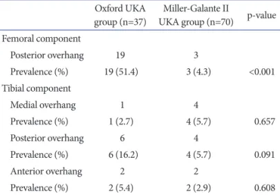
Results: Thirty three of 107 knees (30.8%) had overhang in at least one zone of the femoral or tibial component. In the tibial side, there were no significant differences between the groups in component overhang in each zone. In the femoral side, the Oxford UKA group showed a significantly higher prevalence of the posterior overhang of the femoral component (19/37, 51.4%) than did the M-G II UKA group (3/70, 4.3%; p< 0.001).
However, no significant differences in clinical results were observed between the two groups. There were also no significant differences in clinical results between the overhang and the non-overhang groups.
Conclusions: Posterior overhang of the femoral component was highly prevalent in Oxford UKA patients. However, posterior overhang of the femoral component had no significant relationship with postoperative clinical results in both Oxford and M-G II UKAs at a mean of 48 months follow-up.
Keywords: Knee, Unicompartmental, Arthroplasty, Implant, Overhang pISSN 2234-0726 · eISSN 2234-2451
Knee Surgery & Related Research
Received August 22, 2013; Revised (1st) January 28, 2014;
(2nd) May 11, 2014; Accepted May 15, 2014 Correspondence to: Yong In, MD
Department of Orthopaedic Surgery, Seoul St. Mary’s Hospital, The Catholic University of Korea College of Medicine, 222 Banpo-daero, Seocho-gu, Seoul 137-701, Korea
Tel: +82-2-2258-2838, Fax: +82-2-535-9834 E-mail: [email protected]
Introduction
Although unicompartmental knee arthroplasty (UKA) has been accepted as a reliable procedure for the management of pa- tients with arthritis limited to one compartment of the knee1,2), it is still considered as a technically demanding procedure3). While a good size match between the component and the resected sur-
82
This is an Open Access article distributed under the terms of the Creative Commons Attribution Non-Commercial License (http://creativecommons.org/licenses/by-nc/3.0/) which permits unrestricted non-commercial use, distribution, and reproduction in any medium, provided the original work is properly cited.
Copyright © 2014 KOREAN KNEE SOCIETY www.jksrr.org
The purpose of this study was to compare the prevalence of im- plant overhang between the Oxford and M-G II UKA prostheses and to determine whether overhang was associated with post- operative clinical results. We hypothesized that both prostheses would show similarly low prevalence of implant overhang with successful clinical outcomes.
Materials and Methods
Approval for this retrospective study was obtained from the In- stitutional Review Board of our hospital. Between January 2002 and December 2009, 122 UKAs were performed in 109 patients by a single surgeon for the treatment of medial unicompartmen- tal osteoarthritis of the knee. Forty-six knees were treated with the Oxford phase 3 UKA prosthesis that had a single or twin peg femoral component, and 76 with the M-G II UKA prosthesis. All patients were of Asian ethnicity. The indications for UKA were based on the Kozinn and Scott13) criteria, i.e., medial osteoarthri- tis or medial avascular necrosis, intact anterior cruciate ligament, range of motion larger than 90o, flexion contracture less than 5o, varus deformity less than 10o, and valgus deformity less than 15o. The lateral compartment and patellofemoral joint were well preserved on standing anteroposterior (AP), lateral, and skyline knee radiographs. Patient age was not a contraindication in our study. Four knees (three knees treated with the Oxford UKA prosthesis and one knee treated with the M-G II UKA prosthesis) were revised during the follow-up period and were excluded.
The reasons for failure in the Oxford UKA group were one dis- location of the polyethylene, one femoral component loosening, and one tibial component loosening. The cause of revision in the M-G II UKA group was tibial component loosening. Eleven knees were also excluded due to missing follow-up visits. One hundred and seven knees (37 knees treated with the Oxford UKA prosthesis and 70 knees treated with the M-G II UKA prosthesis) in 95 patients were included in the final radiographic and clini- cal outcome analysis. Among the 37 Oxford UKAs, 30 knees had a single peg femoral component and 7 had a twin peg femoral component. The mean age at operation was 60 years (range, 45 to 75 years) and the mean follow-up period was 48 months (range, 24 to 111 months).
Operation was performed according to the Oxford Unicom- partmental Knee Placement Operating Manual and the M/G Unicompartmental Knee Minimally Invasive Surgical Technique Manual. A minimally invasive technique was used for all knees.
An 8−10 cm medial parapatellar arthrotomy was performed without patellar eversion. The size of the tibial component was
determined by laying metal tibial templates on the excised tibial plateau for both the Oxford and M-G II prostheses. The tibial component should cover the tibial cut surface as much as pos- sible so as to maximize the load distribution. When the tibial component didn’t fit perfectly the resected proximal tibia, we made the maximum amount of effort to fit the anteromedial edge between the implant and the resected proximal tibia. While the size of the femoral component of the Oxford prosthesis was determined using preoperative X-ray templates, the size of the femoral component of the M-G II prosthesis was determined intraoperatively using a femoral cutting guide. The foot of the femoral cutting guide was inserted into the flat surface against the cut distal femoral condyle. A femoral cutting guide having 2−3 mm of the exposed bone above the anterior edge of the guide was considered to be the proper size for M-G II UKA. We inserted the tibial and femoral trial components and finally ensured the size of implant in both the Oxford and M-G II prostheses. The concept and technique for gap balancing were different between the two prostheses. In the Oxford UKA group, gap balancing was achieved by milling the distal femoral bone to obtain an equal extension gap with the flexion gap. However, in the M-G II UKA group, gap balancing was evaluated after bone cutting. A tension gauge with 2 mm thickness was used to ensure that flexion and extension gaps were not too tight in the M-G II UKA group.
Pre- and postoperative tibio-femoral angles were measured on standing knee radiographs. Overhang of the implant was mea- sured in the posterior femoral, anterior tibial, medial tibial, and posterior tibial zones on the AP and lateral knee radiographs.
No reference on criteria for implant overhang after M-G II UKA could be found in the literature. Considering one millimeter of measured error, we defined overhang as a measured distance of larger than 3 mm between the implant and the resected bone edge in any zone6,7,14). Each measurement was performed twice by one independent observer. The average of these measurements was calculated and the prevalence of implant overhang was de- termined. Clinical results were evaluated using the range of knee motion, the Knee Society scores15), and the Western Ontario and McMaster Universities Osteoarthritis Index (WOMAC)16). The knees were divided into two groups according to the type of prosthesis; those treated with the Oxford UKA prosthesis and those with the M-G II UKA prosthesis. The prevalence of im- plant overhang in each location was compared according to the prosthesis type. Clinical results were compared not only between the Oxford UKA and M-G II UKA groups, but also between the overhang group and the non-overhang group at the final follow- up.
The Fisher’s exact test was used to examine the prevalence of overhang of each implant component. The Chi-square test was used to examine categorical demographic data in both groups.
The independent t-test was used to compare continuous demo- graphic variables and clinical results between the groups. Any test was considered statistically significant if the p-value was equal to or less than 0.05. SPSS ver. 13.0 (SPSS Inc., Chicago, IL, USA) was
used to analyze the data.
Results
Of 107 knees, 33 had overhang in at least one location of the femoral or tibial component. The overall prevalence of implant overhang was 30.8%. The prevalence of posterior overhang of the femoral component was significantly higher in the Oxford UKA group than that in the M-G II UKA group (p<0.001) (Table 1). The prevalence of tibial component overhang ranged from 2.7% to 16.2% according to the type of prosthesis and location.
Although the Oxford UKA group showed a higher prevalence of posterior overhang of the tibial component than did the M-G II UKA group, there were no significant differences in the preva- lence of tibial component overhang at each location between the groups (Table 1).
Function as measured by the Knee Society function and Knee score and WOMAC score was compared between the Oxford UKA group and the M-G II UKA group at a minimum follow-up of 24 months (Table 2). Each implant group showed clinical im- provement after operation. Even though the Oxford UKA group showed a significantly higher prevalence of the posterior over- hang of the femoral component than did the M-G II UKA group, there were no significant differences in clinical results between Table 1. Prevalence of Implant Overhang according to the Type of
Prosthesis and Zones
Oxford UKA
group (n=37) Miller-Galante II
UKA group (n=70) p-value Femoral component
Posterior overhang 19 3
Prevalence (%) 19 (51.4) 3 (4.3) <0.001 Tibial component
Medial overhang 1 4
Prevalence (%) 1 (2.7) 4 (5.7) 0.657
Posterior overhang 6 4
Prevalence (%) 6 (16.2) 4 (5.7) 0.091
Anterior overhang 2 2
Prevalence (%) 2 (5.4) 2 (2.9) 0.608
UKA: unicompartmental knee arthroplasty.
Table 2. Comparison between the Oxford UKA Group and the Miller-Galante II UKA Group
Characteristic Oxford UKA group (n=37) Miller-Galante II UKA group (n=70) p-value
Age (yr) 60.3±7.0 (45–71) 60.6±6.9 (49–75) 0.845
Sex (M/F) 5/32 6/64 0.508
BMI (kg/m2) 26.1±3.7 (18.4–36.0) 27.2±3.3 (20.5–34.1) 0.113
Follow-up (mo) 59.3±34.9 (24–110) 43.0±22.1 (24–88) 0.013
Preop TFA (o) Valgus 1.3±2.9 (varus 6.5–valgus 7.4) Valgus 1.5±2.9 (varus 3.4–valgus 10.0) 0.737
Postop TFA (o) Valgus 6.5±3.0 (valgus 1.2–14.5) Valgus 6.3±3.1 (valgus 0.8–14.6) 0.735
Preop ROM (o) 129.1±10.1 (100–140) 127.0±9.6 (105–140) 0.305
Postop ROM (o) 130.3±6.3 (120–140) 132.6±6.2 (120–145) 0.074
Preop Knee Society Knee score 50.0±8.1 (38–70) 49.2±6.3 (39–66) 0.602
Function score 55.8±10.1 (40–80) 51.2±8.3 (20–70) 0.021
Postop Knee Society Knee score 89.5±3.9 (80–95) 89.6±3.6 (82–95) 0.952
Pain score 45.1±3.6 (40–50) 45.7±3.1 (40–50) 0.389
Function score 81.6±5.4 (70–90) 81.6±5.6 (60–90) 0.964
Preop WOMAC score 59.5±9.5 (46.7–86.7) 61.8±6.9 (46.3–82.5) 0.197
Postop WOMAC score 11.9±4.8 (7.5–36.3) 12.2±3.0 (7.1–26.3) 0.726
Values are presented as mean±standard deviation (range).
UKA: unicompartmental knee arthroplasty, BMI: body mass index, Preop: preoperative, TFA: tibio-femoral angle, Postop: postoperative, ROM:
range of motion, WOMAC score: Western Ontario and McMaster Osteoarthritis score.
the two groups.
We compared patients with overhang in at least one location of the femoral or tibial component with patients without overhang.
Table 3 shows the pre- and postoperative clinical data of the over- hang group and non-overhang group regardless of the prosthesis type. At a mean follow-up of 48 months, both overhang group and non-overhang group showed improvements in knee align- ment and clinical outcomes. There were no significant differences in clinical results between the overhang group and non-overhang group.
Discussion
This study revealed the overall prevalence of implant overhang after UKA. The majority of implant overhang occurred in the posterior part of the femoral component of the Oxford UKA prosthesis. Even with the high prevalence of posterior overhang of the femoral component in our UKA study population, we did not find any significant correlation between posterior overhang of the femoral component and clinical results regardless of the presence of overhang or implant type. Mahoney and Kinsey8) re- ported that overhang of the femoral component was highly prev- alent in TKA, and overhang of ≥3 mm in at least one zone was associated with a 90% increase in the odds of clinically important
knee pain at 2 years after surgery. Clarius et al.7) analyzed implant position of the Oxford UKA prosthesis according to the Oxford X-ray rating system. They reported that 17 of 56 knees (30.4%) were overhanging 3 mm or more in the femoral component and it was not correlated with the postoperative Oxford knee score and pain. After UKA, there was no mediolateral overhang of the femoral component because of its narrow shape. We believe that the posterior compartment of the medial side of the knee was forgiving of soft tissue irritation and pain.
Among cases of femoral or tibial overhang, medial tibial over- hang has been considered a problem. Chau et al.6) reported that surgeons must avoid medial tibial overhang of 3 mm or more, as this severely compromised the outcome and might cause ir- ritation of soft tissue and pain. In an in vitro study by Gudena et al.17), they demonstrated that medial collateral ligament load almost doubled from overhang of 2 to 4 mm. When compared with the results of a study by Chau et al.6) which demonstrated a 9% prevalence of medial overhang of the tibial component, the prevalence of medial overhang of the tibial component in our study population was not that high (2.7% in the Oxford UKA group and 5.7% in the M-G II UKA group). With low prevalence of medial overhang of the tibial component, the effect of femoral or tibial component overhang on clinical outcomes was negligible in both the Oxford and M-G II UKA groups. It is believed that Table 3. Comparison between Overhang Group and Non-Overhang Group
Characteristic Overhang group (n=33) Non-overhang group (n=74) p-value
Age (yr) 59.0±6.2 (49–70) 61.2±7.1 (45–75) 0.133
Sex (M/F) 4/29 7/67 0.735
BMI (kg/m2) 26.3±3.5 (18.4–32.3) 27.0±3.5 (20.5–36.0) 0.357
Follow-up (mo) 48.4±28.9 (24–104) 48.8±28.0 (24–111) 0.946
Preop TFA (o) Valgus 0.9±2.9 (varus 6.5–valgus 8.0) Valgus 1.7±2.8 (varus 4.5–valgus 10.0) 0.152
Postop TFA (o) Valgus 6.5±3.7 (valgus 1.0–14.6) Valgus 6.3±2.7 (valgus 0.8–12.7) 0.826
Preop ROM (o) 127.4±10.5 (100–140) 127.8±9.5 (105–140) 0.842
Postop ROM (o) 130.2±6.7 (120–140) 132.5±6.1 (120–145) 0.077
Preop Knee Society Knee score 49.6±8.8 (38–70) 49.4±6.0 (39–64) 0.939
Function score 53.6±8.2 (40–70) 52.4±9.6 (20–80) 0.533
Postop Knee Society Knee score 89.0±3.9 (80–95) 89.8±3.5 (82–95) 0.311
Pain score 44.7±3.7 (40–50) 45.8±3.0 (40–50) 0.086
Function score 82.3±5.7 (70–90) 81.3±5.4 (60–90) 0.391
Preop WOMAC score 59.3±9.2 (46.7–86.7) 61.8±7.2 (46.3–82.5) 0.137
Postop WOMAC score 12.3±5.1 (7.1–36.2) 12.0±2.9 (7.5–26.3) 0.745
Values are presented as mean±standard deviation (range).
BMI: body mass index, Preop: preoperative, TFA: tibio-femoral angle, Postop: postoperative, ROM: range of motion, WOMAC score: Western Ontario and McMaster Osteoarthritis score.
the tibial sizing method using the metal template during surgery was reliable and the shapes and sizes of the tibial components of both the Oxford and M-G II implants were appropriate.
We speculated the reasons for the high prevalence of posterior overhang of the femoral component of the Oxford UKA pros- thesis as follows. First, preoperative X-ray templating for femoral sizing was inaccurate. Bothra et al.18) reported that the use of X- ray template systems in the Oxford UKA lacked reliability. They indicated that variability in prosthetic sizing might be attributable to the difficulty in distinguishing the medial from the lateral fem- oral condyle on lateral radiographs. Second, the medial femoral condyle of a small Asian female patient was smaller than the X- small femoral component of the Oxford UKA prosthesis. Among 19 knees which showed posterior overhang of the femoral component of the Oxford UKA system, 6 knees were implanted with the X-small femoral component (Fig. 1). The M-G II UKA prosthesis offers seven different femoral AP sizing options, and the Oxford UKA prosthesis has five different radii of curvature.
These differences could allow for a limited number of options in choosing the appropriate size of the femoral component for the Oxford UKA. Third, subsequent milling of the femoral condyle during the Oxford UKA in order to balance the flexion-extension gap might have affected posterior overhang of the femoral com- ponent (Fig. 2). Subsequent milling to increase the extension gap resulted in proximal positioning of the femoral component.
During the surgical procedure, it was difficult to identify whether the femoral component was overhanging posteriorly or not. As a technical tip, we recommend that adequate amount of proxi-
mal tibial cut should be performed to decrease subsequent distal femoral milling.
Appropriate implant size matching could be one of the impor- tant factors for a successful outcome of UKA19). To the best of our knowledge, no study has compared the prevalence of implant overhang between Oxford and M-G II UKA. In our study, im- plant overhang had no relation with postoperative clinical results at a mean of 48-month follow-up. In this sense, long-term follow- up studies are needed and we suggest some recommendations to prevent implant overhang. The femoral and tibial components should be flush with all edges of the resected bone as much as possible5,10). If the tibial cut is done as a single piece, this can be helpful for tibial sizing. In cases where the tibial component does not fit perfectly the resected bone, the anteromedial edge should be observed directly between the tibial component and the re- sected proximal tibia to prevent medial overhang of the tibial component. Sometimes, the patient’s height and gender can be helpful to estimate the size of the component20). Also, it may be possible to determine the size of the component from templat- ing using pre-operative radiographs. During UKA procedure, perioperative fluoroscopy may be helpful to determine the size of implant and to prevent implant overhang.
The present study has limitations. First, it was a retrospective study and the number of cases of overhang in each location was too small to provide enough statistical power to the study. In ad- dition, this study lacks data on implant underhang. If we had a larger number of patients in the cohort, patients could have been Fig. 2. Lateral knee radiograph showing posterior overhang of the femo- ral component of the Oxford knee system. During operation, subsequent femoral milling was required in order to balance the flexion and exten- sion gap and a 9 mm bearing was inserted.
Fig. 1. Lateral knee radiograph showing posterior overhang of the femoral and tibial components of the Oxford knee system (Femur size:
X-small, Tibia size: B).
divided into 3 groups; proper fit group, overhang group, and un- derhang group. Despite these limitations, our study population consisted of patients who were operated on by a single surgeon with a mean of 48 months follow-up. Second, common post- operative complaint was of persistent medial knee discomfort.
It could have influenced the clinical results, but it was not clear whether the component overhang caused the pain or tenderness of knee joint7,11). Many other factors could have contributed to the postoperative pain.
Conclusions
In conclusion, posterior overhang of the femoral component was highly prevalent in the Oxford UKA patients. However, the posterior overhang of the femoral component had no significant relationship with postoperative clinical results in both Oxford III and M-G II UKAs at a mean of 48 months follow-up.
Conflict of Interest
No potential conflict of interest relevant to this article was re- ported.
References
1. Naudie D, Guerin J, Parker DA, Bourne RB, Rorabeck CH.
Medial unicompartmental knee arthroplasty with the Miller- Galante prosthesis. J Bone Joint Surg Am. 2004;86:1931-5.
2. Price AJ, Svard U. A second decade lifetable survival analysis of the Oxford unicompartmental knee arthroplasty. Clin Or- thop Relat Res. 2011;469:174-9.
3. Robertsson O, Knutson K, Lewold S, Lidgren L. The routine of surgical management reduces failure after unicompart- mental knee arthroplasty. J Bone Joint Surg Br. 2001;83:45-9.
4. Hitt K, Shurman JR 2nd, Greene K, McCarthy J, Moskal J, Hoeman T, Mont MA. Anthropometric measurements of the human knee: correlation to the sizing of current knee arthroplasty systems. J Bone Joint Surg Am. 2003;85 Suppl 4:115-22.
5. Berger RA, Della Valle CJ. Unicompartmental knee arthro- plasty: indications, techniques, and results. Instr Course Lect. 2010;59:47-56.
6. Chau R, Gulati A, Pandit H, Beard DJ, Price AJ, Dodd CA, Gill HS, Murray DW. Tibial component overhang following unicompartmental knee replacement: does it matter? Knee.
2009;16:310-3.
7. Clarius M, Hauck C, Seeger JB, Pritsch M, Merle C, Aldinger PR. Correlation of positioning and clinical results in Oxford UKA. Int Orthop. 2010;34:1145-51.
8. Mahoney OM, Kinsey T. Overhang of the femoral compo- nent in total knee arthroplasty: risk factors and clinical con- sequences. J Bone Joint Surg Am. 2010;92:1115-21.
9. Bonnin MP, Schmidt A, Basiglini L, Bossard N, Dantony E. Mediolateral oversizing influences pain, function, and flexion after TKA. Knee Surg Sports Traumatol Arthrosc.
2013;21:2314-24.
10. Borus T, Thornhill T. Unicompartmental knee arthroplasty.
J Am Acad Orthop Surg. 2008;16:9-18.
11. Edmondson MC, Isaac D, Wijeratna M, Brink S, Gibb P, Skinner P. Oxford unicompartmental knee arthroplasty:
medial pain and functional outcome in the medium term. J Orthop Surg Res. 2011;6:52.
12. Scott RD, Santore RF. Unicondylar unicompartmental re- placement for osteoarthritis of the knee. J Bone Joint Surg Am. 1981;63:536-44.
13. Kozinn SC, Scott R. Unicondylar knee arthroplasty. J Bone Joint Surg Am. 1989;71:145-50.
14. Kim JG, Kasat NS, Bae JH, Kim SJ, Oh SM, Lim HC. The radiological parameters correlated with the alignment of the femoral component after Oxford phase 3 unicompartmental knee replacement. J Bone Joint Surg Br. 2012;94:1499-505.
15. Insall JN, Dorr LD, Scott RD, Scott WN. Rationale of the Knee Society clinical rating system. Clin Orthop Relat Res.
1989;(248):13-4.
16. Bellamy N, Buchanan WW, Goldsmith CH, Campbell J, Stitt LW. Validation study of WOMAC: a health status instrument for measuring clinically important patient relevant outcomes to antirheumatic drug therapy in patients with osteoarthritis of the hip or knee. J Rheumatol. 1988;15:1833-40.
17. Gudena R, Pilambaraei MA, Werle J, Shrive NG, Frank CB.
A safe overhang limit for unicompartmental knee arthro- plasties based on medial collateral ligament strains: an in vitro study. J Arthroplasty. 2013;28:227-33.
18. Bothra V, Lemon G, Lang D, Smith DM, Ali AM. Reliability of templating in estimating the size of uni-condylar knee ar- throplasty. J Arthroplasty. 2003;18:780-3.
19. Whiteside LA. Making your next unicompartmental knee arthroplasty last: three keys to success. J Arthroplasty. 2005;
20(4 Suppl 2):2-3.
20. Fawzy E, Pandit H, Jenkins C, Dodd CA, Murray DW. De- termination of femoral component size in unicompartmen- tal knee replacement. Knee. 2008;15:403-6.